

Ex vivo T2 relaxation: associations with age-related neuropathology and cognition
- PMID: 24582637
- PMCID: PMC3989898
- DOI: 10.1016/j.neurobiolaging.2014.01.144
Ex vivo T2 relaxation: associations with age-related neuropathology and cognition
Abstract
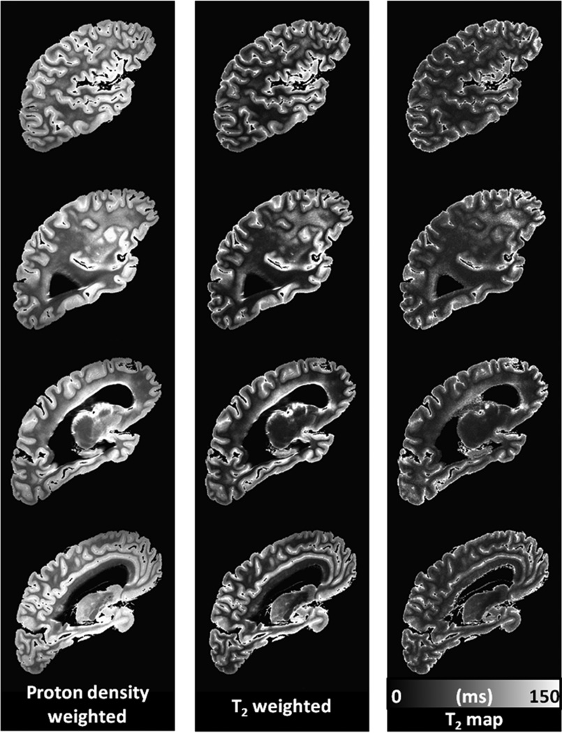
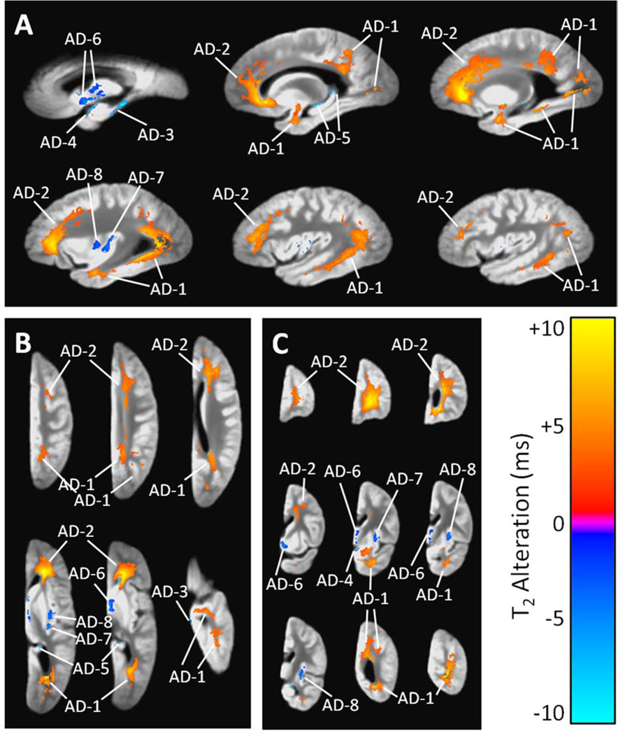
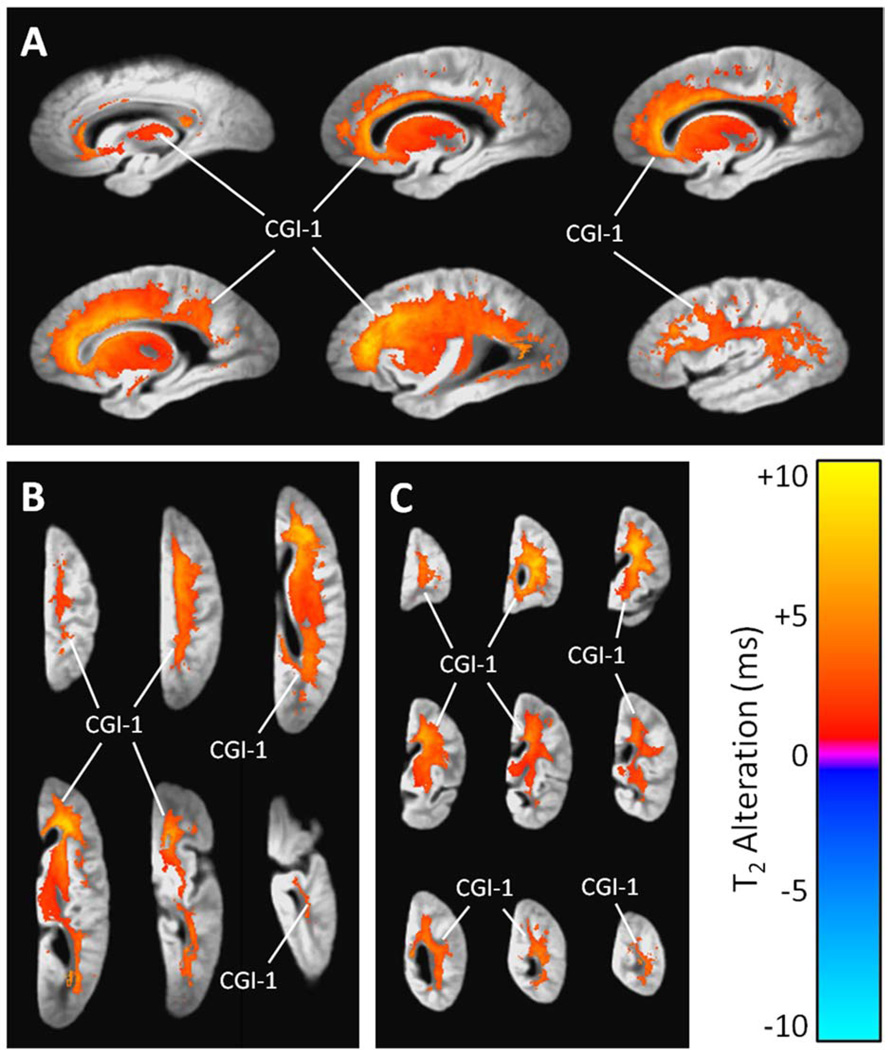
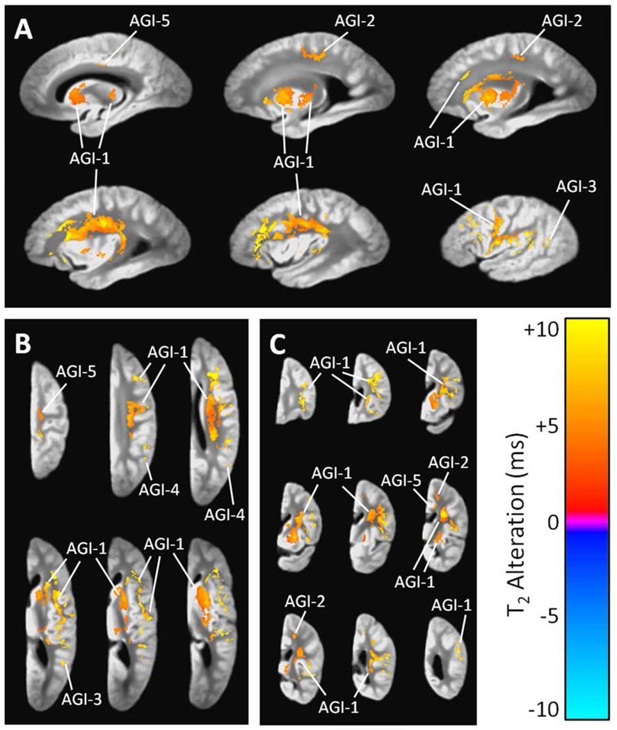
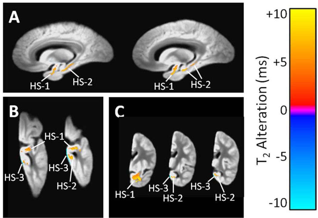
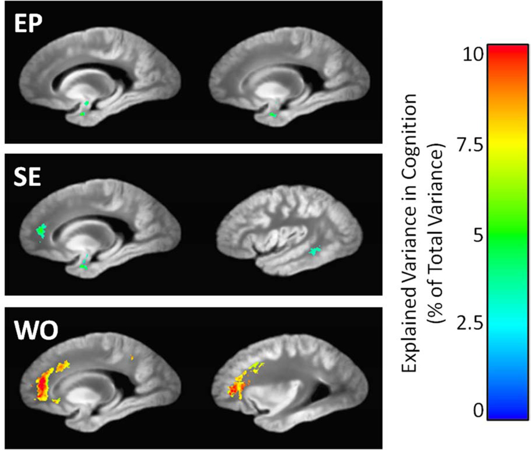
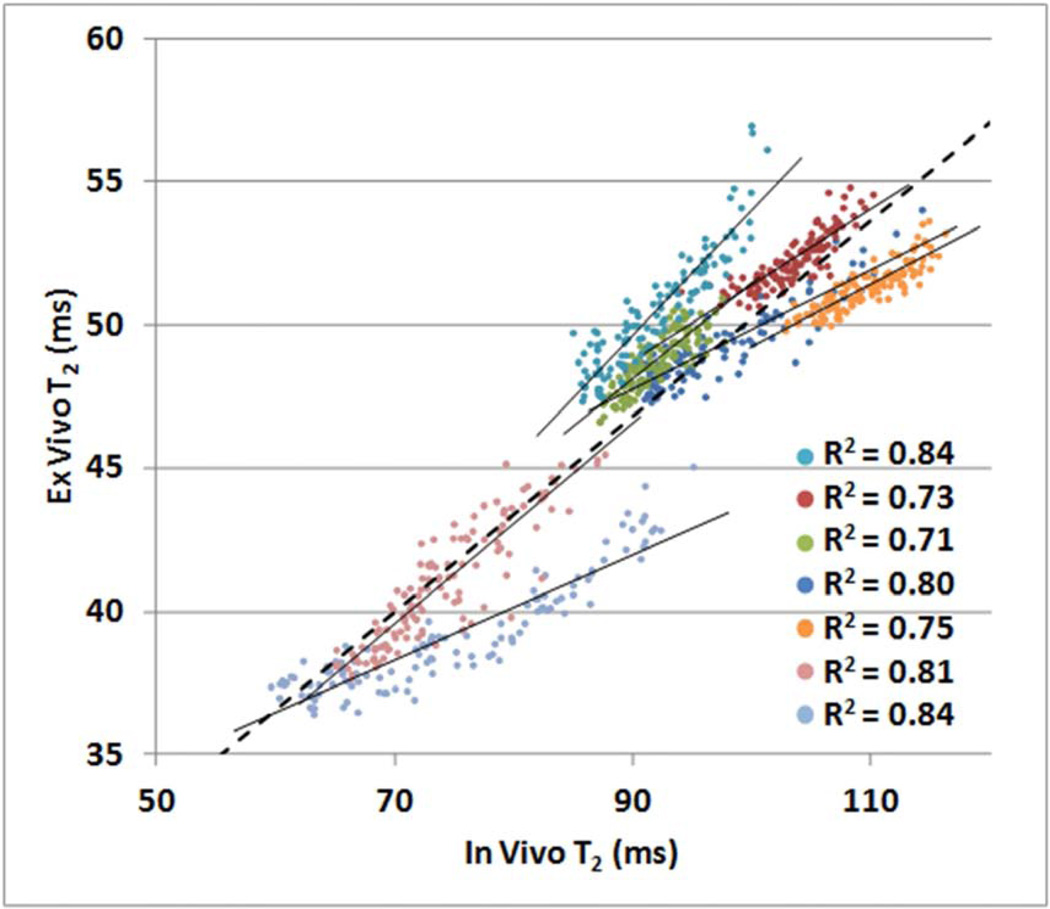
The transverse relaxation time constant, T(2), is sensitive to brain tissue's free water content and the presence of paramagnetic materials such as iron. In this study, ex vivo magnetic resonance imaging was used to investigate alterations in T(2) related to Alzheimer's disease (AD) pathology and other types of neuropathology common in old age, as well as the relationship between T(2) alterations and cognition. Cerebral hemispheres were obtained from 371 deceased older adults. Using fast spin-echo imaging with multiple echo times, T(2) maps were produced and warped to a study-specific template. Hemispheres underwent neuropathologic examination for identification of AD pathology and other common age-related neuropathologies. Voxelwise linear regression was carried out to detect regions of pathology-related T(2) alterations and, in separate analyses, regions in which T(2) alterations were linked to antemortem cognitive performance. AD pathology was associated with T(2) prolongation in white matter of all lobes and T(2) shortening in the basal ganglia and insula. Gross infarcts were associated with T(2) prolongation in white matter of all lobes, and in the thalamus and basal ganglia. Hippocampal sclerosis was associated with T(2) prolongation in the hippocampus and white matter of the temporal lobe. After controlling for neuropathology, T(2) prolongation in the frontal lobe white matter was associated with lower performance in the episodic, semantic, and working memory domains. In addition, voxelwise analysis of in vivo and ex vivo T(2) values indicated a positive relationship between the two, though further investigation is necessary to accurately translate findings of the present study to the in vivo case.
Keywords: Cognition; Gross infarct; Hippocampal sclerosis; MRI; Neuroimaging; Voxelwise.
Copyright © 2014 Elsevier Inc. All rights reserved.
Figures
Similar articles
-
Neuropathologic correlates of regional brain volumes in a community cohort of older adults.Neurobiol Aging. 2015 Oct;36(10):2798-805. doi: 10.1016/j.neurobiolaging.2015.06.025. Epub 2015 Jun 30. Neurobiol Aging. 2015. PMID: 26195068 Free PMC article.
-
Ex vivo MRI transverse relaxation in community based older persons with and without Alzheimer's dementia.Behav Brain Res. 2017 Mar 30;322(Pt B):233-240. doi: 10.1016/j.bbr.2016.09.001. Epub 2016 Sep 3. Behav Brain Res. 2017. PMID: 27596378 Free PMC article.
-
Postmortem MRI: a novel window into the neurobiology of late life cognitive decline.Neurobiol Aging. 2016 Sep;45:169-177. doi: 10.1016/j.neurobiolaging.2016.05.023. Epub 2016 Jun 6. Neurobiol Aging. 2016. PMID: 27459937 Free PMC article.
-
Relation of neuropathology to cognition in persons without cognitive impairment.Ann Neurol. 2012 Oct;72(4):599-609. doi: 10.1002/ana.23654. Ann Neurol. 2012. PMID: 23109154 Free PMC article.
-
Prolongation of magnetic resonance T2 time in hippocampus of human patients marks the presence and severity of Alzheimer's disease.Neurosci Lett. 1992 Jan 6;134(2):187-90. doi: 10.1016/0304-3940(92)90513-7. Neurosci Lett. 1992. PMID: 1589144
Cited by
-
Quantitative magnetic resonance imaging indicates brain tissue alterations in patients after liver transplantation.PLoS One. 2019 Sep 25;14(9):e0222934. doi: 10.1371/journal.pone.0222934. eCollection 2019. PLoS One. 2019. PMID: 31553760 Free PMC article.
-
Inferring protein expression changes from mRNA in Alzheimer's dementia using deep neural networks.Nat Commun. 2022 Feb 3;13(1):655. doi: 10.1038/s41467-022-28280-1. Nat Commun. 2022. PMID: 35115553 Free PMC article.
-
Neuropathologic correlates of regional brain volumes in a community cohort of older adults.Neurobiol Aging. 2015 Oct;36(10):2798-805. doi: 10.1016/j.neurobiolaging.2015.06.025. Epub 2015 Jun 30. Neurobiol Aging. 2015. PMID: 26195068 Free PMC article.
-
Associations of amygdala volume and shape with transactive response DNA-binding protein 43 (TDP-43) pathology in a community cohort of older adults.Neurobiol Aging. 2019 May;77:104-111. doi: 10.1016/j.neurobiolaging.2019.01.022. Epub 2019 Jan 31. Neurobiol Aging. 2019. PMID: 30784812 Free PMC article.
-
Shared proteomic effects of cerebral atherosclerosis and Alzheimer's disease on the human brain.Nat Neurosci. 2020 Jun;23(6):696-700. doi: 10.1038/s41593-020-0635-5. Epub 2020 May 18. Nat Neurosci. 2020. PMID: 32424284 Free PMC article.
References
-
- Ardekani BA, Guckemus S, Bachman A, Hoptman MJ, Wojtaszek M, Nierenberg J. Quantitative comparison of algorithms for inter-subject registration of 3D volumetric brain MRI scans. J. Neurosci. Methods. 2005;142:67–76. - PubMed
-
- Arfanakis K, Gui M, Tamhane A, Carew J. Investigating the Medial Temporal Lobe in Alzheimer’s Disease and Mild Cognitive Impairment, with Turboprop Diffusion Tensor Imaging, MRI-volumetry, and T2-relaxometry. Brain Imaging Behav. 2007;1:11–21.
-
- Auriel E, Bornstein NM, Berenyi E, Varkonyi I, Gabor M, Majtenyi K, Szepesi R, Goldberg I, Lampe R, Csiba L. Clinical, radiological and pathological correlates of leukoaraiosis. Acta Neurol. Scand. 2011;123:41–47. - PubMed
-
- Bartzokis G, Cummings JL, Sultzer D, Henderson VW, Nuechterlein KH, Mintz J. White matter structural integrity in healthy aging adults and patients with Alzheimer disease: a magnetic resonance imaging study. Arch. Neurol. 2003;60:393–398. - PubMed
Publication types
MeSH terms
Grants and funding
LinkOut - more resources
Full Text Sources
Other Literature Sources
Medical
