Acute chlorine gas exposure produces transient inflammation and a progressive alteration in surfactant composition with accompanying mechanical dysfunction
- PMID: 24582687
- PMCID: PMC4361901
- DOI: 10.1016/j.taap.2014.02.006
Acute chlorine gas exposure produces transient inflammation and a progressive alteration in surfactant composition with accompanying mechanical dysfunction
Abstract
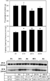
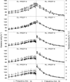
Acute Cl2 exposure following industrial accidents or military/terrorist activity causes pulmonary injury and severe acute respiratory distress. Prior studies suggest that antioxidant depletion is important in producing dysfunction, however a pathophysiologic mechanism has not been elucidated. We propose that acute Cl2 inhalation leads to oxidative modification of lung lining fluid, producing surfactant inactivation, inflammation and mechanical respiratory dysfunction at the organ level. C57BL/6J mice underwent whole-body exposure to an effective 60ppm-hour Cl2 dose, and were euthanized 3, 24 and 48h later. Whereas pulmonary architecture and endothelial barrier function were preserved, transient neutrophilia, peaking at 24h, was noted. Increased expression of ARG1, CCL2, RETLNA, IL-1b, and PTGS2 genes was observed in bronchoalveolar lavage (BAL) cells with peak change in all genes at 24h. Cl2 exposure had no effect on NOS2 mRNA or iNOS protein expression, nor on BAL NO3(-) or NO2(-). Expression of the alternative macrophage activation markers, Relm-α and mannose receptor was increased in alveolar macrophages and pulmonary epithelium. Capillary surfactometry demonstrated impaired surfactant function, and altered BAL phospholipid and surfactant protein content following exposure. Organ level respiratory function was assessed by forced oscillation technique at 5 end expiratory pressures. Cl2 exposure had no significant effect on either airway or tissue resistance. Pulmonary elastance was elevated with time following exposure and demonstrated PEEP refractory derecruitment at 48h, despite waning inflammation. These data support a role for surfactant inactivation as a physiologic mechanism underlying respiratory dysfunction following Cl2 inhalation.
Keywords: Alternative activation; Bronchoalveolar lavage; Nitric oxide; Pulmonary mechanics; Respiratory impedance.
Copyright © 2014 Elsevier Inc. All rights reserved.
Figures


Similar articles
-
Lung injury and oxidative stress induced by inhaled chlorine in mice is associated with proinflammatory activation of macrophages and altered bioenergetics.Toxicol Appl Pharmacol. 2023 Feb 15;461:116388. doi: 10.1016/j.taap.2023.116388. Epub 2023 Jan 20. Toxicol Appl Pharmacol. 2023. PMID: 36690086 Free PMC article.
-
Prolonged injury and altered lung function after ozone inhalation in mice with chronic lung inflammation.Am J Respir Cell Mol Biol. 2012 Dec;47(6):776-83. doi: 10.1165/rcmb.2011-0433OC. Epub 2012 Aug 9. Am J Respir Cell Mol Biol. 2012. PMID: 22878412 Free PMC article.
-
Acute respiratory changes and pulmonary inflammation involving a pathway of TGF-β1 induction in a rat model of chlorine-induced lung injury.Toxicol Appl Pharmacol. 2016 Oct 15;309:44-54. doi: 10.1016/j.taap.2016.08.027. Epub 2016 Aug 30. Toxicol Appl Pharmacol. 2016. PMID: 27586366
-
Inflammatory mechanisms of pulmonary injury induced by mustards.Toxicol Lett. 2016 Feb 26;244:2-7. doi: 10.1016/j.toxlet.2015.10.011. Epub 2015 Oct 23. Toxicol Lett. 2016. PMID: 26478570 Free PMC article. Review.
-
The guardians of pulmonary harmony: alveolar macrophages orchestrating the symphony of lung inflammation and tissue homeostasis.Eur Respir Rev. 2024 May 29;33(172):230263. doi: 10.1183/16000617.0263-2023. Print 2024 Apr 30. Eur Respir Rev. 2024. PMID: 38811033 Free PMC article. Review.
Cited by
-
Bronchoscopic lung lavage and exogenous surfactant successfully reverse respiratory failure after severe chlorine exposure: A pediatric case report.Clin Case Rep. 2025 Jan 14;13(1):e9302. doi: 10.1002/ccr3.9302. eCollection 2025 Jan. Clin Case Rep. 2025. PMID: 39810993 Free PMC article.
-
Exposure to Sodium Hypochlorite or Cigarette Smoke Induces Lung Injury and Mechanical Impairment in Wistar Rats.Inflammation. 2022 Aug;45(4):1464-1483. doi: 10.1007/s10753-022-01625-0. Epub 2022 May 2. Inflammation. 2022. PMID: 35501465
-
Toxic effects of chlorine gas and potential treatments: a literature review.Toxicol Mech Methods. 2021 May;31(4):244-256. doi: 10.1080/15376516.2019.1669244. Epub 2019 Oct 1. Toxicol Mech Methods. 2021. PMID: 31532270 Free PMC article. Review.
-
Disease-modifying treatment of chemical threat agent-induced acute lung injury.Ann N Y Acad Sci. 2020 Nov;1480(1):14-29. doi: 10.1111/nyas.14438. Epub 2020 Jul 29. Ann N Y Acad Sci. 2020. PMID: 32726497 Free PMC article. Review.
-
Development of a clinical assay to measure chlorinated tyrosine in hair and tissue samples using a mouse chlorine inhalation exposure model.Anal Bioanal Chem. 2021 Mar;413(6):1765-1776. doi: 10.1007/s00216-020-03146-x. Epub 2021 Jan 28. Anal Bioanal Chem. 2021. PMID: 33511457 Free PMC article.
References
-
- Allen GB, Leclair T, Cloutier M, Thompson-Figueroa J, Bates JHT. The response to recruitment worsens with progression of lung injury and fibrin accumulation in a mouse model of acid aspiration. AJP - Lung Physiol. 2007;292:L1580–L1589. - PubMed
-
- Amin SD, Majumdar A, Alkana P, Walkey AJ, O’Connor GT, Suki B. Modeling the effects of stretch-dependent surfactant secretion on lung recruitment during variable ventilation. J Biomed Sci and Eng. 2013;6:61–70.
-
- Ban M, Hettich D. Effect of Th2 cytokine antagonist treatments on chemical-induced allergic response in mice. J. Appl. Toxicol. 2005;25:239–247. - PubMed
Publication types
MeSH terms
Substances
Grants and funding
- R01 CA132624/CA/NCI NIH HHS/United States
- R01 HL074115/HL/NHLBI NIH HHS/United States
- AR055073/AR/NIAMS NIH HHS/United States
- T32 ES007148/ES/NIEHS NIH HHS/United States
- HL086621/HL/NHLBI NIH HHS/United States
- P30 ES005022/ES/NIEHS NIH HHS/United States
- ES007148/ES/NIEHS NIH HHS/United States
- ES0050022E/ES/NIEHS NIH HHS/United States
- U54 AR055073/AR/NIAMS NIH HHS/United States
- ES005022/ES/NIEHS NIH HHS/United States
- HL074115/HL/NHLBI NIH HHS/United States
- R01 ES004738/ES/NIEHS NIH HHS/United States
- CA132624/CA/NCI NIH HHS/United States
- R01 HL086621/HL/NHLBI NIH HHS/United States
LinkOut - more resources
Full Text Sources
Other Literature Sources
Medical
Research Materials
Miscellaneous

