Review
. 2014 Jul;115(1):52-67.
doi: 10.1016/j.pbiomolbio.2014.02.006. Epub 2014 Feb 28.

Functions of crystallins in and out of lens: roles in elongated and post-mitotic cells

Affiliations
Review

Functions of crystallins in and out of lens: roles in elongated and post-mitotic cells

Christine Slingsby et al. Prog Biophys Mol Biol. 2014 Jul.

Abstract

The vertebrate lens evolved to collect light and focus it onto the retina. In development, the lens grows through massive elongation of epithelial cells possibly recapitulating the evolutionary origins of the lens. The refractive index of the lens is largely dependent on high concentrations of soluble proteins called crystallins. All vertebrate lenses share a common set of crystallins from two superfamilies (although other lineage specific crystallins exist). The α-crystallins are small heat shock proteins while the β- and γ-crystallins belong to a superfamily that contains structural proteins of uncertain function. The crystallins are expressed at very high levels in lens but are also found at lower levels in other cells, particularly in retina and brain. All these proteins have plausible connections to maintenance of cytoplasmic order and chaperoning of the complex molecular machines involved in the architecture and function of cells, particularly elongated and post-mitotic cells. They may represent a suite of proteins that help maintain homeostasis in such cells that are at risk from stress or from the accumulated insults of aging.

Keywords: Chaperone; Crystallins; Epithelial cell; Lens; Retina; Stress.


Figures

Figure 1
Figure 1. Lens formation is associated with cell elongation
A: Steps in the embryonic development of the vertebrate lens. (Adapted from (Wistow, 1993))
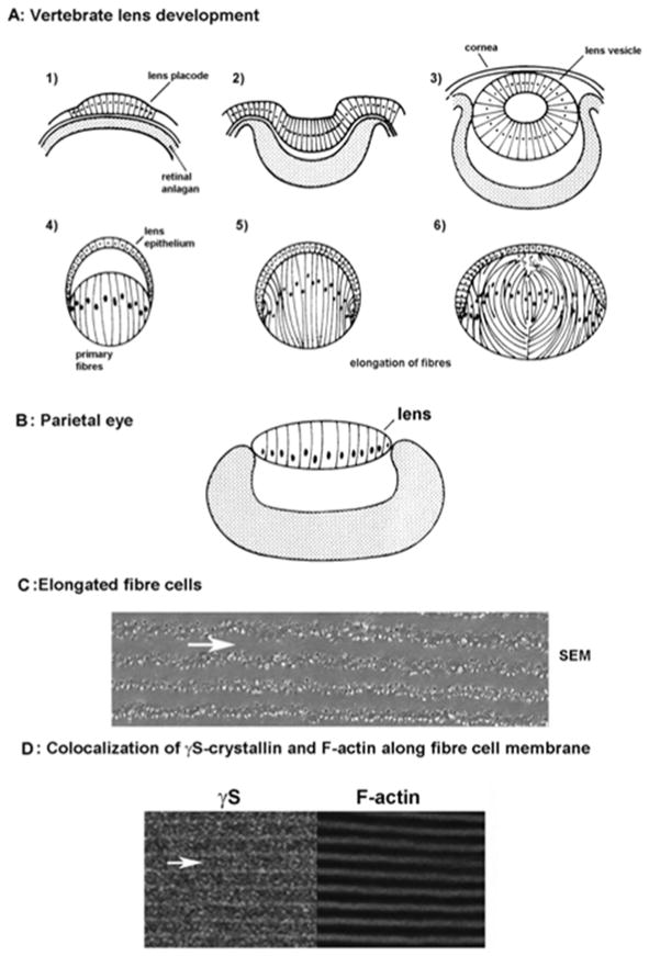
  1. Epithelial cells overlying the retinal anlagen elongate to form the lens placode

  2. Elongation continues as the optic vesicle invaginates.

  3. Formation of the lens vesicle and cornea

  4. Within the lens vesicle, primary fiber cells elongate

  5. Fibres fill the lens

  6. New layers of secondary fibres form throughout life through differentiation and elongation of equatorial epithelial cells.

B: Schematic structure of the lizard parietal eye (adapted from (Eakin, 1973)). A monolayer of elongated cells forms the simple cellular lens. C: Elongation and intercalation of mature fiber cells. Scanning electron micrograph of a fiber cells from adult mouse lens. Arrow indicates the longitudinal axis of a fiber cell. Extensive cell-cell contacts are formed through intercalation of complex junctions. (For methods, see (Fan et al., 2012)) D: Co-localization of γS-crystallin and F-actin along the plasma membranes of mature mouse fiber cells (adapted from (Fan et al., 2012)).
Figure 2
Figure 2. Modular assembly of β- and γ-crystallins from Greek key motifs and a “paired domain” interface
The two columns on the left show structures of the β- and γ-crystallins currently in the PDB Protein Data Bank. The two columns on the right show calculated dimer structures of the β-crystallins, with their PDB identifiers, along with their arrangements within the crystal lattices. A γ-crystallin monomer has four Greek key motifs that form two similar domains organized about a pseudo twofold axis. The first motif of each domain is shown in dark blue and the second motif is in cyan. The cyan motifs provide the conserved tyrosine and tryptophan corners, as well as hydrophobic residues that form the “paired domain” interface as well as some conserved side chains including glutamines. γ-crystallin is monomeric because the “paired domain” interface is intra-chain. The same interface is present in all the resolved β-crystallins. βB2-crystallin is different as the linker is extended so the “paired domain” interface forms between two different chains which creates a domain swapped dimer. In βB1, βB3 and βA4-crystallin, the subunit domains are paired as in γ-crystallins, and so these dimers need an additional interface. This new interface is the same as that observed in the βB2 tetramer formed in the crystal lattice. The third column shows the calculated biological assemblies as calculated by PISA (Krissinel and Henrick, 2007). The fourth column shows the location in the crystal lattice of the conserved dimers highlighted in blue from the surrounding grey crystal lattice. In the case of βB3-crystallin, the calculated assembly unit is a trimer, but the conserved dimer interface is formed in the crystal lattice. This figure is adapted from Figure 5 and SFigure 1 from Slingsby et al (Slingsby et al., 2013).
Figure 3
Figure 3. Changes to side chains that perturb assembly interactions
Four domains are shown from βB2 tetramer, with the “paired domains” painted in uniform colors. In a domain swapped dimer, the “paired domains” come from different chains, but are from the same chain in other β-crystallin homodimers and in γ-crystallins. In βB2 tetramer (shown) the four domains at the dimer-dimer interface are also from different chains, whereas in other β-crystallin homodimers, they come from two chains. Two glutamines colored in CPK are appended to the paired N- and C-terminal lavender domains showing their side-by-side location close to the local 2-fold between the domains. The side chain topologically equivalent to Ser129 in βB1 is shown (misty rose) appended to both C-terminal domains showing that a single mutation will clash with itself at this interface in a homo-oligomer.

References

    1. Acunzo J, Katsogiannou M, Rocchi P. Small heat shock proteins HSP27 (HspB1), alphaB-crystallin (HspB5) and HSP22 (HspB8) as regulators of cell death. Int J Biochem Cell Biol. 2012;44:1622–31. - PubMed
    1. Anderson DH, Stern WH, Fisher SK, Erickson PA, Borgula GA. The onset of pigment epithelial proliferation after retinal detachment. Invest Ophthalmol Vis Sci. 1981;21:10–6. - PubMed
    1. Andley UP. Crystallins in the eye: Function and pathology. Prog Retin Eye Res. 2007;26:78–98. - PubMed
    1. Aquilina JA, Benesch JL, Bateman OA, Slingsby C, Robinson CV. Polydispersity of a mammalian chaperone: mass spectrometry reveals the population of oligomers in alphaB-crystallin. Proc Natl Acad Sci U S A. 2003;100:10611–6. - PMC - PubMed
    1. Arrigo AP. Human small heat shock proteins: protein interactomes of homo- and hetero-oligomeric complexes: an update. FEBS Lett. 2013;587:1959–69. - PubMed

Publication types

LinkOut - more resources