. 2014 May;50(5):468-77.
doi: 10.1016/j.oraloncology.2014.02.004. Epub 2014 Feb 28.

Inhibitors of NF-kappaB reverse cellular invasion and target gene upregulation in an experimental model of aggressive oral squamous cell carcinoma

Affiliations

Inhibitors of NF-kappaB reverse cellular invasion and target gene upregulation in an experimental model of aggressive oral squamous cell carcinoma

Jeff Johnson et al. Oral Oncol. 2014 May.

Abstract

Background: Oral squamous cell carcinoma (OSCC) is diagnosed in 640,000 patients yearly with a poor (50%) 5-year survival rate that has not changed appreciably in decades.

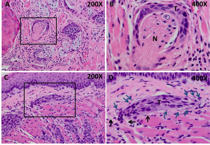
Paitents and methods: To investigate molecular changes that drive OSCC progression, cDNA microarray analysis was performed using human OSCC cells that form aggressive poorly differentiated tumors (SCC25-PD) in a murine orthotopic xenograft model compared to cells that produce well-differentiated tumors (SCC25-WD).

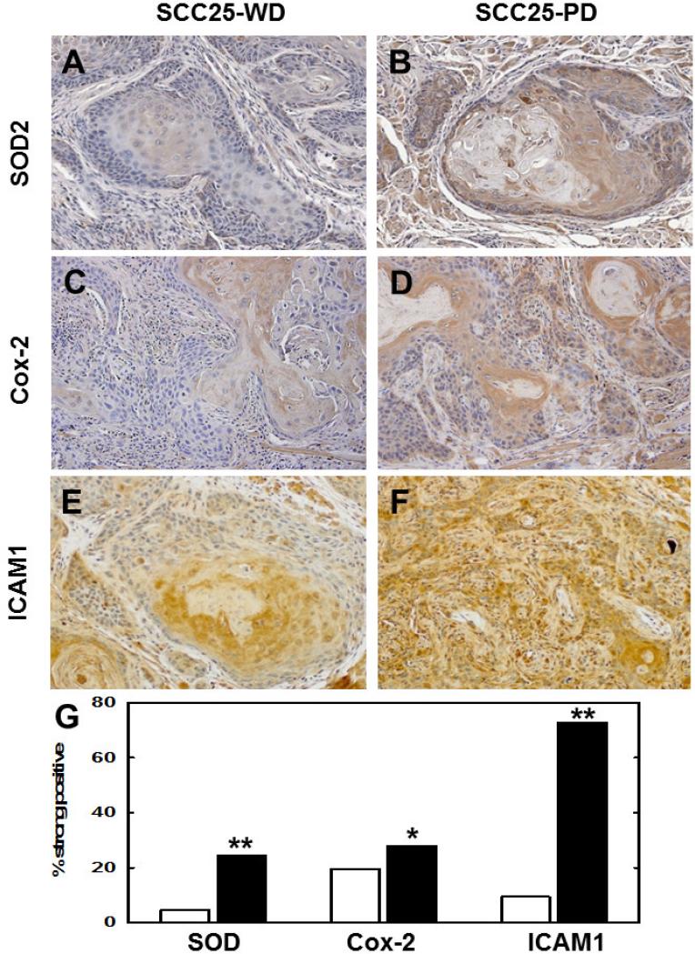
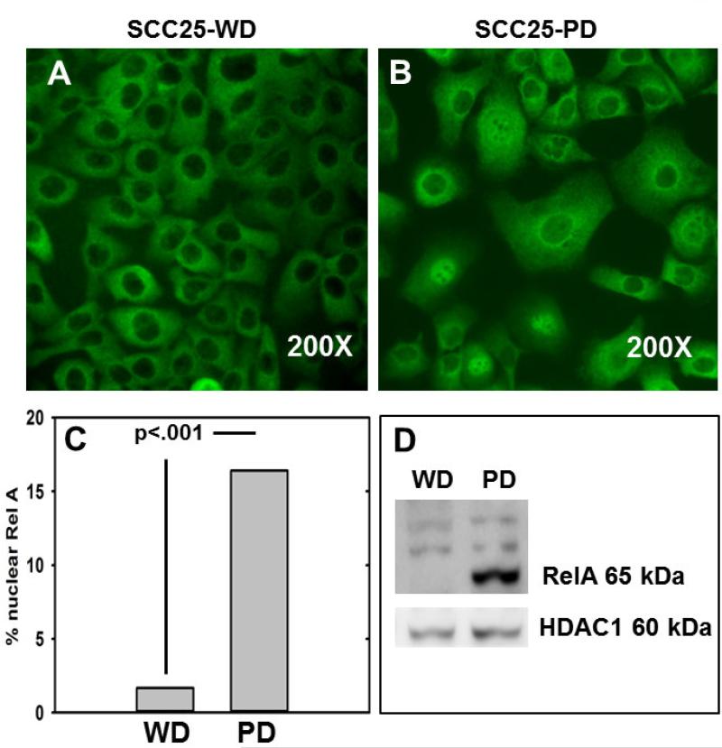
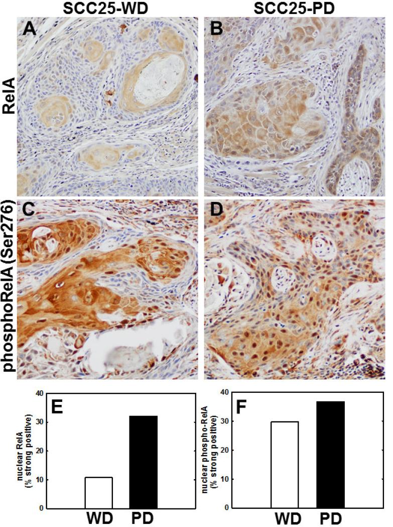
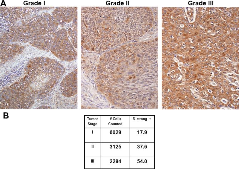
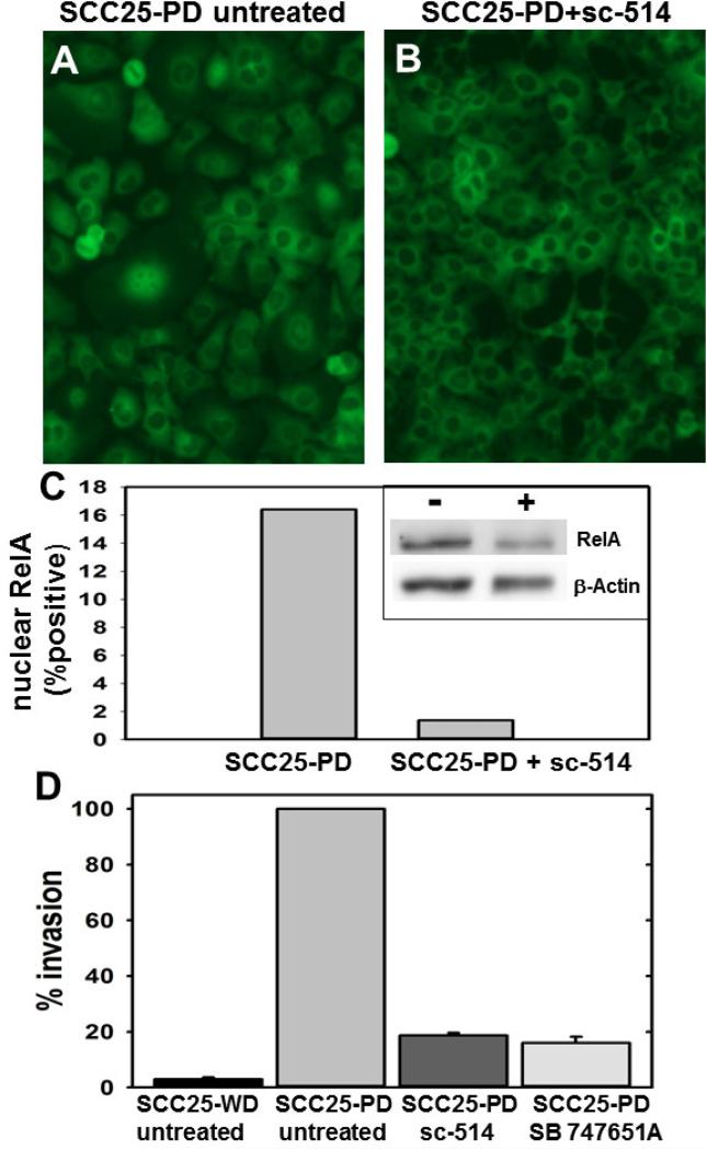
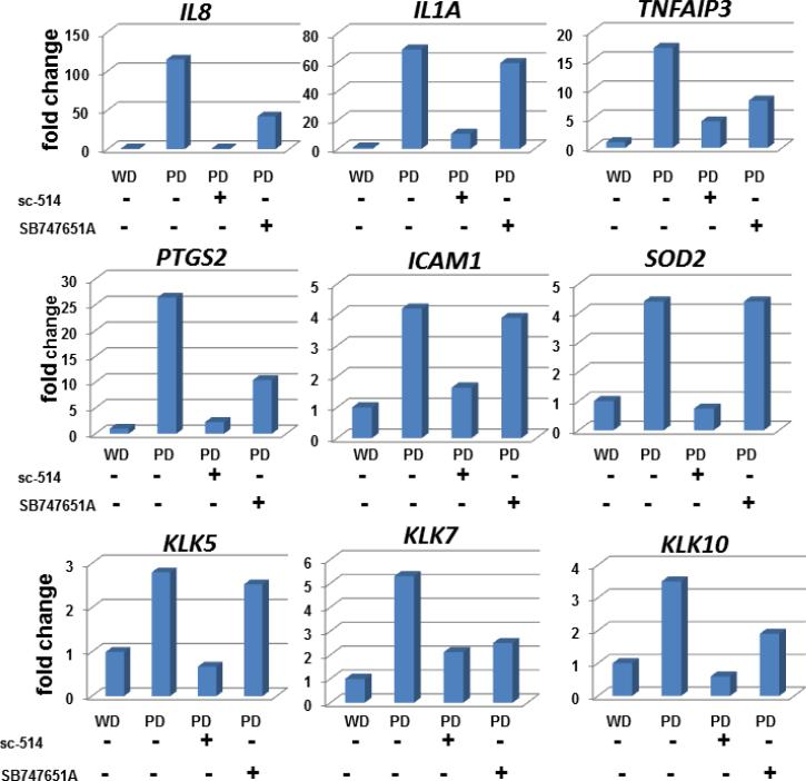
Results: As this analysis revealed that 59 upregulated genes were NF-κB target genes, the role of NF-κB activation in alteration of the transcriptional profile was evaluated. The mRNA and protein upregulation of a panel NF-κB target genes was validated by real-time qPCR and immunohistochemistry. Additionally, nuclear translocation of RelA was greatly increased in SCC25-PD, increased nuclear RelA was observed in oral tumors initiated with SCC25-PD compared with tumors initiated by SCC25-WD, and nuclear RelA correlated with stage of disease on two human OSCC tissue microarrays. Treatment of SCC25-PD cells with the IKKβ-inhibitor sc-514, that effectively prevents RelA phosphorylation on Ser 536, reversed nuclear-translocation of RelA and strongly inhibited NF-κB gene activation. Furthermore, blocking the phosphorylation of RelA using the MSK1/2 inhibitor SB 747651A significantly reduced the mRNA upregulation of a subset of target genes. Treatment with sc-514 or SB747651A markedly diminished cellular invasiveness.

Conclusions: These studies support a model wherein NF-κB is constitutively active in aggressive OSCC, while blocking the NF-κB pathway reduces NF-κB target gene upregulation and cellular invasiveness.

Keywords: Immunohistochemistry; Invasion; NF-kappaB (NF-κB); Oral cancer; Transcriptional regulation.


Figures

Figure 1
Figure 1. Perineural and vascular invasion in SCC25-PD tumors
(A,B) Representative H&E stained section showing tumor cords surrounding nerve bundle (boxed area). (T) - Tumor, (N) - nerve. Panel A – 200X magnification, Panel B – 400X magnification. (C,D) H&E stained section showing vascular invasion of tumor cord. (T) – tumor, black arrows – red blood cells, blue arrows – vascular endothelial cells. Panel C – 200X magnification, Panel D – 400X magnification.
Figure 2
Figure 2. Immunohistochemical validation of upregulated NF-κB target genes
Representative sections from murine orthotopic (tongue) SCC25-WD tumors (left column) and SCC25-PD tumors (right column) were stained with antibodies directed against (A,B) SOD2, (C,D) Cox-2 or (E,F) ICAM1. Magnification 200X. (G) Quantitation of tumor staining for each antigen in SCC25-WD tumors (white bar) and SCC25-PD tumors (black bar). For quantitation of tumor staining, stained slides were scanned on an Aperio ScanScope slide scanner and a minimum of 2500 cells from at least 10 tumor areas were scored on a standard 0 to 3 point scale using algorithm macros developed on the ImageScope software for quantitative analysis according to the manufacturer's instructions.”Strong positive” is defined as 2+ or 3+. (Detailed quantitation results are shown in Supplementary Table 1.) The automated quantitation results were consistent with those obtained by manual counting methods. (*) p<.05; (**) p<.001, relative to the paired SCC25-WD tumor.
Figure 3
Figure 3. Enhanced RelA nuclear translocation in SCC25-PD
(A,B) Immunofluorescence evaluation of RelA in (A) SCC25-WD cells and (B) SCC25-PD cells. (C) Quantitation of nuclear RelA staining. (D) Representative western blot of nuclear extracts from SCC25-WD (WD) and SCC25-PD (PD), as indicated, probed with antibodies directed against RelA (upper panel) and HDAC1 (lower panel). Experiment was performed in triplicate.
Figure 4
Figure 4. Nuclear staining for RelA and phospho-RelA(Ser276) is enhanced in SCC25-PD tumors relative to SCC25-WD tumors
(A,B) Immunohistochemical analysis of tumor section from orthotopic (tongue) mouse xenograft model initiated with (A) SCC25-WD cells or (B) SCC25-PD cells stained with antibodies directed against RelA. (C,D) Immunohistochemical analysis of tumor section from orthotopic murine xenograft initiated with (C) SCC25-WD cells and (D) SCC25-PD cells stained with antibodies directed against phospho-RelA(Ser276). Magnification – 200X. (E) Quantitation of nuclear RelA staining. (F) Quantitation of nuclear phospho-RelA(Ser276) staining. (Detailed quantitation results are shown in Supplementary Table 2).
Figure 5
Figure 5. Immunohistochemical analysis of RelA staining in human tongue tumors
(A) Representative image fields from staged human tongue SCC tissue microarray stained with antibodies directed against RelA. (B) Quantitation of staining as described in Materials and Methods. %strong positive is the sum of the percentage of cells staining 2+ and 3+.
Figure 6
Figure 6. Inhibitors reverse nuclear translocation of RelA and block invasion
(A,B) Cells were treated as indicated and processed for immuno-fluorescence microscopy with antibodies directed against RelA. Experiment was performed in triplicate. (A) SCC25-PD cells, untreated. (B) SCC25-PD cells treated with sc-514 (10 μM, 25 h). (C) Graph showing percentage of cells with positive nuclear RelA in control and sc-514-treated SCC25-PD cells, as indicated. (inset) Western blot of nuclear extracts of control (-) and sc-514-treated (+) SCC25-PD cells probed with antibodies directed against RelA (upper panel) and β-actin (lower panel). (D) Analysis of invasion. Cells were untreated or treated, as indicated, prior to addition to a Boyden chamber containing Matrigel as described in Experimental Procedures. Invading cells were enumerated and results shows as % of invasion relative to untreated SCC25-PD cells (designated 100%). (sc-514, 10 uM; SB 747651A, 20 uM)
Figure 7
Figure 7. Effect of NF-κB pathway inhibitors on target gene upregulation
Quantitative rtPCR was used to determine mRNA levels for the indicated genes in (column 1) untreated SCC25-WD cells, (column 2) untreated SCC25-PD cells, (column 3) SCC25-PD cells treated with sc-514 (10 μM, 25 h), and (column 4) SCC25-PD cells treated with SB747651A (20μM, 25 h). Graphs show fold change relative to untreated SCC25-WD cells (designated as ‘1’). Experiments were performed in triplicate.

References

    1. Sano D, Myers JN. Metastasis of squamous cell carcinoma of the oral tongue. Can. Met. Rev. 2007;26(3-4):645–62. - PubMed
    1. Rusthoven K, Ballonoff A, Raben D, Chen C. Poor prognosis in patients with stage I and II oral tongue squamous cell carcinoma. Cancer. 2008;112(2):345–51. - PubMed
    1. Yan M, Xu Q, Zhang P, Zhou XJ, Zhang ZY, Chen WT. Correlation of NF-κB signal pathway with tumor metastasis of human head and neck squamous cell carcinoma. BMC Cancer. 2010;10:437. - PMC - PubMed
    1. Myers JN, Holsinger FC, Jasser SA, Bekele BN, Fidler IJ. An orthotopic nude mouse model of oral tongue squamous cell carcinoma. Clin Cancer Res. 2002;8:293–8. - PubMed
    1. Ziober AF, Patel KR, Alawi F, Gimotty P, Weber RS, Feldman MM, Chalian AA, Weinstein GS, Hunt J, Ziober BL. Identification of a gene signature for rapid screening of oral squamous cell carcinoma. Clin Cancer Res. 2006;12(20 Pt 1):5960–71. - PubMed

Publication types