. 2014 May 16:267:67-82.
doi: 10.1016/j.neuroscience.2014.02.023. Epub 2014 Feb 26.

μ-Opioid receptor inhibition of substance P release from primary afferents disappears in neuropathic pain but not inflammatory pain

Affiliations

μ-Opioid receptor inhibition of substance P release from primary afferents disappears in neuropathic pain but not inflammatory pain

W Chen et al. Neuroscience. .

Abstract

Opiate analgesia in the spinal cord is impaired during neuropathic pain. We hypothesized that this is caused by a decrease in μ-opioid receptor inhibition of neurotransmitter release from primary afferents. To investigate this possibility, we measured substance P release in the spinal dorsal horn as neurokinin 1 receptor (NK1R) internalization in rats with chronic constriction injury (CCI) of the sciatic nerve. Noxious stimulation of the paw with CCI produced inconsistent NK1R internalization, suggesting that transmission of nociceptive signals by the injured nerve was variably impaired after CCI. This idea was supported by the fact that CCI produced only small changes in the ability of exogenous substance P to induce NK1R internalization or in the release of substance P evoked centrally from site of nerve injury. In subsequent experiments, NK1R internalization was induced in spinal cord slices by stimulating the dorsal root ipsilateral to CCI. We observed a complete loss of the inhibition of substance P release by the μ-opioid receptor agonist [D-Ala(2), NMe-Phe(4), Gly-ol(5)]-enkephalin (DAMGO) in CCI rats but not in sham-operated rats. In contrast, DAMGO still inhibited substance P release after inflammation of the hind paw with complete Freund's adjuvant and in naïve rats. This loss of inhibition was not due to μ-opioid receptor downregulation in primary afferents, because their colocalization with substance P was unchanged, both in dorsal root ganglion neurons and primary afferent fibers in the dorsal horn. In conclusion, nerve injury eliminates the inhibition of substance P release by μ-opioid receptors, probably by hindering their signaling mechanisms.

Keywords: inflammation; internalization; neurokinin 1 receptor; neuropathic pain; opioid receptor; substance P.


Conflict of interest statement

The authors declare no competing financial interests.

Figures
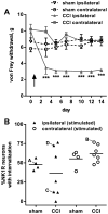
Figure 1
Figure 1. NK1R internalization induced by hind paw clamp after CCI
A. Responses of the rats to von Frey hair stimulation of the hind paws were followed every 2 days for 14 days after CCI (n = 7 rats) or sham surgery (n = 4). Two-way ANOVA revealed a significant effect of CCI (p < 0.0001), time (p = 0.0001), interaction (p < 0.0001) and subject matching (p = 0.0018). Dunnett’s multiple comparison test: *** p<0.001 compared with CCI contralateral. B. On day 14, rats were anesthetized and both hind paws were clamped sequentially for 30 s. Ten min later, the spinal cord was extracted and NK1R internalization was quantified in lamina 1 of spinal segments L4-L5. Letters next to the symbols indicate data from rats corresponding to the panels in Fig. 2.
Figure 2
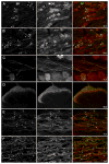
Figure 2. Confocal images of NK1R neurons in lamina I after CCI and paw clamp
Rats received unilateral sham surgery (A) or CCI (B–D), and 7 days later both paws were clamped to evoke substance P release. Images were taken from sections of the L4 spinal segment ipsilateral (A, C, D) or contralateral (B) to the operated paw. Paw clamp produced NK1R internalization in numerous cells ipsilaterally to sham surgery (A) and contralaterally to CCI (B); however, ipsilaterally to CCI some rats presented abundant NK1R internalization (D) whereas others showed little internalization (C). The percentage of NK1R lamina 1 neurons with internalization in these rats in given in Fig. 2B, where the letters next to the symbols correspond to the same rats as the panels of this figure. Main panels: images taken with a 10x objective; voxel size of 830 × 830 × 5983 nm, 1 confocal plane. Insets: images taken with a 63x objective of the lamina I neurons indicated by the frames in the main panels; voxel size of 132 × 132 × 383 nm, 3–6 confocal planes. Neurons with NK1R internalization are indicated with “*” and neurons without internalization by “o”, with the symbol placed over the nucleus. Scale bars indicate 100 μm in the main panels and 10 μm in the insets.
Figure 3
Figure 3. Concentration-responses for substance P to induce NK1R internalization after CCI
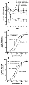
A, B. After CCI or sham surgery (n = 3 rats per group), responses of the rats to von Frey hair stimulation of the hind paws were followed every 2 days for 14 days. Two-way ANOVA of all data in panels A and B revealed a significant effect of CCI (p = 0.0253), time (p < 0.0001), interaction (p = 0.0006) and subject matching (p < 0.0001). Dunnett’s post-hoc tests compared CCI ipsilateral to CCI contralateral (* p < 0.05, *** p < 0.001) or CCI ipsilateral to sham ipsilateral (†† p < 0.01). C, D. On day 14, rats were sacrificed and used to prepare spinal cord slices (6 slices/rat). Each slice was incubated for 10 min with the concentrations of substance P indicated, so that a full concentration-response was obtained for each rat. Points represent the mean ± SEM of 3 rats / group. Curves correspond to non-linear regression fittings to a dose-response function. Fitting was done simultaneously for the two sets of data in each panel (the ‘bottom’ and ‘top’ parameters were constrained to be the same), and Akaike’s Information Criterion was used to test whether the two curves had the same or different EC50. The probability that the EC50s are different is 99.67% for the curves in panel B (ipsilateral) and 99.05% for the curves in panel C (contralateral). EC50 values were: sham ipsilateral, 5.6 nM (95% CI = 2.9–10.5 nM); CCI ipsilateral, 22.0 nM (95% CI = 11.8–40.9 nM); sham contralateral, 6.0 nM (95% CI = 3.7–9.7 nM); CCI contralateral, 14.6 nM (95% CI = 9.1–23.4 nM).
Figure 4
Figure 4. Loss of DAMGO inhibition of substance P release after CCI
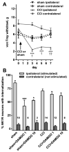
A. Responses of the rats to von Frey hair stimulation in the hind paws were followed every 2 days for 7 days after unilateral CCI (n = 10 rats) or sham surgery (n = 10 rats). Two-way ANOVA revealed a significant effect of CCI-side (p = 0.0002), time (p < 0.0001), interaction (p < 0.0001) and subject matching (p < 0.0001). Tukey’s post-hoc tests compared CCI ipsilateral to CCI contralateral (*** p < 0.001) or to sham ipsilateral († p < 0.05, †† p < 0.01). B. On day 7, 2–3 spinal cord slices (L4-L5 segments) were prepared from each rat (10 with sham surgery and 14 with CCI): one was used served as control and the others were superfused with DAMGO (1 μM or 10 μM). The dorsal root ipsilateral to CCI or sham surgery was stimulated electrically (3000 pulses at 100 Hz) to elicit substance P release. Peptidase inhibitors (captopril 10 μM and thiorphan 10 μM) were present in the superfusate to decrease substance P degradation. NK1R internalization was quantified in lamina I. Numbers represent the number of slices (n). Two-way ANOVA restricted to the ipsilateral dorsal horn revealed a significant effect of DAMGO (p < 0.0001), no significant effect of CCI (p = 0.32) and significant interaction (p = 0.0041). Tukey’s post-hoc test revealed significant inhibition by DAMGO only in sham-operated rats (* p = 0.012, *** p < 0.0001).
Figure 5
Figure 5. DAMGO inhibition of substance P release after CFA
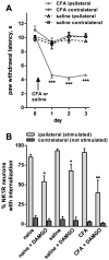
A. Responses of the rats to thermal stimulation of the hind paws were followed daily for 3 days after injecting one hind paw with CFA (n = 3 rats) or saline (n = 5 rats). Two-way ANOVA revealed a significant effect of CFA (p < 0.0001), time (p < 0.0001), interaction (p < 0.0001) but not subject matching (p = 0.0815). Tukey’s post-hoc tests compared CFA ipsilateral to CFA contralateral or to saline ipsilateral (*** p < 0.001). B. Three days after the injection in the hindpaw, two spinal cord slices with the dorsal root ipsilateral to the injection were prepared from each rat (and also from 4 naïve rats). That root was electrically stimulated (3000 pulses at 100 Hz) while the slice was superfused with aCSF containing peptidase inhibitors (captopril 10 μM and thiorphan 10 μM). One of the slices from each rat was also superfused with 1 μM DAMGO, while the other slice from that rat served as a control. NK1R internalization was quantified in lamina 1, and found to be negligible in the contralateral side. One-way ANOVA restricted to data from the ipsilateral dorsal horn revealed a significant effect of DAMGO (p < 0.0001), no significant effect of CFA (p = 0.139), and no significant interaction (p = 0.266). Sidak’s multiple comparison test revealed significant inhibition by DAMGO: * p < 0.05, ** p < 0.01.
Figure 6
Figure 6. Confocal images of NK1R neurons in lamina I - DAMGO after CCI or CFA
Rats received sham surgery (A, B), CCI (C, D) or CFA in the hind paw (E, F). Seven days after sham or CCI, or 3 days after CFA, spinal cord slices prepared from these rats were electrically stimulated at the ipsilateral dorsal root to induce substance P release. In the absence of DAMGO, dorsal root stimulation produced NK1R internalization in most lamina I cells in the sham (A), CCI (C) and CFA-injected rats (E). However, 1 μM DAMGO inhibited the NK1R internalization evoked by dorsal root stimulation in the sham (B) and the CFA-injected rats (F), but not in the CCI rats (D). The percentage of NK1R lamina 1 neurons with internalization in these rats is given in Figs. 3B and 4B. Main panels: images taken with a 10x objective; voxel size of 830 × 830 × 5983 nm, 1 confocal plane. Insets: images taken with a 63x objective of the lamina I neurons indicated by the frames in the main panels; voxel size of 132 × 132 × 383 nm, 3–7 confocal planes. Neurons with NK1R internalization are indicated with “*” and neurons without internalization by “o”, with the symbol placed over the nucleus. Scale bars indicate 100 μm in the main panels and 10 μm in the insets.
Figure 7
Figure 7. Quantification of substance P and MOR colocalization in DRG and spinal cord
A. Responses of 3 rats to von Frey hair stimulation in the hind paws were followed every 2 days for 6 days after unilateral CCI. Two-way ANOVA revealed a significant effect of CCI (p = 0.0182), time (p < 0.0001), interaction (p = 0.0036) and subject matching (p = 0.0198). Sidak’s post-hoc tests compared CCI ipsilateral to CCI contralateral: * p < 0.05, ** p < 0.01, *** p < 0.001. B. In the L4-L5 DRG from those 3 rats or from 4 naïve rats (“control”), colocalization of substance P (SP) with MORs was measured by counting cell bodies with each label and with both labels; n = 12 confocal stacks from control rats, n = 9 confocal stacks from CCI rats (3 per rat). C. In the dorsal horn of the same rats, colocalization of SP with MORs was measured as the Pearson’s correlation coefficient of the intensity of these two labels in 12 (control) or 9 (CCI) confocal stacks (3 per rat).
Figure 8
Figure 8. Colocalization of substance P and MORs in DRG and spinal cord in naïve and CCI rats
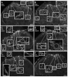
Naïve rats or rats that received CCI 7 days earlier were euthanized and fixed. Sections from the L4 or L5 DRG and L4-L5 spinal segments were labeled with antibodies recognizing substance P (SP, green) and MOR (red). A, B. DRG from a naïve rat (A, 20x objective, 4 optical sections) or a CCI rat (B, ipsilateral to CCI, 20x objective, 6 optical sections); arrows indicate neuron bodies with SP/MOR colocalization. C. DRG ipsilateral to CCI (63x objective, 19 optical sections); SP and MOR colocalize in numerous primary afferent fibers. D. Dorsal horn ipsilateral to CCI (coronal section, segment L5, 10x objective, 1 optical section); SP and MOR labels are present mostly in laminae I and II. E, F. Laminae I–II (sagittal sections, segment L4, 63x objective, 6 optical sections) from a naïve rat (E) or a CCI rat (F, ipsilateral to CCI); arrowheads bracket fibers with SP/MOR colocalization.

Similar articles

Cited by

References

    1. Abbadie C, Trafton J, Liu H, Mantyh PW, Basbaum AI. Inflammation increases the distribution of dorsal horn neurons that internalize the neurokinin-1 receptor in response to noxious and non-noxious stimulation. J Neurosci. 1997;17:8049–8060. - PMC - PubMed
    1. Adelson DW, Lao L, Zhang G, Kim W, Marvizón JC. Substance P release and neurokinin 1 receptor activation in the rat spinal cord increases with the firing frequency of C-fibers. Neuroscience. 2009;161:538–553. - PMC - PubMed
    1. Aimone LD, Jones SL, Gebhart GF. Stimulation-produced descending inhibition from the periaqueductal gray and nucleus raphe magnus in the rat: mediation by spinal monoamines but not opioids. Pain. 1987;31:123–136. - PubMed
    1. Allen BJ, Li J, Menning PM, Rogers SD, Ghilardi J, Mantyh PW, Simone DA. Primary afferent fibers that contribute to increased substance P receptor internalization in the spinal cord after injury. J Neurophysiol. 1999;81:1379–1390. - PubMed
    1. Allen BJ, Rogers SD, Ghilardi JR, Menning PM, Kuskowski MA, Basbaum AI, Simone DA, Mantyh PW. Noxious cutaneous thermal stimuli induce a graded release of endogenous substance P in the spinal cord: imaging peptide action in vivo. J Neurosci. 1997;17:5921–5927. - PMC - PubMed

Publication types

MeSH terms