

Overexpression of FABP3 inhibits human bone marrow derived mesenchymal stem cell proliferation but enhances their survival in hypoxia
- PMID: 24583397
- PMCID: PMC4049325
- DOI: 10.1016/j.yexcr.2014.02.015
Overexpression of FABP3 inhibits human bone marrow derived mesenchymal stem cell proliferation but enhances their survival in hypoxia
Abstract
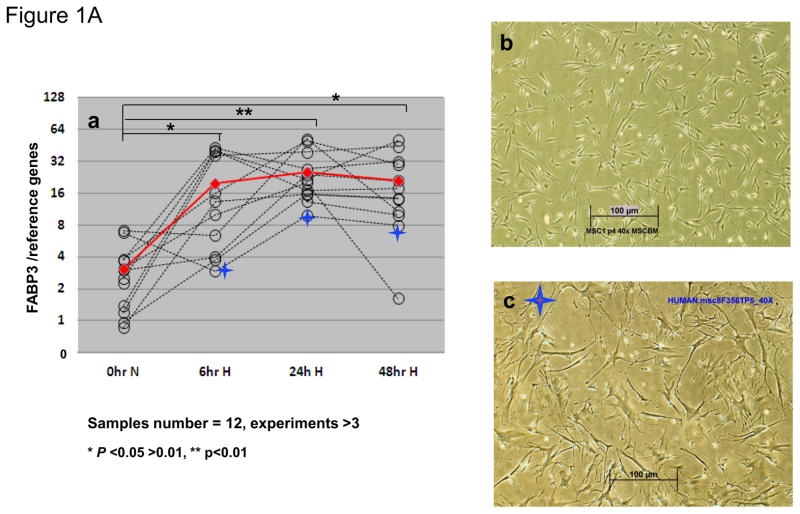
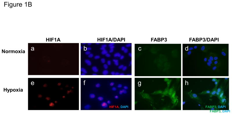
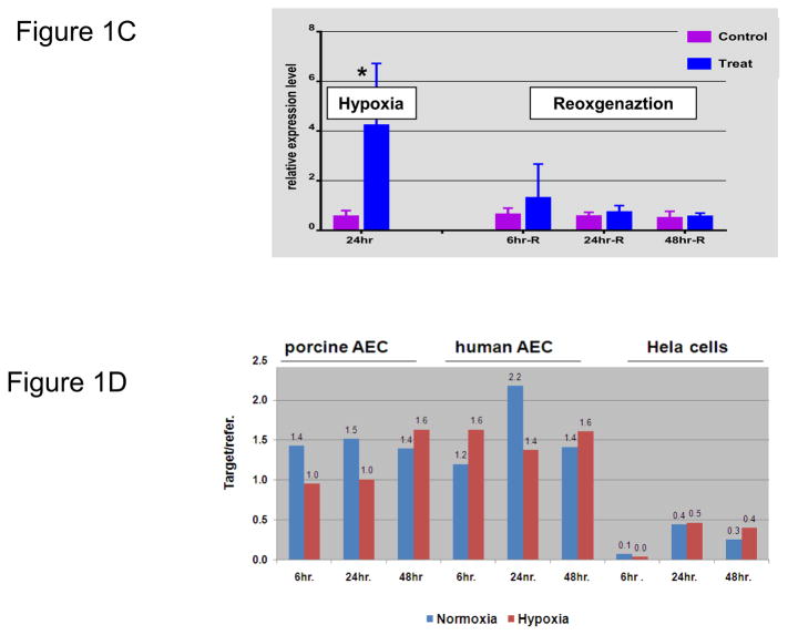
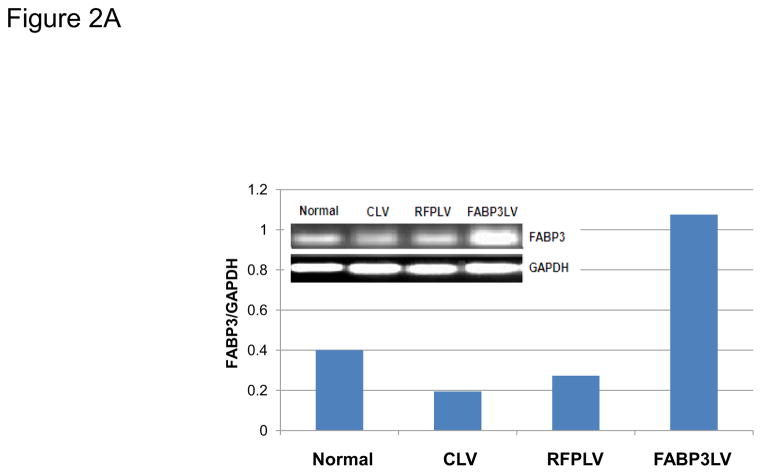
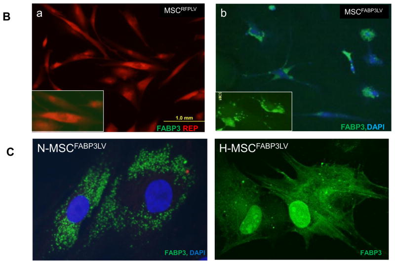
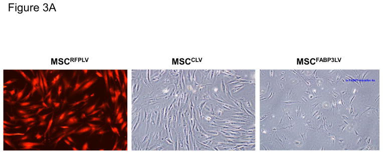
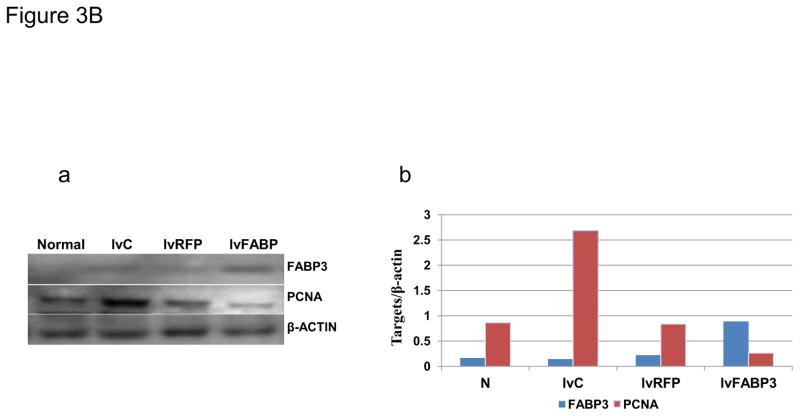
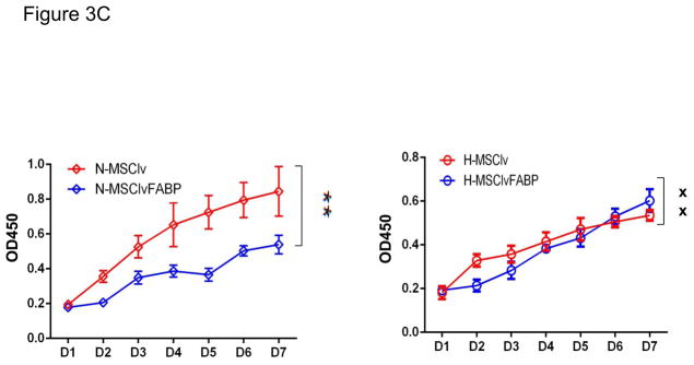
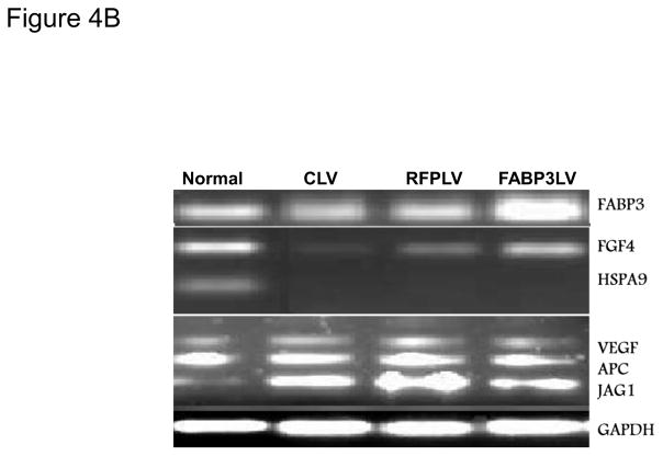
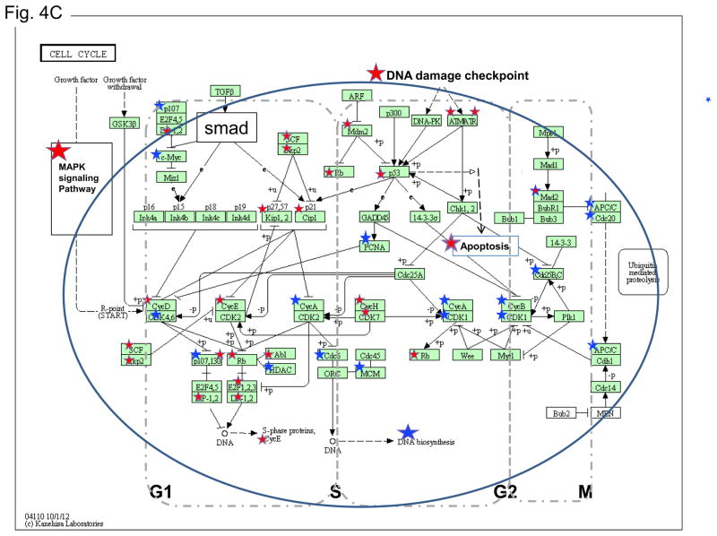
Studying the proliferative ability of human bone marrow derived mesenchymal stem cells in hypoxic conditions can help us achieve the effective regeneration of ischemic injured myocardium. Cardiac-type fatty acid binding protein (FABP3) is a specific biomarker of muscle and heart tissue injury. This protein is purported to be involved in early myocardial development, adult myocardial tissue repair and responsible for the modulation of cell growth and proliferation. We have investigated the role of FABP3 in human bone marrow derived mesenchymal stem cells under ischemic conditions. MSCs from 12 donors were cultured either in standard normoxic or modified hypoxic conditions, and the differential expression of FABP3 was tested by quantitative (RT)PCR and western blot. We also established stable FABP3 expression in MSCs and searched for variation in cellular proliferation and differentiation bioprocesses affected by hypoxic conditions. We identified: (1) the FABP3 differential expression pattern in the MSCs under hypoxic conditions; (2) over-expression of FABP3 inhibited the growth and proliferation of the MSCs; however, improved their survival in low oxygen environments; (3) the cell growth factors and positive cell cycle regulation genes, such as PCNA, APC, CCNB1, CCNB2 and CDC6 were all down-regulated; while the key negative cell cycle regulation genes TP53, BRCA1, CASP3 and CDKN1A were significantly up-regulated in the cells with FABP3 overexpression. Our data suggested that FABP3 was up-regulated under hypoxia; also negatively regulated the cell metabolic process and the mitotic cell cycle. Overexpression of FABP3 inhibited cell growth and proliferation via negative regulation of the cell cycle and down-regulation of cell growth factors, but enhances cell survival in hypoxic or ischemic conditions.
Keywords: Cell cycle; Cell proliferation; FABP3; Human mesenchymal stem cell; Hypoxia.
Published by Elsevier Inc.
Conflict of interest statement
None.
Figures

Similar articles
-
Peptide hormone ELABELA promotes rat bone marrow-derived mesenchymal stem cell proliferation and migration by manipulating the cell cycle through the PI3K/AKT pathway under the hypoxia and ischemia microenvironment.Stem Cell Res Ther. 2022 Jan 28;13(1):32. doi: 10.1186/s13287-021-02691-1. Stem Cell Res Ther. 2022. PMID: 35090551 Free PMC article.
-
Hypoxic culture enhances the expansion of rat bone marrow-derived mesenchymal stem cells via the regulatory pathways of cell division and apoptosis.In Vitro Cell Dev Biol Anim. 2018 Oct;54(9):666-676. doi: 10.1007/s11626-018-0281-3. Epub 2018 Aug 22. In Vitro Cell Dev Biol Anim. 2018. PMID: 30136033
-
Hypoxia-Induced Mesenchymal Stromal Cells Exhibit an Enhanced Therapeutic Effect on Radiation-Induced Lung Injury in Mice due to an Increased Proliferation Potential and Enhanced Antioxidant Ability.Cell Physiol Biochem. 2017;44(4):1295-1310. doi: 10.1159/000485490. Epub 2017 Nov 29. Cell Physiol Biochem. 2017. PMID: 29183009
-
Zinc and hypoxic preconditioning: a strategy to enhance the functionality and therapeutic potential of bone marrow-derived mesenchymal stem cells.Mol Cell Biochem. 2022 Dec;477(12):2735-2749. doi: 10.1007/s11010-022-04468-3. Epub 2022 May 24. Mol Cell Biochem. 2022. PMID: 35610401
-
The role of hypoxia in bone marrow-derived mesenchymal stem cells: considerations for regenerative medicine approaches.Tissue Eng Part B Rev. 2010 Apr;16(2):159-68. doi: 10.1089/ten.TEB.2009.0296. Tissue Eng Part B Rev. 2010. PMID: 19698058 Review.
Cited by
-
Evaluation of the cardioprotective effects of crystalloid del Nido cardioplegia solution via a rapid and accurate cardiac marker: heart-type fatty acid-binding protein.Turk J Med Sci. 2020 Jun 23;50(4):999-1006. doi: 10.3906/sag-2002-53. Turk J Med Sci. 2020. PMID: 32394686 Free PMC article. Clinical Trial.
-
Heart-Type Fatty Acid-Binding Protein (H-FABP) and its Role as a Biomarker in Heart Failure: What Do We Know So Far?J Clin Med. 2020 Jan 7;9(1):164. doi: 10.3390/jcm9010164. J Clin Med. 2020. PMID: 31936148 Free PMC article. Review.
-
Early weaning of calves after different dietary regimens affects later rumen development, growth, and carcass traits in Hanwoo cattle.Asian-Australas J Anim Sci. 2017 Oct;30(10):1425-1434. doi: 10.5713/ajas.17.0315. Epub 2017 Jul 6. Asian-Australas J Anim Sci. 2017. PMID: 28728406 Free PMC article.
-
Crohn's disease patient serum changes protein expression in a human mesenchymal stem cell model in a linear relationship to patients' disease stage and to bone mineral density.J Clin Transl Endocrinol. 2018 Jun 19;13:26-38. doi: 10.1016/j.jcte.2018.06.002. eCollection 2018 Sep. J Clin Transl Endocrinol. 2018. PMID: 30003044 Free PMC article.
-
Transcriptomic and Proteomic Analyses Reveal the Potential Mode of Action of Chondrocyte Sheets in Hyaline Cartilage Regeneration.Int J Mol Sci. 2019 Dec 24;21(1):149. doi: 10.3390/ijms21010149. Int J Mol Sci. 2019. PMID: 31878307 Free PMC article.
References
-
- Bartsch T, Brehm M, Zeus T, Kogler G, Wernet P, Strauer BE. Transplantation of autologous mononuclear bone marrow stem cells in patients with peripheral arterial disease (the TAM-PAD study) Clin Res Cardiol. 2007;96:891–899. - PubMed
-
- Edelberg JM, Tang L, Hattori K, Lyden D, Rafii S. Young adult bone marrow-derived endothelial precursor cells restore aging-impaired cardiac angiogenic function. Circ Res. 2002;90:E89–93. - PubMed
-
- Strauer BE, Brehm M, Zeus T, Kostering M, Hernandez A, Sorg RV, Kogler G, Wernet P. Repair of infarcted myocardium by autologous intracoronary mononuclear bone marrow cell transplantation in humans. Circulation. 2002;106:1913–1918. - PubMed
Publication types
MeSH terms
Substances
Grants and funding
LinkOut - more resources
Full Text Sources
Other Literature Sources
Research Materials
Miscellaneous
