. 2014 Jul;21(7):1071-80.
doi: 10.1038/cdd.2014.26. Epub 2014 Feb 28.

G0/G1 switch gene 2 has a critical role in adipocyte differentiation

Affiliations

G0/G1 switch gene 2 has a critical role in adipocyte differentiation

H Choi et al. Cell Death Differ. 2014 Jul.

Abstract

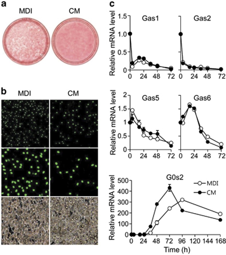
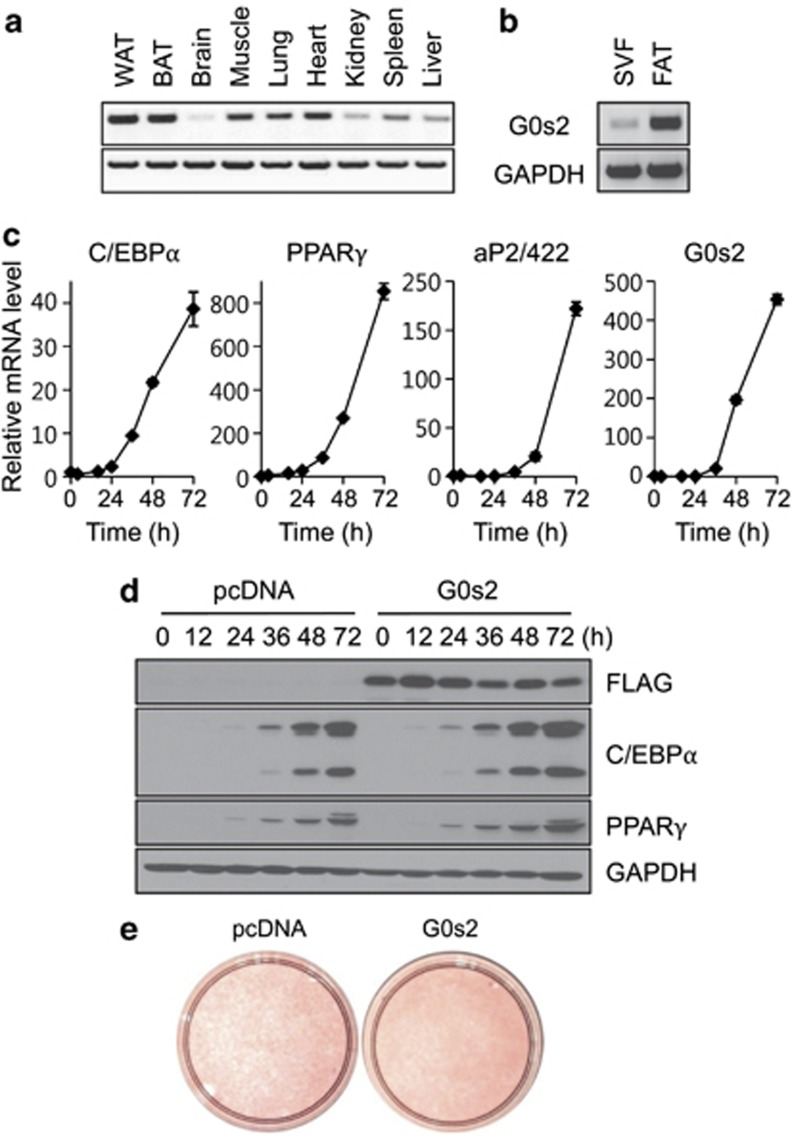
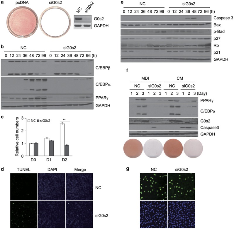
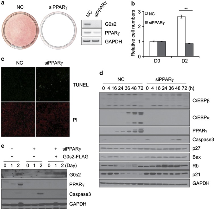
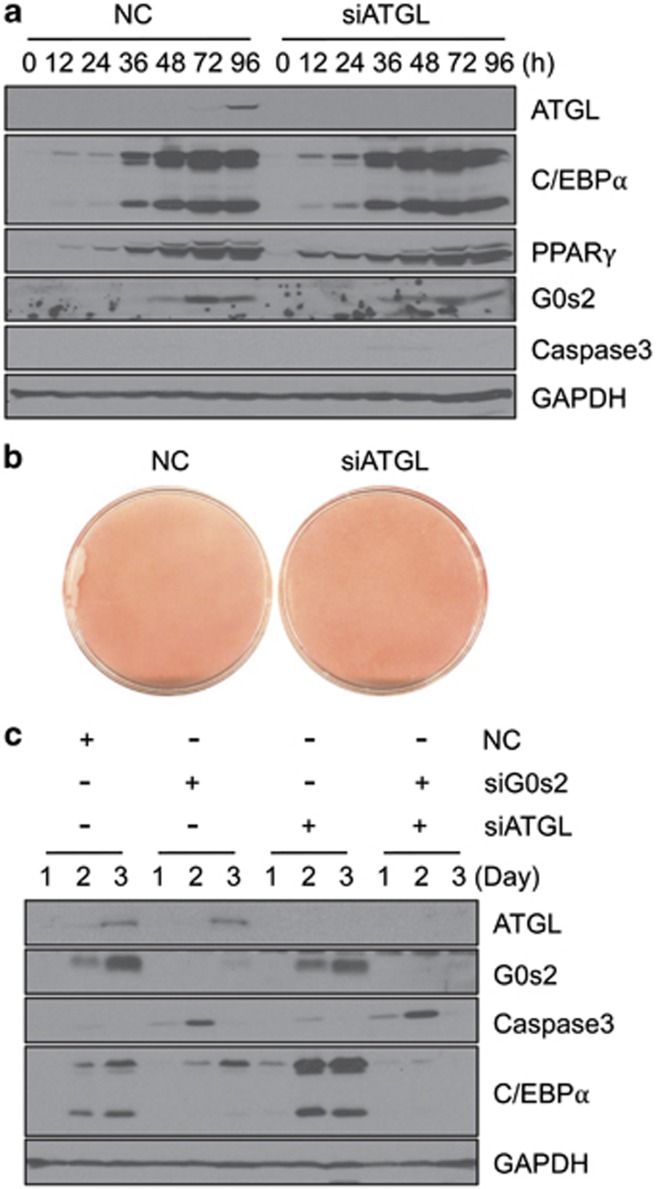
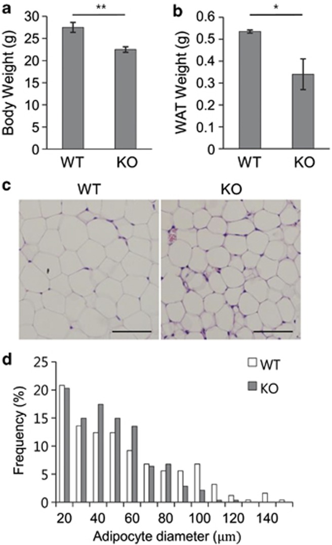
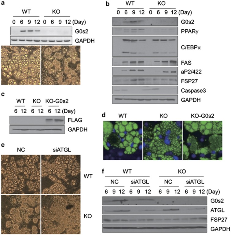
Mouse 3T3-L1 preadipocytes differentiate into adipocytes when treated with 3-isobutyl-1-methylxanthine, dexamethasone, and insulin. Although mechanisms of adipogenesis, including transcriptional cascades, are understood, it is still unclear how clonally expanded cells eventually enter the terminal differentiation program. From gene expression profile studies, we identified G0/G1 switch gene 2 (G0s2) as a novel regulator of adipogenesis. The gene was found to be expressed at a higher level in white and brown adipose tissues, and it was induced in 3T3-L1 cells by hormonal treatment. Importantly, G0s2 expression was closely associated with the transition from mitotic clonal expansion to terminal differentiation. Knockdown of G0s2 expression with siRNA inhibited adipocyte differentiation, whereas constitutive overexpression of G0s2 accelerated differentiation of preadipocytes to mature adipocytes. Expression of G0s2 was found to be regulated by peroxisome proliferator-activated receptor γ (PPARγ), which is a well-known regulator of adipocyte differentiation. Absence of either PPARγ or G0s2 expression resulted in apoptotic pathway activation before terminal differentiation. To determine whether G0s2 has a role in vivo, G0s2-knockout mice were generated. The knockout mice were normal in appearance, but they had less adipose mass than wild-type littermates. Mouse embryonic fibroblast cells from G0s2-deficient mice exhibited impaired adipogenesis and contained unusually small intracellular lipid droplets, suggesting that G0s2 has a role in lipid droplet formation. Our studies demonstrate that G0s2 has an important role in adipogenesis and accumulation of triacylglycerol.


Figures

Figure 1
Figure 1
The effect of treatment with differentiation medium or CM on differentiation of 3T3-L1 preadipocytes. (a) Post-confluent 3T3-L1 preadipocytes cells were treated with differentiation medium containing 3-isobutyl-1-methylxanthine, dexamethasone, and insulin (MDI) or CM. Cells were then stained with Oil red-O on day 5. (b) Immunofluorescence analysis of bromodeoxyuridine (BrdU)-labeled adipocytes. 3T3-L1 cells were induced by MDI or CM and then BrdU was pulsed for 2 h (from 16 to 18 h after initiation of differentiation). The time period of BrdU pulse corresponds to the S phase in cells induced by the standard protocol. Representative images of fluorescence microscopy with magnification × 20 (upper), × 40 (middle), and of light microscopy with magnification × 20 (lower). (c) Preadipocyte 3T3-L1 cells were induced with MDI or CM. Cells were harvested at 0, 4, 16, 24, 36, 48, 72, 96, and 168 h after induction. Total RNA was isolated, and the expression levels of Gas1, Gas2, Gas5, Gas6, and G0s2 were determined by quantitative PCR. All reactions were performed in triplicate. Data represent the mean±S.D.
Figure 2
Figure 2
G0s2 is predominantly expressed in fat tissues and its overexpression enhances adipocyte differentiation. (a) Tissue distribution of G0s2 expression by reverse transcriptase-PCR analysis. (b) G0s2 expression in stromal vascular fraction (SVF) and fat cell fraction (FAT). (c) G0s2 expression during adipogenesis was validated by quantitative real-time PCR. C/EBPα, PPARγ, and aP2/422 were used as adipocyte differentiation markers. (d) 3T3-L1 cells expressing control vector (pcDNA) or G0s2-FLAG expression vector were transfected by electroporation and then induced to differentiate. Protein collected at indicated times after induction was used to assay levels of G0s2, C/EBPα, and PPARγ expression. GAPDH was used as loading control. (e) Oil red-O staining after complete differentiation (day 4)
Figure 3
Figure 3
Knockdown of G0s2 by siRNA inhibits adipocyte differentiation and leads to apoptosis. (a) Knockdown of G0s2 abrogates 3T3-L1 cell differentiation. Oil red-O staining of adipocytes with negative control (NC) and G0s2-knockdown (siG0s2) at day 5. The knockdown efficiency by siRNA was assessed by reverse transcriptase-PCR at day 2 after differentiation. (b) C/EBPβ, C/EBPα, and PPARγ protein levels were measured in the NC and siG0s2 cells by western blotting analysis, using GAPDH as loading control. (c) Cell counts were determined at days 0, 1, and 2 after induction. Cells were induced with differentiation medium containing 3-isobutyl-1-methylxanthine, dexamethasone, and insulin (MDI) after transfection of siRNA, and the cells were counted. **P<0.01. Data represent the mean±S.D. (d) Cells treated with negative control (NC) or knockdown of G0s2 (siG0s2) were fixed 48 h after induction and then stained with DAPI (4,6-diamidino-2-phenylindole). They were analyzed by fluorescence microscopy or for DNA fragmentation by TUNEL. (e) Cells were harvested at the indicated times, and western blotting analysis of caspase 3, Bax, p-Bad, p27, Rb, or p21 expression levels during 3T3-L1 differentiation was performed using GAPDH as loading control. (f) Knockdown of G0s2 upon CM treatment did not induce 3T3-L1 differentiation. Cells treated with negative control (NC) or knockdown of G0s2 (siG0s2) were induced either by MDI or CM and were analyzed by western blotting and Oil red-O staining. (g) Bromodeoxyuridine (BrdU) incorporation analysis in the cells treated with siG0s2. BrdU was pulsed for 2 h (from 16 to 18 h after initiation of differentiation), and cells were observed by fluorescence microscopy with magnification × 40
Figure 4
Figure 4
Knockdown of PPARγ by siRNA inhibits differentiation of 3T3-L1 preadipocytes and induces apoptosis during adipogenesis. (a) Effects of PPARγ knockdown was assessed by Oil red-O staining and reverse transcriptase-PCR. NC, Negative control, siPPARγ, PPARγ-knockdown. (b) Cell numbers were determined on days 0 and 2 after differentiation. **P<0.01. Data represent the mean±S.D. (c) Cells with negative control (NC) and knockdown of PPARγ (siPPARγ) were fixed 48 h after induction and then stained with DAPI (4,6-diamidino-2-phenylindole). They were analyzed by fluorescence microscopy or for DNA fragmentation by TUNEL. (d) Protein expression of the adipogenesis-associated targets C/EBPβ, C/EBPα, and PPARγ and apoptosis-related genes caspase 3, Bax, p27, and p21. (e) 3T3-L1 cells stably expressing control vector or G0s2-FLAG were transfected with siPPARγ, and effects on protein expression were examined
Figure 5
Figure 5
ATGL gene knockdown does not induce apoptosis in 3T3-L1 preadipocytes. (a) ATGL expression was suppressed in 3T3-L1 preadipocytes by siRNA, and then cells were differentiated into adipocytes. Cells were harvested at the indicated times, followed by western blotting analysis of ATGL, G0s2, C/EBPα, PPARγ, caspase 3, and GAPDH expression. (b) Oil red-O staining after differentiation (day 5). (c) 3T3-L1 cells were transfected with siRNAs targeting G0s2 and/or ATGL, followed by differentiation into adipocytes. Levels of ATGL, G0s2, caspase 3, C/EBPα, and GAPDH proteins were analyzed in immunoblots at the indicated days
Figure 6
Figure 6
Fat development is impaired in G0s2-knockout mice. (a) Body weight and (b) epididymal fat pad from white adipose tissue (WAT) were evaluated in G0s2-knockout mice and wild-type control mice at the age of 12 weeks. Data represent the mean±S.D. *P<0.05, **P<0.01. (b) Hematoxylin/eosin-stained WAT sections from wild-type and G0s2-knockout mice. Scale bar=100 μm. (c) Distribution of adipocyte size in epididymal WAT from wild-type or G0s2-knockout mice on normal diet (n=3 per group). (d) The diameter of epididymal white adipocytes was determined by ImageJ, and >500 adipocytes were examined for each group. All data are expressed as means±S.D.
Figure 7
Figure 7
Adipocyte differentiation is inhibited in G0s2-knockout MEF cells, and ectopic expression of G0s2 in G0s2-knockout MEF restores adipogenesis. (a) MEFs from wild-type (WT) or G0s2-knockout (KO) mice were induced for adipocyte differentiation. Expression of G0s2 was analyzed by reverse transcriptase-PCR, and microscopy was done at day 12. (b) Western blotting analysis of G0s2 and adipocyte-specific proteins in WT and KO MEFs. (c) KO MEFs were transfected with G0s2-FLAG-overexpression vector. Western blotting analysis of expression in WT containing empty vector (WT), G0s2-KO containing empty vector (KO), or rescued G0s2-KO (KO-G0s2) was performed with anti-FLAG antibody. (d) BODIPY staining of WT, G0s2-KO, and KO-G0s2 MEF cells. The fluorescent dyes BODIPY and DAPI (4,6-diamidino-2-phenylindole) were used to stain the lipids contained in droplets of differentiated fat cells and nuclei of existing cells. (e) WT or KO MEFs were transfected with siRNA directed against ATGL, and microscopy ( × 20) was performed. (f) Western blotting analysis was carried out in WT or KO MEFs after ATGL siRNA transfection

Similar articles

Cited by

References

    1. Kopelman PG. Obesity as a medical problem. Nature. 2000;404:635–643. - PubMed
    1. Gregoire FM, Smas CM, Sul HS. Understanding adipocyte differentiation. Physiol Rev. 1998;78:783–809. - PubMed
    1. Spiegelman BM, Flier JS. Adipogenesis and obesity: rounding out the big picture. Cell. 1996;87:377–389. - PubMed
    1. Rosen ED, Spiegelman BM. Molecular regulation of adipogenesis. Annu Rev Cell Dev Biol. 2000;16:145–171. - PubMed
    1. Green H, Kehinde O. An established preadipose cell line and its differentiation in culture. II. Factors affecting the adipose conversion. Cell. 1975;5:19–27. - PubMed

Publication types

LinkOut - more resources