. 2014 Jun;21(6):956-66.
doi: 10.1038/cdd.2014.15. Epub 2014 Feb 28.

Endogenous c-Myc is essential for p53-induced apoptosis in response to DNA damage in vivo

Affiliations

Endogenous c-Myc is essential for p53-induced apoptosis in response to DNA damage in vivo

T J Phesse et al. Cell Death Differ. 2014 Jun.

Abstract

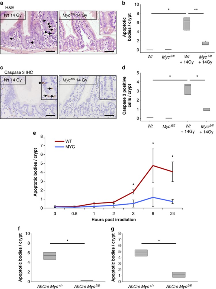
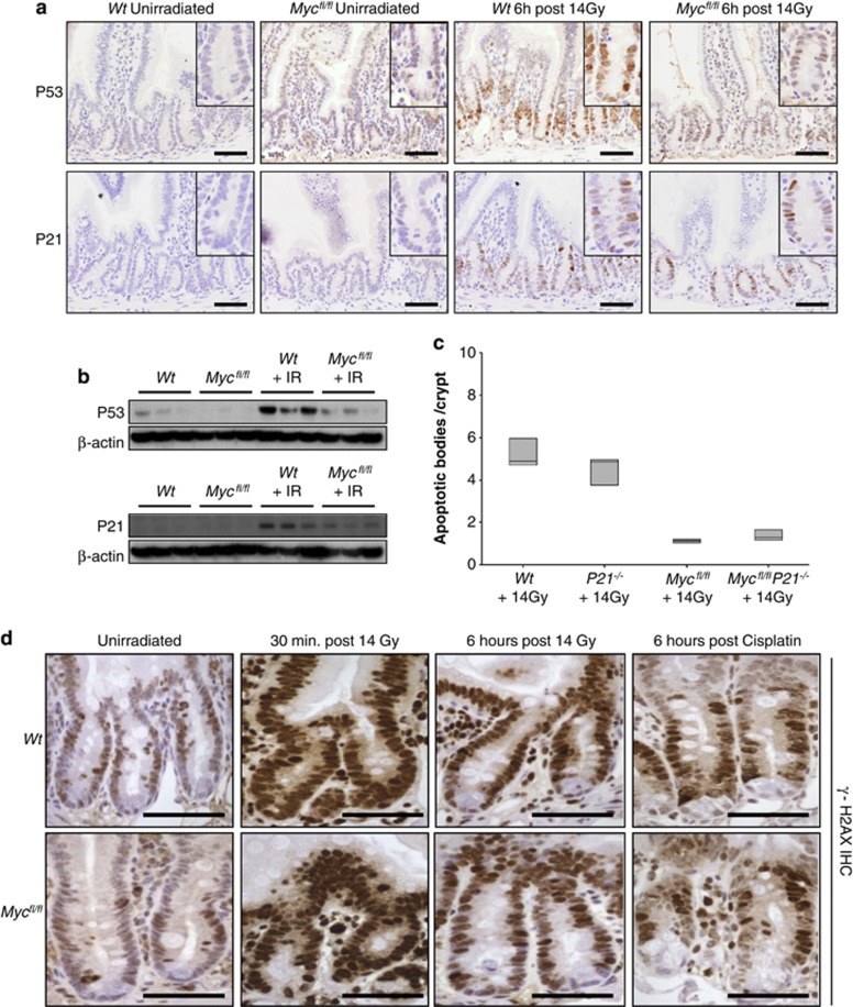
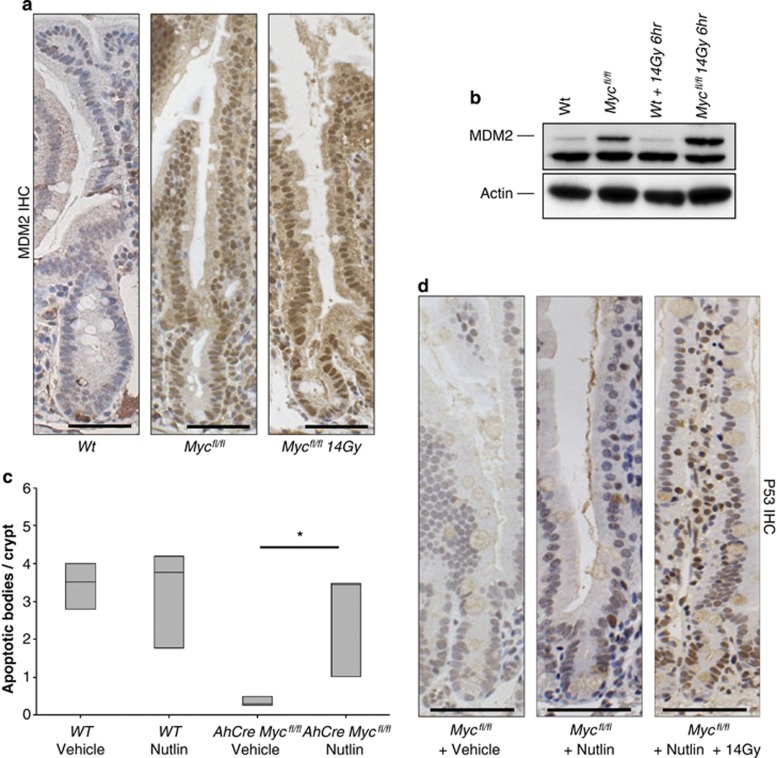
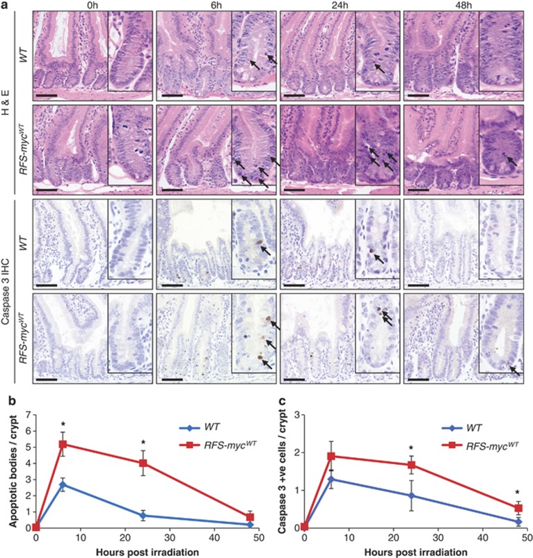
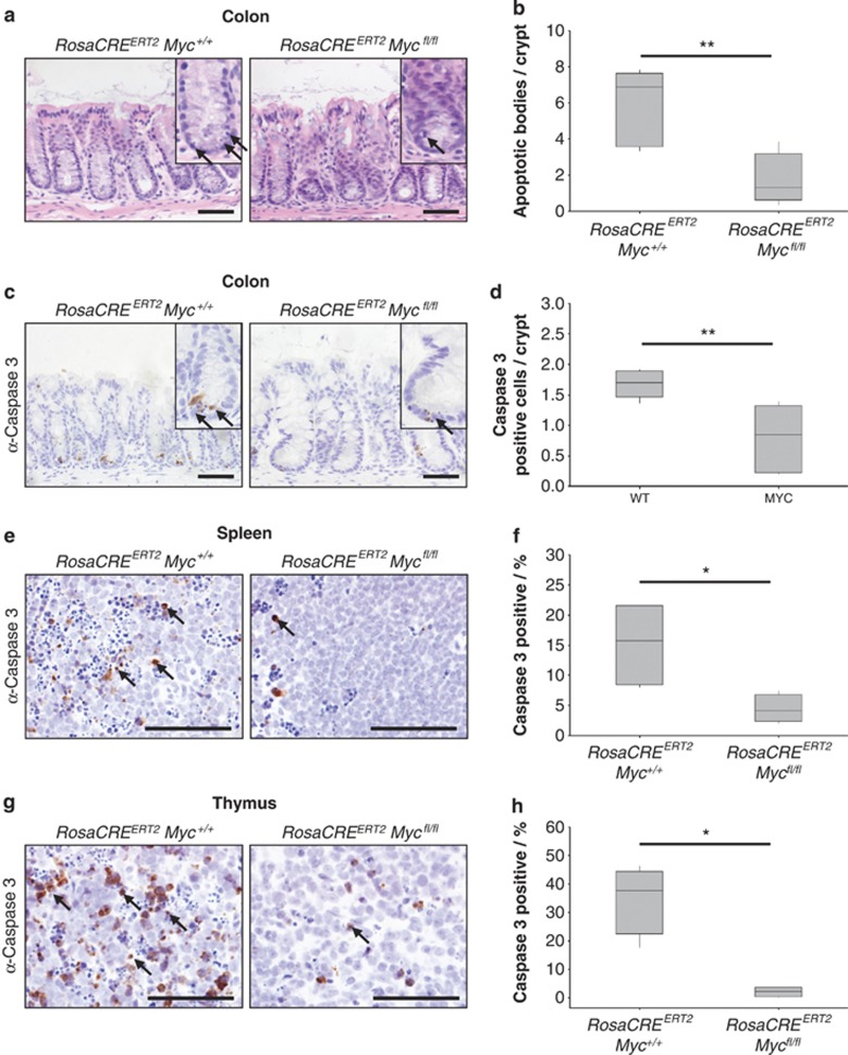
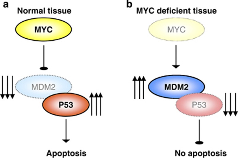
Recent studies have suggested that C-MYC may be an excellent therapeutic cancer target and a number of new agents targeting C-MYC are in preclinical development. Given most therapeutic regimes would combine C-MYC inhibition with genotoxic damage, it is important to assess the importance of C-MYC function for DNA damage signalling in vivo. In this study, we have conditionally deleted the c-Myc gene in the adult murine intestine and investigated the apoptotic response of intestinal enterocytes to DNA damage. Remarkably, c-Myc deletion completely abrogated the immediate wave of apoptosis following both ionizing irradiation and cisplatin treatment, recapitulating the phenotype of p53 deficiency in the intestine. Consistent with this, c-Myc-deficient intestinal enterocytes did not upregulate p53. Mechanistically, this was linked to an upregulation of the E3 Ubiquitin ligase Mdm2, which targets p53 for degradation in c-Myc-deficient intestinal enterocytes. Further, low level overexpression of c-Myc, which does not impact on basal levels of apoptosis, elicited sustained apoptosis in response to DNA damage, suggesting c-Myc activity acts as a crucial cell survival rheostat following DNA damage. We also identify the importance of MYC during DNA damage-induced apoptosis in several other tissues, including the thymus and spleen, using systemic deletion of c-Myc throughout the adult mouse. Together, we have elucidated for the first time in vivo an essential role for endogenous c-Myc in signalling DNA damage-induced apoptosis through the control of the p53 tumour suppressor protein.


Figures

Figure 1
Figure 1
MYC-deficient crypts do not undergo apoptosis following treatment with DNA-damaging agents. (a) H&E staining of wild type (AhCre+ Myc+/+) and MYC deficient (AhCre+ Mycfl/fl) intestines 6 h following 14 Gy irradiation, arrows show apoptotic figures in wild-type mice. Scale bars=50 μm. (b) Scoring of apoptotic figures from H&E sections shows a significant decrease in apoptosis in MYC-deficient mice following 14 Gy irradiation compared with wild type (* Wt versus Wt + 14 Gy, P =0.04, Mann Whitney n=3 versus 6, ** Wt + 14 Gy versus Myc + 14 Gy, P =0.0041, Mann Whitney n=6 versus 5). (c) Immunohistochemical staining for cleaved (‘active') Caspase 3 was performed on intestinal sections of wild type and MYC-deficient mice. Scale bars=50 μm. (d) Quantification of these sections revealed a significant decrease in the number of Caspase-3-positive cells in MYC-deficient mice following 14 Gy irradiation compared with wild type (* Wt versus Wt +14 Gy and Wt + 14 Gy versus Myc + 14 Gy, P=0.04, Mann Whitney n=3) (e) Graph showing that MYC is essential for the induction of apoptosis following 14 Gy irradiation. Each time point represents at least three mice, illustrating significantly lower levels of apoptosis in MYC-deficient mice at all time points after 2 h (* wt versus Myc, P=0.04, Mann Whitney n=3. Error bars are S.D.). (f) Scoring of apoptotic figures from H&E sections shows a significant decrease in apoptosis in MYC-deficient mice compared with wild type following lower levels of irradiation (5 Gy) (* wt versus Myc, P=0.04, Mann Whitney n=3). (g) Scoring of apoptotic figures from H&E sections shows a significant decrease in apoptosis in MYC-deficient mice following 10 mg/kg cisplatin treatment compared with wild type (* wt versus Myc, P=0.04, Mann Whitney n=3)
Figure 2
Figure 2
Myc deletion prevents P53 accumulation after DNA damage. (a) IHC for P53 or P21 in wildtype (AhCre Myc+/+) or MYC-deficient (AhCre Mycfl/f) mice 6 h after 14 Gy γ-irradiation. Note the induction of nuclear P53 following irradiation in wild-type mice that is attenuated in MYC-deficient enterocytes, and the upregulation of P21 following irradiation in both wild type and MYC-deficient enterocytes. Scale bars=50 μm. (b) Immunoblotting shows a marked increase in P53 levels following irradiation that is not seen in MYC-deficient intestinal extracts (top panels). Immunoblotting shows a marked increase in P21 levels following irradiation in both wild type and MYC-deficient intestinal extracts (bottom panels). (c) Scoring of apoptotic cells per crypt on the genotypes indicated 6 h after exposure to 14 Gy. AhCre+ Mycfl/fl P21−/− mice display the same lack of apoptotic response to 14 Gy irradiation as the MYC-deficient mice, illustrating that the induction of P21 in AhCre Mycfl/f mice is not responsible for the failure to upregulate P53 and induce apoptosis (AhCre Mycfl/fl versus AhCre+ Mycfl/fl P21−/−, P=0.7656, Mann Whitney n=5 versus 3). (d) Immunohistochemistry for γ-H2AX in wild-type (AhCre Myc+/+) or MYC-deficient (AhCre Mycfl/f) mice following 14 Gy γ-irradiation or cisplatin treatment. The large upregulation of γ-H2AX 30 min after irradiation is beginning to clear by 6 h after irradiation as DNA damage is repaired. This expression pattern is observed in both wild type and MYC-deficient crypts (and cisplatin treated), illustrating that MYC-deficient enterocytes are able to sense DNA damage stimuli. Scale bars=50 μm
Figure 3
Figure 3
Myc deletion causes an accumulation of MDM2. (a) MDM2 IHC showing no expression in intestinal crypts of wild type (AhCre Myc+/+) and a large increase of MDM2 expression in MYC-deficient (AhCre Mycfl/fl) mice in both nonirradiated and 14 Gy irradiated mice. Scale bars=25 μm. (b) Immunoblotting for MDM2 shows a marked increase in MDM2 protein levels in both nonirradiated and 14 Gy irradiated mice MYC-deficient intestines. (c) MYC-deficient mice treated with Nutlin exhibited a full restoration of the apoptotic response (* wt versus Myc, P =0.04, Mann Whitney n=3), which was not observed in MYC-deficient mice treated with vehicle. Note this restoration of apoptosis correlates with an induction of P53 in MYC-deficient mice treated with Nutlin. (d) Immunohistochemistry for P53 demonstrating that P53 now accumulates in nutlin-treated MYC-deficient intestines following gamma irradiation but not vehicle-treated or nonirradiated MYC-deficient intestines. Scale bars=50 μm
Figure 4
Figure 4
Deregulated MYC expression increases DNA damage-induced apoptotic response. (a) H&E staining (top panels) and caspase 3 IHC (bottom panels) of wild type (AhCre Rosa26+/+) and Myc transgene expressing (RFS-mycWT) small intestines 0, 6, 24 and 48 h following 5 Gy irradiation, arrows indicate apoptotic bodies and caspase 3-positive cells. Scale bars=50 μm. (b) Scoring of apoptotic bodies from H&E sections shows a significant increase in apoptosis in small intestines overexpressing MYC at 6 h (* WT versus RFS-mycWT, P=0.0184, Mann Whitney n=5 versus 3) and 24 h (* WT versus RFS-mycWT, P=0.04, Mann Whitney n=3) following 5 Gy irradiation (Error bars are standard deviation). (c) Scoring of caspase 3-positive cells shows a significant increase in apoptosis in small intestines overexpressing MYC at 24 h (* WT versus RFS-mycWT, P=0.04, Mann Whitney n=3) and 48 h (* WT versus RFS-mycWT, P=0.04, Mann Whitney n=3) following 5 Gy irradiation (Error bars are S.D.)
Figure 5
Figure 5
MYC deletion prevents DNA damage-induced apoptosis in multiple tissues. (a) H&E staining of wild type (RosaCreERT2 Myc+/+) and MYC-deficient (RosaCreERT2 Mycfl/fl) colons 6 h following 14 Gy irradiation, arrows show apoptotic bodies. Scale bars=50 μm. (b) Scoring of apoptotic bodies from H&E sections shows a significant decrease in apoptosis in MYC-deficient colons following 14 Gy irradiation compared with wild type (** wt versus Myc, P=0.0059, Mann Whitney n=5 versus 7). (c) Caspase 3 IHC staining of wild type (RosaCreERT2 Myc+/+) and MYC-deficient (RosaCreERT2 Mycfl/fl) colons 6 h following 14 Gy irradiation, arrows indicate Caspase 3-positive cells. Scale bars=50 μm. (d) Scoring of Caspase 3-positive cells (percentage of total cells: n>500) shows a significant decrease in apoptotic cells in MYC-deficient colons (** wt versus Myc, P=0.0059, Mann Whitney n=5versus7). (e) Caspase 3 IHC staining of wild type (RosaCreERT2 Myc+/+) and MYC-deficient (RosaCreERT2 Mycfl/fl) spleens 6 h following 14 Gy irradiation, arrows indicate Caspase 3-positive cells. Scale bars=50 μm. (f) Scoring of Caspase 3-positive cells (percentage of total cells: n>500) shows a significant decrease in apoptotic cells in MYC-deficient spleens (* wt versus Myc, P=0.0152, Mann Whitney n=4). (g) Caspase 3 IHC staining of wild type (RosaCreERT2 Myc+/+) and MYC-deficient (RosaCreERT2 Mycfl/fl) thymus 6 h following 14 Gy irradiation, arrows indicate Caspase 3-positive cells. Scale bars=50 μm. (h) Scoring of Caspase 3-positive cells (percentage of total cells: n>500) shows a significant decrease in apoptotic cells in MYC-deficient thymus (* wt versus Myc, P=0.0152, Mann Whitney n=4)
Figure 6
Figure 6
Model of MYC regulation of apoptosis in response to DNA damage in the intestine. (a) In wild type, MYC-proficient mice, the intestinal epithelial cells respond to DNA damage by upregulating MYC, which then inhibits MDM2. This allows P53 levels to increase and induce apoptosis. (b) When MYC is deleted from the intestinal epithelial cells they can no longer inhibit MDM2 in response to DNA damage, and consequently P53 is not upregulated resulting in reduced apoptosis

References

    1. Askew DS, Ashmun RA, Simmons BC, Cleveland JL. Constitutive c-myc expression in an IL-3-dependent myeloid cell line suppresses cell cycle arrest and accelerates apoptosis. Oncogene. 1991;6:1915–1922. - PubMed
    1. Evan GI, Wyllie AH, Gilbert CS, Littlewood TD, Land H, Brooks M, et al. Induction of apoptosis in fibroblasts by c-myc protein. Cell. 1992;69:119–128. - PubMed
    1. Hoffman B, Liebermann DA. Apoptotic signaling by c-MYC. Oncogene. 2008;27:6462–6472. - PubMed
    1. Hermeking H, Eick D. Mediation of c-Myc-induced apoptosis by p53. Science. 1994;265:2091–2093. - PubMed
    1. Pelengaris S, Khan M, Evan GI. Suppression of Myc-induced apoptosis in beta cells exposes multiple oncogenic properties of Myc and triggers carcinogenic progression. Cell. 2002;109:321–334. - PubMed

Publication types