

TR4 nuclear receptor functions as a tumor suppressor for prostate tumorigenesis via modulation of DNA damage/repair system
- PMID: 24583925
- PMCID: PMC4043245
- DOI: 10.1093/carcin/bgu052
TR4 nuclear receptor functions as a tumor suppressor for prostate tumorigenesis via modulation of DNA damage/repair system
Abstract
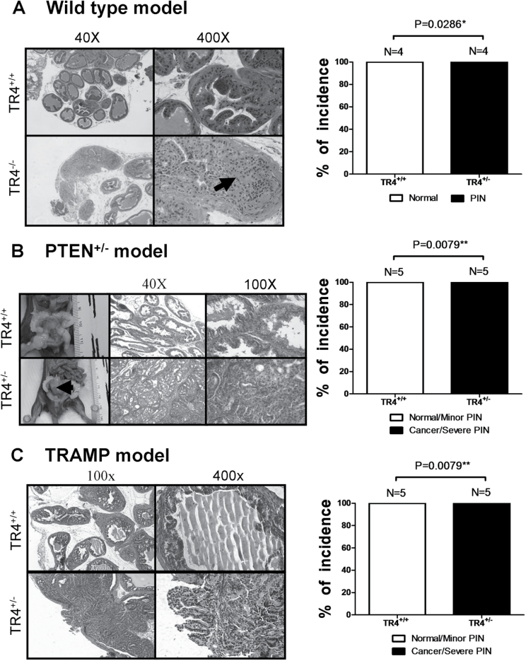
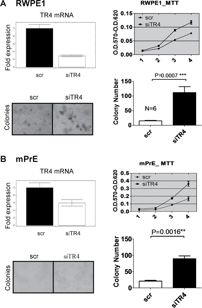
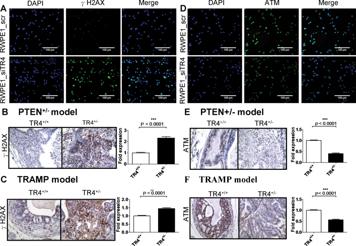
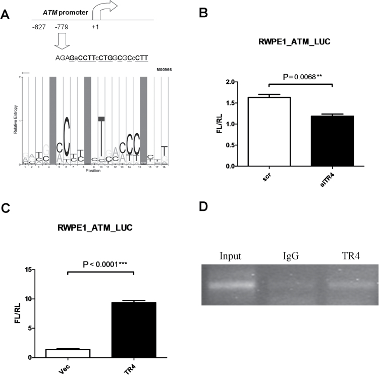
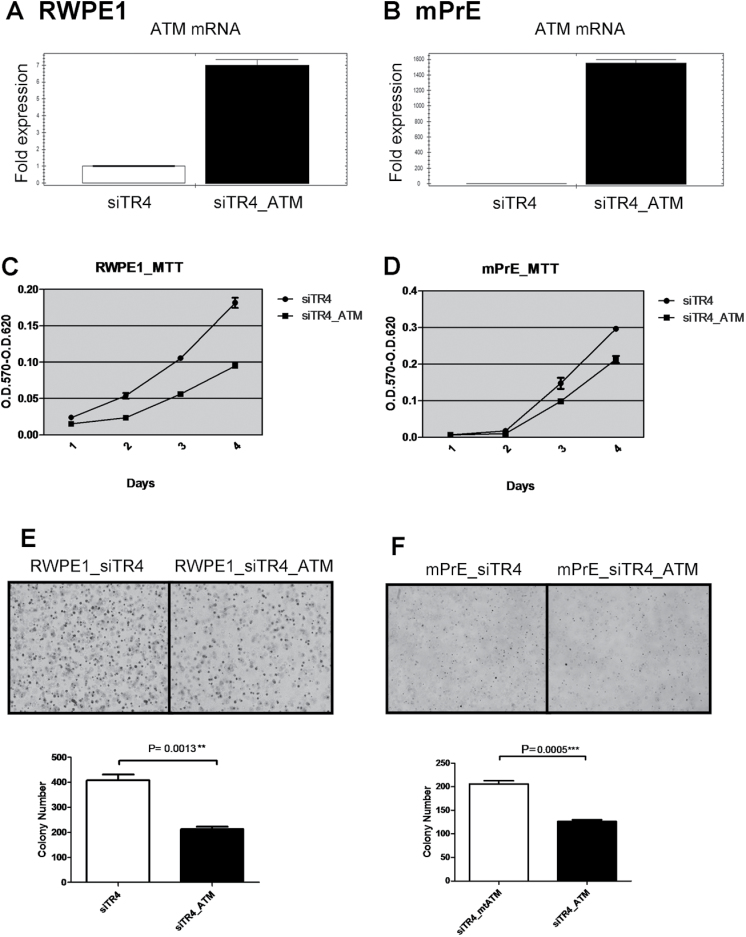
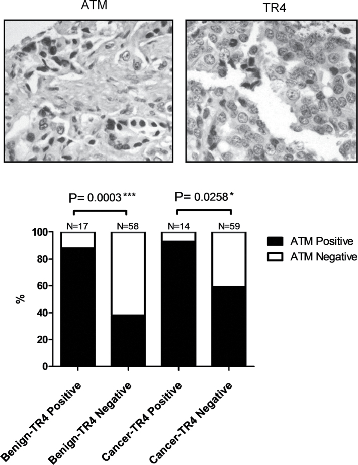
Testicular nuclear receptor 4 (TR4), a member of the nuclear receptor superfamily, plays important roles in metabolism, fertility and aging. The linkage of TR4 functions in cancer progression, however, remains unclear. Using three different mouse models, we found TR4 could prevent or delay prostate cancer (PCa)/prostatic intraepithelial neoplasia development. Knocking down TR4 in human RWPE1 and mouse mPrE normal prostate cells promoted tumorigenesis under carcinogen challenge, suggesting TR4 may play a suppressor role in PCa initiation. Mechanism dissection in both in vitro cell lines and in vivo mice studies found that knocking down TR4 led to increased DNA damage with altered DNA repair system that involved the modulation of ATM expression at the transcriptional level, and addition of ATM partially interrupted the TR4 small interfering RNA-induced tumorigenesis in cell transformation assays. Immunohistochemical staining in human PCa tissue microarrays revealed ATM expression is highly correlated with TR4 expression. Together, these results suggest TR4 may function as a tumor suppressor to prevent or delay prostate tumorigenesis via regulating ATM expression at the transcriptional level.
© The Author 2014. Published by Oxford University Press. All rights reserved. For Permissions, please email: journals.permissions@oup.com.
Figures
Similar articles
-
TR4 nuclear receptor increases prostate cancer invasion via decreasing the miR-373-3p expression to alter TGFβR2/p-Smad3 signals.Oncotarget. 2015 Jun 20;6(17):15397-409. doi: 10.18632/oncotarget.3778. Oncotarget. 2015. PMID: 25980442 Free PMC article.
-
Recent advances in the study of testicular nuclear receptor 4.J Zhejiang Univ Sci B. 2013 Mar;14(3):171-7. doi: 10.1631/jzus.B1200357. J Zhejiang Univ Sci B. 2013. PMID: 23463759 Free PMC article.
-
Differential roles of PPARγ vs TR4 in prostate cancer and metabolic diseases.Endocr Relat Cancer. 2014 Jun;21(3):R279-300. doi: 10.1530/ERC-13-0529. Epub 2014 Mar 12. Endocr Relat Cancer. 2014. PMID: 24623743 Review.
-
TR4 nuclear receptor promotes prostate cancer metastasis via upregulation of CCL2/CCR2 signaling.Int J Cancer. 2015 Feb 15;136(4):955-64. doi: 10.1002/ijc.29049. Epub 2014 Jul 14. Int J Cancer. 2015. PMID: 24975468
-
TR4 Nuclear Receptor Different Roles in Prostate Cancer Progression.Front Endocrinol (Lausanne). 2015 May 27;6:78. doi: 10.3389/fendo.2015.00078. eCollection 2015. Front Endocrinol (Lausanne). 2015. PMID: 26074876 Free PMC article. Review.
Cited by
-
The Differential Effects of Anti-Diabetic Thiazolidinedione on Prostate Cancer Progression Are Linked to the TR4 Nuclear Receptor Expression Status.Neoplasia. 2015 Apr;17(4):339-47. doi: 10.1016/j.neo.2015.02.005. Neoplasia. 2015. PMID: 25925376 Free PMC article.
-
Homeostatic nuclear RAGE-ATM interaction is essential for efficient DNA repair.Nucleic Acids Res. 2017 Oct 13;45(18):10595-10613. doi: 10.1093/nar/gkx705. Nucleic Acids Res. 2017. PMID: 28977635 Free PMC article.
-
Use of miR‑145 and testicular nuclear receptor 4 inhibition to reduce chemoresistance to docetaxel in prostate cancer.Oncol Rep. 2021 Mar;45(3):963-974. doi: 10.3892/or.2021.7925. Epub 2021 Jan 5. Oncol Rep. 2021. PMID: 33650661 Free PMC article.
-
TR4 Nuclear Receptor Alters the Prostate Cancer CD133+ Stem/Progenitor Cell Invasion via Modulating the EZH2-Related Metastasis Gene Expression.Mol Cancer Ther. 2015 Jun;14(6):1445-53. doi: 10.1158/1535-7163.MCT-14-0971. Epub 2015 Apr 1. Mol Cancer Ther. 2015. PMID: 25833838 Free PMC article.
-
TR4 nuclear receptor increases prostate cancer invasion via decreasing the miR-373-3p expression to alter TGFβR2/p-Smad3 signals.Oncotarget. 2015 Jun 20;6(17):15397-409. doi: 10.18632/oncotarget.3778. Oncotarget. 2015. PMID: 25980442 Free PMC article.
References
-
- Siegel R., et al. (2011). Cancer statistics, 2011: the impact of eliminating socioeconomic and racial disparities on premature cancer deaths. Cancer J. Clin., 61, 212–236 - PubMed
-
- Hoffman R.M., et al. (2001). Racial and ethnic differences in advanced-stage prostate cancer: the Prostate Cancer Outcomes Study. J. Natl Cancer Inst., 93, 388–395 - PubMed
-
- Lichtenstein P., et al. (2000). Environmental and heritable factors in the causation of cancer–analyses of cohorts of twins from Sweden, Denmark, and Finland. N. Engl. J. Med., 343, 78–85 - PubMed
-
- Struewing J.P., et al. (1997). The risk of cancer associated with specific mutations of BRCA1 and BRCA2 among Ashkenazi Jews. N. Engl. J. Med., 336, 1401–1408 - PubMed
-
- Chavarro J.E., et al. (2008). A prospective study of trans-fatty acid levels in blood and risk of prostate cancer. Cancer Epidemiol. Biomarkers Prev., 17, 95–101 - PubMed
Publication types
MeSH terms
Substances
Grants and funding
LinkOut - more resources
Full Text Sources
Other Literature Sources
Medical
Molecular Biology Databases
Research Materials
Miscellaneous
