. 2014 Mar 3:5:3413.
doi: 10.1038/ncomms4413.

Astrocytic laminin regulates pericyte differentiation and maintains blood brain barrier integrity

Affiliations

Astrocytic laminin regulates pericyte differentiation and maintains blood brain barrier integrity

Yao Yao et al. Nat Commun. .

Abstract

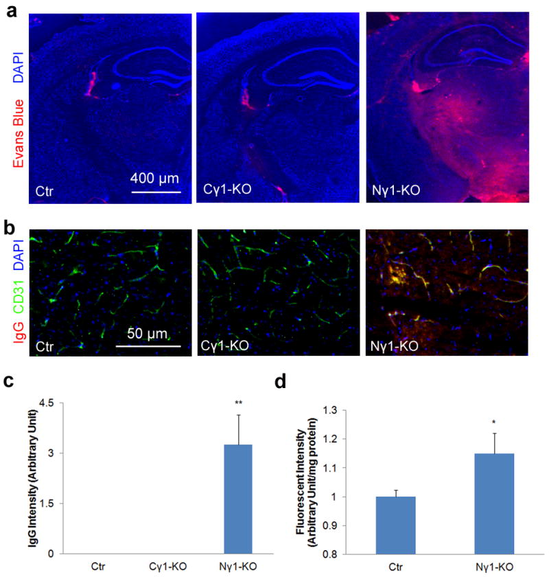
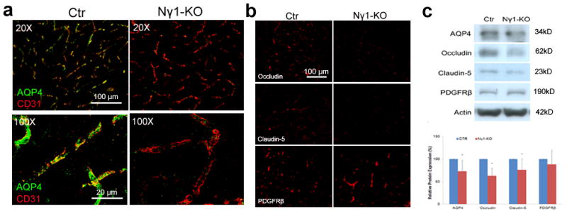
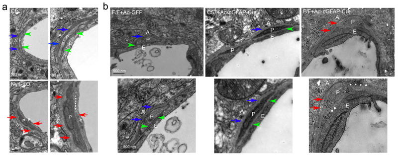
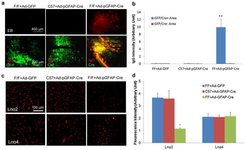
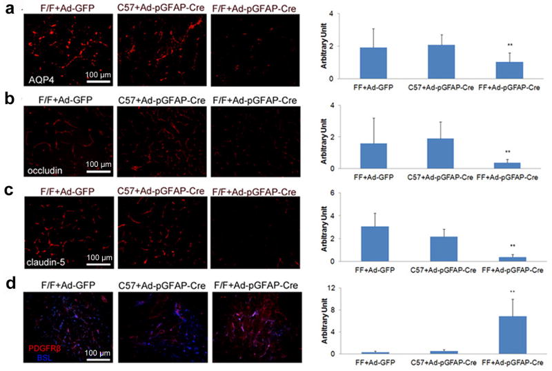
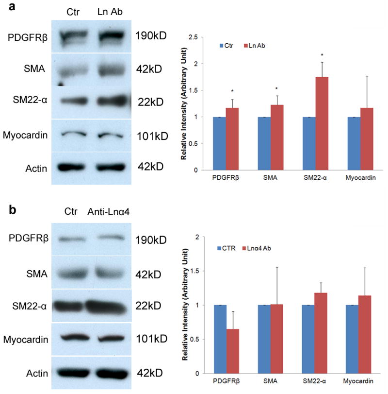
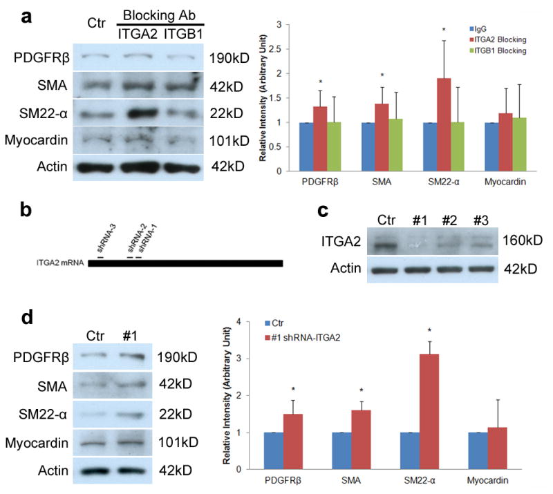
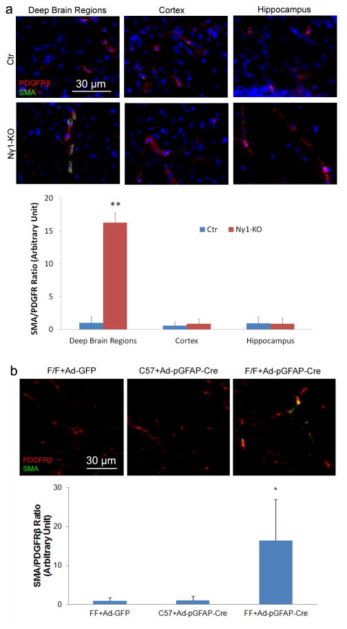
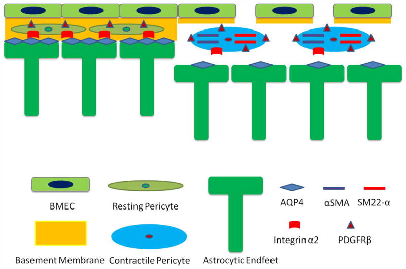
Blood brain barrier (BBB) breakdown is not only a consequence of but also contributes to many neurological disorders, including stroke and Alzheimer's disease. How the basement membrane (BM) contributes to the normal functioning of the BBB remains elusive. Here we use conditional knockout mice and an acute adenovirus-mediated knockdown model to show that lack of astrocytic laminin, a brain-specific BM component, induces BBB breakdown. Using functional blocking antibody and RNAi, we further demonstrate that astrocytic laminin, by binding to integrin α2 receptor, prevents pericyte differentiation from the BBB-stabilizing resting stage to the BBB-disrupting contractile stage, and thus maintains the integrity of BBB. Additionally, loss of astrocytic laminin decreases aquaporin-4 (AQP4) and tight junction protein expression. Altogether, we report a critical role for astrocytic laminin in BBB regulation and pericyte differentiation. These results indicate that astrocytic laminin maintains the integrity of BBB through, at least in part, regulation of pericyte differentiation.


Conflict of interest statement

Competing financial interests

The authors declare no competing financial interests.

Figures

Figure 1
Figure 1
BBB integrity is compromised in Nγ1-KO mice. Mice were injected with Evans blue intraperitoneally 12 hours before the mice were sacrificed. After perfusion, brain sections were used to examine Evans blue directly under fluorescent microscopy and mouse IgG by immunohistochemistry. (a and b) Evans blue (a) and mouse IgG (b) infiltrate the brain parenchyma of Nγ1-KO but not Ctr or Cγ1-KO mice. (c) Quantification of mouse IgG intensity in the brain parenchyma of Nγ1-KO mice. Data are quantified using 9 sections from 3 animals. (d) Quantification of FITC-Dextran in the brain parenchyma. FITC-Dextran (500 kDa) was injected intravenously 12 hours before mice were sacrificed. After perfusion, brains were collected and homogenized. Infiltrated FITC-Dextran was quantified using a fluorescence plate reader at 485 nm (n=3). Scale bars represent 400 μm (a) and 50 μm (b). Data are shown as mean ± sd. *p<0.05 and **p<0.001 versus the Ctrs by student’s t-test.
Figure 2
Figure 2
Individual BBB components are affected in the Nγ1-KO brains. (a) Immunohistochemistry shows that astrocytic endfeet marker AQP4 is expressed at high level in Ctr mice, but dramatically reduced in Nγ1-KO mice. High power confocal Z-projection images show that AQP4 co-localizes with endothelial cell marker CD31 in Ctr but not Nγ1-KO brains. (b) Occludin and claudin-5, but not PDGFRβ, are significantly decreased in Nγ1-KO brains. (c) Representative Western blot of AQP4, occludin, claudin-5, PDGFRβ, and actin using striatum lysates from Ctr and Nγ1-KO mice. Full blots of these proteins are shown in Supplementary Figure 14a. Western blot was quantified by densitometry. All bands were normalized to actin (n=6). Scale bars represent 100 μm (low magnification images in a and b) and 20 μm (high magnification images in a). Data are shown as mean ± sd. *p<0.05 versus the Ctrs by student’s t-test.
Figure 3
Figure 3
Ultrastructural changes in Nγ1-KO and laminin-knockdown brains. (a) In Ctr mice, the BM between astrocytes and pericytes (blue arrows) and that between pericytes and endothelial cells (green arrowheads) are compact and pericytes are tightly attached to endothelial cells. In the Nγ1-KO mice, however, pericytes change their morphology and both BMs show a patchy and diffused pattern (red arrows). Moreover, the endothelial membrane integrity is compromised at some places (white stars) in Nγ1-KO mice. (b) In Ctr mice (F/F+Ad-GFP and C57+Ad-pGFAP-Cre), pericytes are surrounded by nicely aligned and compact BMs secreted by both astrocytes (blue arrows) and endothelial cells (green arrowheads). In astrocytic laminin-knockdown mice (F/F+Ad-pGFAP-Cre), on the other hand, pericytes change their morphology and both BMs become patchy and diffuse (red arrows). In addition, the BMs are hard to define at some places and the membrane integrity is severely compromised in these astrocytic laminin-knockdown mice (white stars). A: Astrocytes; P: Pericytes; E: Endothelia. Scale bars represent 500 nm (low magnification images in a on the left and b), and 100 nm (high magnification images in a on the right).
Figure 4
Figure 4
Acute ablation of astrocytic laminin leads to BBB breakdown. Mice were injected with adenovirus as indicated. Seven days after injection, the brains were collected and analyzed. (a) Evans blue and mouse IgG infiltrate into the brain parenchyma of F/F mice with Ad-pGFAP-Cre injection, but not the Ctrs---F/F mice with Ad-GFP injection or C57Bl6 mice with Ad-pGFAP-Cre injection. GFP (for Ad-GFP injected brains) and Cre (for Ad-pGFAP-Cre injected brains) were used to reveal infected regions. (b) Quantification of IgG intensity in GFP/Cre positive and negative areas in adenovirus-injected mouse brains. Data are quantified using 9 sections from 3 animals per group. (c) Laminin is specifically knocked down in astrocytes by adenoviruses. Seven days after adenoviral injection, brains were collected and immunostained for laminin α2 (Lnα2, marker for astrocytic laminin) and α4 (Lnα4, marker for endothelial laminin). Our data show that the adenoviruses we used specifically knocked down astrocytic laminin without affecting endothelial laminin. (d) Quantification of Lnα2 and Lnα4 intensity in the brain parenchyma of the adenovirus-injected mice. Data are quantified using 6 sections from 3 animals per group. Scale bars represent 400 μm (EB in a), 200 μm (IgG in a) and 100 μm (c). Data are shown as mean ± sd. *p<0.05 and **p<0.001 versus the Ctrs by student’s t-test.
Figure 5
Figure 5
Acute ablation of astrocytic laminin affects individual BBB components. Mice were injected with adenovirus as indicated. Seven days after injection, the brains were collected and analyzed. (a) Immunohistochemistry shows that astrocytic endfeet marker AQP4 is significantly decreased in F/F mice with Ad-pGFAP-Cre injection, but not in the Ctrs--- F/F mice with Ad-GFP injection or C57 mice with Ad-pGFAP-Cre injection. AQP4 fluorescence intensity was quantified using 12–13 random sections from 3 or more mice per grop. (b and c) Immunohistochemistry shows that occludin (b) and claudin-5 (c) are significantly decreased in F/F mice with Ad-pGFAP-Cre injection, but not in the Ctrs. The fluorescence intensity of these proteins was quantified using 9–13 random sections from at least 3 mice per group. (d) Immunohistochemistry shows that the pericyte marker PDGFRβ is significantly increased in F/F mice with Ad-pGFAP-Cre injection, but not in the Ctrs. PDGFRβ fluorescence intensity was quantified using 11–12 random sections from at least 3 mice per group. Scale bars represent 100 μm. Data are shown as mean ± sd. **p<0.001 versus the Ctrs by student’s t-test.
Figure 6
Figure 6
Laminin-111 but not laminin-α4 blocking antibody affects pericyte differentiation. (a) Immunoblots show that laminin-111 blockage (Ln Ab) significantly enhances the expression of PDGFRβ, SMA, and SM22-α, but not myocardin in pericytes. Full blots of these proteins are shown in Supplementary Figure 14b. Rabbit IgG treated cells were used as a Ctr. All bands were normalized to actin (n=5–6). (b) Immunoblots show that laminin-α4 blockage (Anti-Lnα4) does not change the expression of PDGFRβ, SMA, SM22-α, or myocardinin in pericytes. Full blots of these proteins are shown in Supplementary Figure 14c. Rabbit IgG treated cells were used as a Ctr. All bands were normalized to actin (n=3). Data are shown as mean ± sd. *p<0.05 versus the Ctrs by student’s t-test.
Figure 7
Figure 7
Astrocytic laminin mediates pericyte differentiation via integrin α2. (a) Immunoblots show that integrin α2 blockage (ITGA2) but not integrin β1 blockage significantly increases the expression of PDGFRβ, SMA, and SM22-α, but not myocardin in pericytes. Full blots of these proteins are shown in Supplementary Figure 14d. Rabbit IgG treated cells were used as a Ctr. All bands were normalized to actin (n=6). (b) Schematic diagram of shRNA designed to target ITGA2 mRNA. (c) Immunoblot analysis shows that all three ITGA2-specific shRNAs (#1–3) dramatically reduce ITGA2 at protein level and ITGA2-specific shRNA-3 (#1) is the most efficient one. Full blots of ITGA2 and actin are shown in Supplementary Figure 14e. Scramble shRNA was used as a Ctr. (d) Immunoblot analysis shows that transduction of pericytes with lenti-shRNA-1 (#1) significantly enhances the expression of PDGFRβ, SMA, and SM22-α, but does not affect myocardin level. Full blots of these proteins are shown in Supplementary Figure 14f. Scramble shRNA was used as a Ctr. All bands were normalized to actin (n=4–5). Data are shown as mean ± sd. *p<0.05 versus the Ctrs by student’s t-test.
Figure 8
Figure 8
Loss of astrocytic laminin increases the expression of SMA by capillary pericytes in vivo. Immunohistochemistry analysis shows significant enhanced SMA expression in PDGFRβ positive capillary pericytes in deep brain regions of Nγ1-KO mice (a). In the Ctr brain or the cortex and hippocampus of Nγ1-KO mice, however, no SMA positive capillary pericytes are observed (a). In addition, acute ablation of astrocytic laminin by intracranial injection of adenoviruses significantly elevates the expression of SMA in PDGFRβ positive pericytes (b). The quantification was performed by calculating the ratio of SMA intensity to PDGFRβ intensity in 9–10 random sections from at least 3 mice per group. Scale bars represent 30 μm. Data are shown as mean ± sd. *p<0.05 and **p<0.01 versus Ctrs by student’s t-test.
Figure 9
Figure 9
Proposed model of how astrocytic laminin affects pericyte differentiation and BBB integrity. Under physiological conditions, pericytes are embedded in the BMs produced by both astrocytes and endothelial cells. They are in the resting stage, signified by the low expression levels of PDGFRβ and contractile proteins (SMA and SM-22α). When astrocytic laminin is lost during pathological conditions, pericytes hypertrophy and the expression levels of PDGFRβ and contractile proteins are enhanced, switching pericytes to their contractile stage. This process is mediated by integrin α2. This transition changes the function of pericytes from BBB stabilization to BBB disruption. In addition, lack of astrocytic laminin also leads to reduction of AQP4 expression at astrocytic endfeet. Together these effects result in decreased expression of tight junction proteins on endothelial cells, leading to BBB breakdown and subsequent damage.

Similar articles

Cited by

References

    1. Persidsky Y, Ramirez SH, Haorah J, Kanmogne GD. Blood-brain barrier: structural components and function under physiologic and pathologic conditions. J Neuroimmune Pharmacol. 2006;1:223–236. - PubMed
    1. Zipser BD, et al. Microvascular injury and blood-brain barrier leakage in Alzheimer’s disease. Neurobiol Aging. 2007;28:977–986. - PubMed
    1. van Vliet EA, et al. Blood-brain barrier leakage may lead to progression of temporal lobe epilepsy. Brain. 2007;130:521–534. - PubMed
    1. Wardlaw JM, et al. Lacunar stroke is associated with diffuse blood-brain barrier dysfunction. Ann Neurol. 2009;65:194–202. - PubMed
    1. Guillemin GJ, Brew BJ. Microglia, macrophages, perivascular macrophages, and pericytes: a review of function and identification. J Leukoc Biol. 2004;75:388–397. - PubMed

Publication types

MeSH terms