

Leukocyte infiltration and activation of the NLRP3 inflammasome in white adipose tissue following thermal injury
- PMID: 24584061
- PMCID: PMC4166573
- DOI: 10.1097/CCM.0000000000000209
Leukocyte infiltration and activation of the NLRP3 inflammasome in white adipose tissue following thermal injury
Abstract
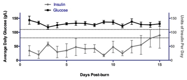
Objectives: Severe thermal injury is associated with extreme and prolonged inflammatory and hypermetabolic responses, resulting in significant catabolism that delays recovery or even leads to multiple organ failure and death. Burned patients exhibit many symptoms of stress-induced diabetes, including hyperglycemia, hyperinsulinemia, and hyperlipidemia. Recently, the nucleotide-binding domain, leucine-rich family (NLR), pyrin-containing 3 (NLRP3) inflammasome has received much attention as the sensor of endogenous "danger signals" and mediator of "sterile inflammation" in type II diabetes. Therefore, we investigated whether the NLRP3 inflammasome is activated in the adipose tissue of burned patients, as we hypothesize that, similar to the scenario observed in chronic diabetes, the cytokines produced by the inflammasome mediate insulin resistance and metabolic dysfunction.
Design: Prospective cohort study.
Setting: Ross Tilley Burn Centre & Sunnybrook Research Institute.
Patients: We enrolled 76 patients with burn sizes ranging from 1% to 70% total body surface area. All severely burned patients exhibited burn-induced insulin resistance and hyperglycemia.
Interventions: None.
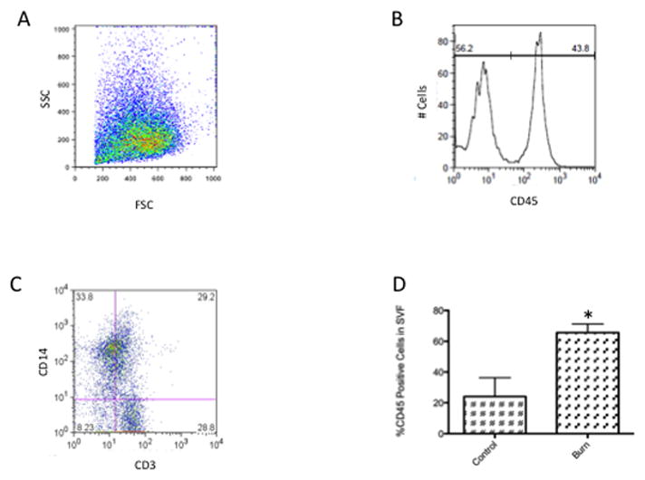
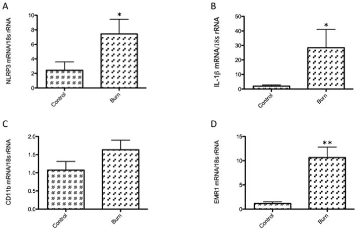
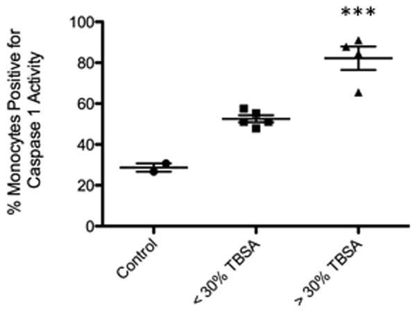
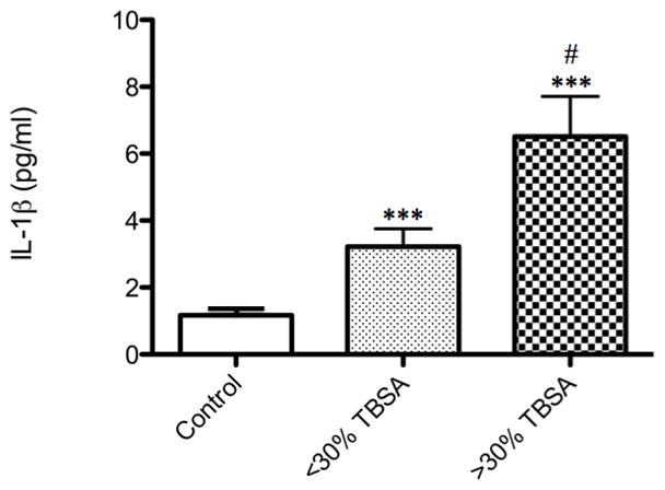
Measurements and main results: We examined the adipose tissue of control and burned patients and found, via flow cytometry and gene expression studies, increased infiltration of leukocytes-especially macrophages-and evidence of inflammasome priming and activation. Furthermore, we observed increased levels of interleukin-1β in the plasma of burned patients when compared to controls.
Conclusions: In summary, our study is the first to show activation of the inflammasome in burned humans, and our results provide impetus for further investigation of the role of the inflammasome in burn-induced hypermetabolism and, potentially, developing novel therapies targeting this protein complex for the treatment of stress-induced diabetes.
Figures
Comment in
-
Remote inflammatory and metabolic impact of acute injury.Crit Care Med. 2014 Jun;42(6):1539-40. doi: 10.1097/CCM.0000000000000292. Crit Care Med. 2014. PMID: 24836786 No abstract available.
Similar articles
-
Apelin inhibits the activation of the nucleotide-binding domain and the leucine-rich, repeat-containing family, pyrin-containing 3 (NLRP3) inflammasome and ameliorates insulin resistance in severely burned rats.Surgery. 2015 Jun;157(6):1142-52. doi: 10.1016/j.surg.2015.01.011. Epub 2015 Mar 25. Surgery. 2015. PMID: 25817096
-
Remote inflammatory and metabolic impact of acute injury.Crit Care Med. 2014 Jun;42(6):1539-40. doi: 10.1097/CCM.0000000000000292. Crit Care Med. 2014. PMID: 24836786 No abstract available.
-
Lower NLRP3 inflammasome activity in NAG-1 transgenic mice is linked to a resistance to obesity and increased insulin sensitivity.Obesity (Silver Spring). 2014 May;22(5):1256-63. doi: 10.1002/oby.20638. Epub 2013 Dec 5. Obesity (Silver Spring). 2014. PMID: 24124102 Free PMC article.
-
Regulation and Function of the Nucleotide Binding Domain Leucine-Rich Repeat-Containing Receptor, Pyrin Domain-Containing-3 Inflammasome in Lung Disease.Am J Respir Cell Mol Biol. 2016 Feb;54(2):151-60. doi: 10.1165/rcmb.2015-0231TR. Am J Respir Cell Mol Biol. 2016. PMID: 26418144 Free PMC article. Review.
-
The NLRP3 inflammasome regulates adipose tissue metabolism.Biochem J. 2020 Mar 27;477(6):1089-1107. doi: 10.1042/BCJ20190472. Biochem J. 2020. PMID: 32202638 Review.
Cited by
-
White Adipose Tissue Browning: A Double-edged Sword.Trends Endocrinol Metab. 2016 Aug;27(8):542-552. doi: 10.1016/j.tem.2016.06.006. Epub 2016 Jul 5. Trends Endocrinol Metab. 2016. PMID: 27397607 Free PMC article. Review.
-
Accumulation of myeloid lineage cells is mapping out liver fibrosis post injury: a targetable lesion using Ketanserin.Exp Mol Med. 2018 Jul 19;50(7):1-13. doi: 10.1038/s12276-018-0118-x. Exp Mol Med. 2018. PMID: 30026607 Free PMC article.
-
Impaired Immune Response in Elderly Burn Patients: New Insights Into the Immune-senescence Phenotype.Ann Surg. 2016 Jul;264(1):195-202. doi: 10.1097/SLA.0000000000001408. Ann Surg. 2016. PMID: 26649579 Free PMC article.
-
Modulation of Burn Hypermetabolism in Preclinical Models.Cureus. 2023 Jan 8;15(1):e33518. doi: 10.7759/cureus.33518. eCollection 2023 Jan. Cureus. 2023. PMID: 36779088 Free PMC article. Review.
-
Systemic immune response of burns from the acute to chronic phase.Acute Med Surg. 2024 Jun 18;11(1):e976. doi: 10.1002/ams2.976. eCollection 2024 Jan-Dec. Acute Med Surg. 2024. PMID: 38894736 Free PMC article. Review.
References
Publication types
MeSH terms
Substances
Grants and funding
LinkOut - more resources
Full Text Sources
Other Literature Sources
Medical
