

Seroprevalences of Toxoplasma gondii and Neospora caninum in pet rabbits in Japan
- PMID: 24584081
- PMCID: PMC4108769
- DOI: 10.1292/jvms.13-0632
Seroprevalences of Toxoplasma gondii and Neospora caninum in pet rabbits in Japan
Abstract
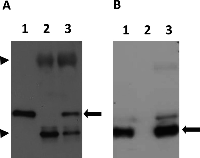
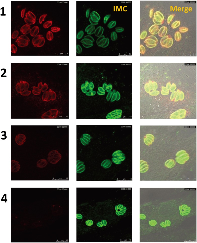
The potential contamination of Toxoplasma gondii and Neospora caninum oocysts in the human environment is a concern from the public health viewpoint. However, estimation of their seroprevalences in humans cannot be performed in a manner that distinguishes between oocysts and tissue cysts as a source of infection. Rabbits are considered popular pet animals in Japan that can acquire natural infections by the aforementioned parasites only through the ingestion of oocysts. Therefore, this study was conducted to estimate the seroprevalences of T. gondii and N. caninum in pet rabbits in Japan as an indicator of the possible oocyst contamination in the environment surrounding human beings. Serum samples of 337 rabbits were examined by different serological methods. Enzyme-linked immunosorbent assays were performed to measure the titer of IgG and IgM antibodies. Samples revealed to be seropositive by ELISA were further analyzed by a latex agglutination test, Western blotting and an indirect immunofluorescence assay. The rates of seropositivity for T. gondii were 0.89% (3/337) and 0.29% (1/337) in IgG and IgM ELISA, respectively. SAG1 and SAG2 were detected as major antigens by the positive rabbit sera in Western blotting associated with strong staining observed by IFA in T. gondii tachyzoites. Regarding N. caninum, none of the serum samples showed a specific reaction in both Western blotting and the IFA. The results of this study indicate low seroprevalences of toxoplasmosis and neosporosis in pet rabbits in Japan, suggesting low oocyst contamination in the human environment.
Figures

Similar articles
-
Seroprevalence, risk factors, and serological cross-reactivity for diagnosis of Toxoplasma gondii and Neospora caninum infections in goats in India.Microb Pathog. 2022 Dec;173(Pt A):105780. doi: 10.1016/j.micpath.2022.105780. Epub 2022 Sep 17. Microb Pathog. 2022. PMID: 36122852
-
Evaluation of Toxoplasma gondii and Neospora caninum infections in sheep from Uberlândia, Minas Gerais State, Brazil, by different serological methods.Vet Parasitol. 2011 Feb 10;175(3-4):252-9. doi: 10.1016/j.vetpar.2010.10.017. Epub 2010 Oct 16. Vet Parasitol. 2011. PMID: 21075529
-
Evaluation of serological tests for the diagnosis of Neospora caninum infection in dogs: optimization of cut off titers and inhibition studies of cross-reactivity with Toxoplasma gondii.Vet Parasitol. 2007 Feb 28;143(3-4):234-44. doi: 10.1016/j.vetpar.2006.08.028. Epub 2006 Sep 12. Vet Parasitol. 2007. PMID: 16973287
-
Seroprevalence of Toxoplasma gondii and Neospora caninum infections in cattle in Mongolia.Vet Parasitol Reg Stud Reports. 2018 Dec;14:11-17. doi: 10.1016/j.vprsr.2018.08.001. Epub 2018 Aug 18. Vet Parasitol Reg Stud Reports. 2018. PMID: 31014714
-
Identification of Oocyst-Driven Toxoplasma gondii Infections in Humans and Animals through Stage-Specific Serology-Current Status and Future Perspectives.Microorganisms. 2021 Nov 13;9(11):2346. doi: 10.3390/microorganisms9112346. Microorganisms. 2021. PMID: 34835471 Free PMC article. Review.
Cited by
-
Seroprevalence of Neospora caninum in pet cats, dogs and rabbits from urban areas of Poland.BMC Vet Res. 2024 Jan 31;20(1):37. doi: 10.1186/s12917-024-03894-3. BMC Vet Res. 2024. PMID: 38297328 Free PMC article.
-
Lactate dehydrogenase in Toxoplasma gondii controls virulence, bradyzoite differentiation, and chronic infection.PLoS One. 2017 Mar 21;12(3):e0173745. doi: 10.1371/journal.pone.0173745. eCollection 2017. PLoS One. 2017. PMID: 28323833 Free PMC article.
-
Dembo polymerase chain reaction technique for detection of bovine abortion, diarrhea, and respiratory disease complex infectious agents in potential vectors and reservoirs.J Vet Sci. 2018 May 31;19(3):350-357. doi: 10.4142/jvs.2018.19.3.350. J Vet Sci. 2018. PMID: 29284216 Free PMC article.
-
Wild Rabbit Exposure to Leishmania infantum, Toxoplasma gondii, Anaplasma phagocytophilum and Babesia caballi Evidenced by Serum and Aqueous Humor Antibody Detection.Microorganisms. 2021 Dec 17;9(12):2616. doi: 10.3390/microorganisms9122616. Microorganisms. 2021. PMID: 34946216 Free PMC article.
-
Prevalence of Toxoplasma gondii and other intestinal parasites in cats in Tokachi sub-prefecture, Japan.J Vet Med Sci. 2018 Jun 29;80(6):960-967. doi: 10.1292/jvms.17-0713. Epub 2018 May 2. J Vet Med Sci. 2018. PMID: 29731476 Free PMC article.
References
-
- Barr B. C., Conrad P. A., Sverlow K. W., Tarantal A. F., Hendrickx A. G.1994. Experimental fetal and transplacental Neospora infection in the nonhuman primate. Lab. Invest. 71: 236–242 - PubMed
-
- Boyer K., Hill D., Mui E., Wroblewski K., Karrison T., Dubey J. P., Sautter M., Noble A. G., Withers S., Swisher C., Heydemann P., Hosten T., Babiarz J., Lee D., Meier P., McLeod R.2011. Unrecognized ingestion of Toxoplasma gondii oocysts leads to congenital toxoplasmosis and causes epidemics in North America. Clin. Infect. Dis. 53: 1081–1089. doi: 10.1093/cid/cir667 - DOI - PMC - PubMed
Publication types
MeSH terms
Substances
LinkOut - more resources
Full Text Sources
Other Literature Sources