Review
. 2015 Jan;30(1):15-30.
doi: 10.1007/s00467-013-2706-2. Epub 2014 Mar 1.

ARPKD and early manifestations of ADPKD: the original polycystic kidney disease and phenocopies

Affiliations
Review

ARPKD and early manifestations of ADPKD: the original polycystic kidney disease and phenocopies

Carsten Bergmann. Pediatr Nephrol. 2015 Jan.

Abstract

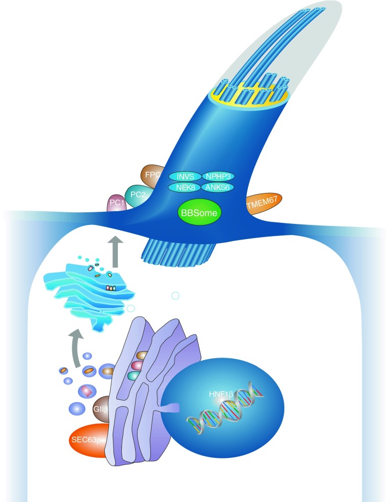
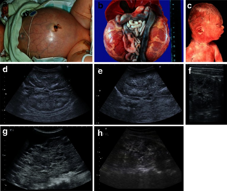
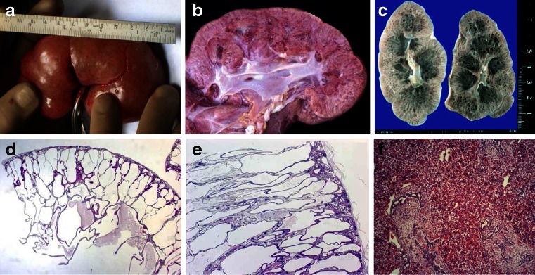
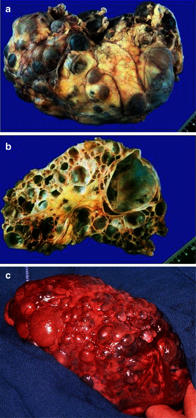
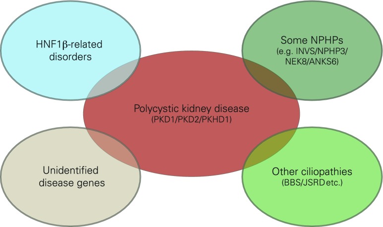
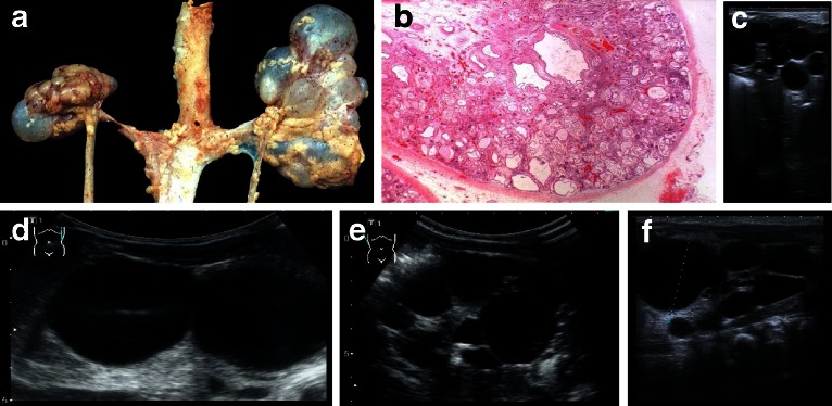
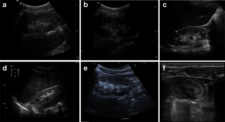
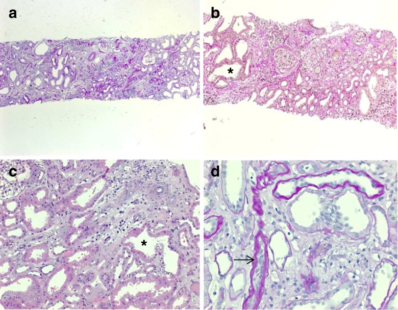
Renal cysts are clinically and genetically heterogeneous conditions. Polycystic kidney disease (PKD) is common and its characterization has paved the way for the identification of a growing number of cilia-related disorders (ciliopathies) of which most show cystic kidneys. While the recessive form of PKD (ARPKD) virtually always presents in childhood, early onset can, in some instances, also occur in the dominant form (ADPKD). Both ADPKD genes (PKD1 and PKD2) can also be inherited in a recessive way, making the story more complex with evidence for a dosage-sensitive network. Several phenocopies are known, and mutations in HNF1ß or genes that typically cause other ciliopathies, such as nephronophthisis, Bardet-Biedl, Joubert syndrome and related disorders, can mimic PKD. An accurate genetic diagnosis is crucial for genetic counseling, prenatal diagnostics, and the clinical management of patients and their families. The increasing number of genes that have to be considered in patients with cystic kidney disease is challenging to address by conventional techniques and largely benefits from next-generation sequencing-based approaches. The parallel analysis of targeted genes considerably increases the detection rate, allows for better interpretation of identified variants, and avoids genetic misdiagnoses.


Figures

Fig. 1
Fig. 1
Schematic diagram of a primary cilium and associated processes. Polycystic kidney disease (PKD) is controlled by a defined network of different genes/proteins discussed in this review. Cilia are small antennae that detect a variety of different extracellular stimuli and orchestrate multiple signaling pathways with nuclear trafficking of some molecules [e.g., C-termini of polycystin-1 (PC1) and fibrocystin/polyductin (FPC)]. The inner ciliary structure is defined by the axoneme that is composed of nine microtubule doublets derived from the basal body and mother centriole of the centrosome. Along this microtubule core, the transport of proteins in the anterograde direction toward the tip of the cilium and in the retrograde direction towards the cell body is organized by an elaborate process called intraflagellar transport
Fig. 2
Fig. 2
ah Autosomal recessive polycystic kidney disease (ARPKD). a Baby with distended abdomen due to voluminous kidneys that led to respiratory problems and early demise. b Abdominal situs of a perinatally demised ARPKD patient with symmetrically enlarged kidneys that maintained their reniform configuration. c Potter’s phenotype with distinctive facial features due to a lack of amniotic fluid. dh Renal ultrasound scans of babies and young children with ARPKD. Symmetrically enlarged echogenic kidneys with fusiform dilations of collecting ducts and distal tubules are arranged radially throughout the renal parenchyma from medulla to cortex
Fig. 3
Fig. 3
af Microscopic and macroscopic appearance of autosomal recessive polycystic kidney disease (ARPKD). a Considerably enlarged kidney with retained reniform configuration (patient passed away at the age  of  2.5 months). b, c Cross sections of ARPKD kidneys reveal cortical extension of fusiform or cylindrical spaces arranged radially throughout the renal parenchyma from medulla to cortex. d, e Microscopic appearance of fusiform dilations of renal collecting ducts and distal tubuli lined by columnar or cuboidal epithelium. These dilated collecting ducts run perpendicular to the renal capsule. f Obligatory hepatobiliary changes in ARPKD subsumed as ductal plate malformation and characterized by dysgenesis of the hepatic portal triad with hyperplastic biliary ducts and congenital hepatic fibrosis
Fig. 4
Fig. 4
Typical sonographic images of autosomal dominant polycystic kidney disease (ADPKD) in different patients of adolescent age
Fig. 5
Fig. 5
a, c Macroscopic appearance of advanced-stage autosomal dominant polycystic kidney disease (ADPKD) showing enlarged kidneys with multiple cysts that almost completely destroyed and replaced the renal parenchyma. b On cut section, multiple cysts in the cortex and medulla can be seen that vary considerably in size and appearance, from a few millimeters to diameters of many centimeters
Fig. 6
Fig. 6
Several phenocopies of polycystic kidney disease (PKD) are known and mutations in the transcription factor hepatocyte nuclear factor 1 beta (HNF1ß) or genes that typically cause other ciliopathies, such as nephronophthisis (NPHP), Bardet-Biedl (BBS), and Joubert syndrome and related disorders (JSRD), can mimic PKD in some cases
Fig. 7
Fig. 7
a Cystic kidney dysplasia due to early embryonic maldevelopment (Potter type II). Both kidneys have lost their reniform structure. The histologic (b) and ultrasonographic (cf) pattern is irregular, with excessive connective tissue in between cysts of various sizes and at different locations
Fig. 8
Fig. 8
af Sonographic appearance of patients of different ages with nephronophthisis (NPHP). All patients show a typical NPHP pattern with normal or small-sized kidneys, enhanced echogenicity of the renal cortex, and reduced cortico-medullary differentiation (progressive with renal failure); cysts (e.g., patient in c) are usually a late sign and occur secondarily after patients have progressed to end-stage renal disease and are typically located at the cortico-medullary junction. a, b 10-year-old boy with homozygous NPHP1 deletion, serum creatinine of 3 mg/dl, glomerular filtration rate of 20 ml/min and mild arterial hypertension. c, d 10- and 15-year-old patients with progressed clinical course. e 14-year-old boy with Senior–Loken syndrome carrying a homozygous deletion of the NPHP1 gene. Peritoneal dialysis was started recently. f 2-day-old male patient with sonographic evidence of NPHP, polyuria, and systemic hypertension
Fig. 9
Fig. 9
ad Renal histology of a 14-year-old female with juvenile NPHP carrying a homozygous NPHP1 deletion. Chronic tubulo-interstitial alterations with tubular basement membrane disintegration and thickening, focal tubular atrophy (arrow in d), cystic dilated tubuli (asterisk in b and c), and disproportionate tubulo-interstitial fibrosis with some inflammatory cells were observed

References

    1. Osathanondh V, Potter EL. Pathogenesis of polycystic kidneys. Survey of results of microdissection. Arch Pathol. 1964;77:510–512. - PubMed
    1. Badano JL, Mitsuma N, Beales PL, Katsanis N. The ciliopathies: an emerging class of human genetic disorders. Annu Rev Genomics Hum Genet. 2006;7:125–148. doi: 10.1146/annurev.genom.7.080505.115610. - DOI - PubMed
    1. Bergmann C. Ciliopathies. Eur J Pediatr. 2012;171(9):1285–1300. doi: 10.1007/s00431-011-1553-z. - DOI - PMC - PubMed
    1. Harris PC, Torres VE (2009) Polycystic kidney disease. Annu Rev Med 60:321–337 - PMC - PubMed
    1. Kaariainen H, Koskimies O, Koskimies O. Dominant and recessive polycystic kidney disease in children: evaluation of clinical features and laboratory data. Pediatr Nephrol. 1988;2(3):296–302. doi: 10.1007/BF00858681. - DOI - PubMed

MeSH terms

LinkOut - more resources