. 2014 Mar 3:4:4258.
doi: 10.1038/srep04258.

Inhibition of post-translational N-glycosylation by HRD1 that controls the fate of ABCG5/8 transporter

Affiliations

Inhibition of post-translational N-glycosylation by HRD1 that controls the fate of ABCG5/8 transporter

Shingo Suzuki et al. Sci Rep. .

Abstract

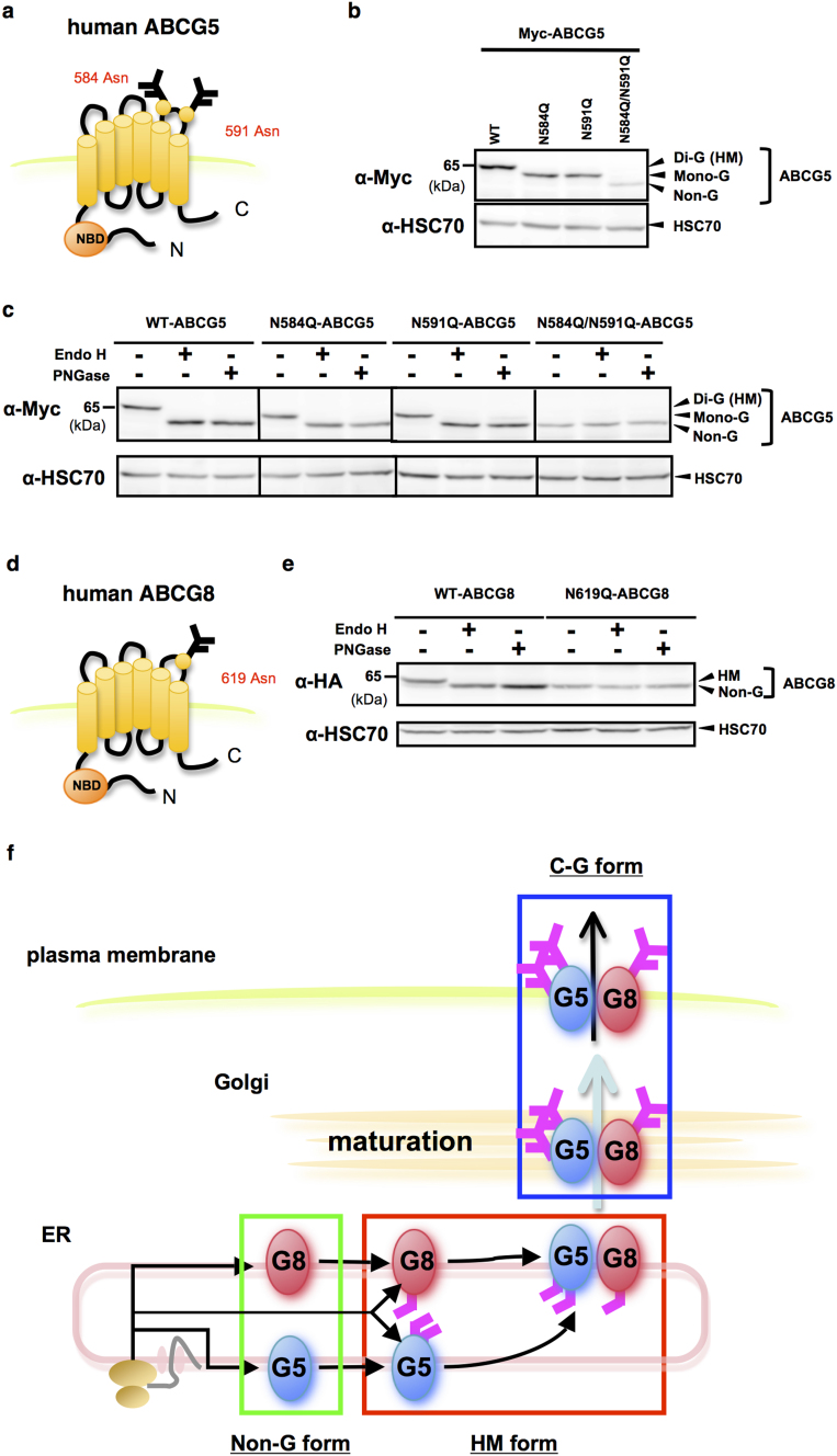
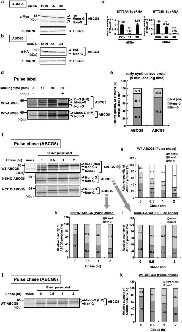
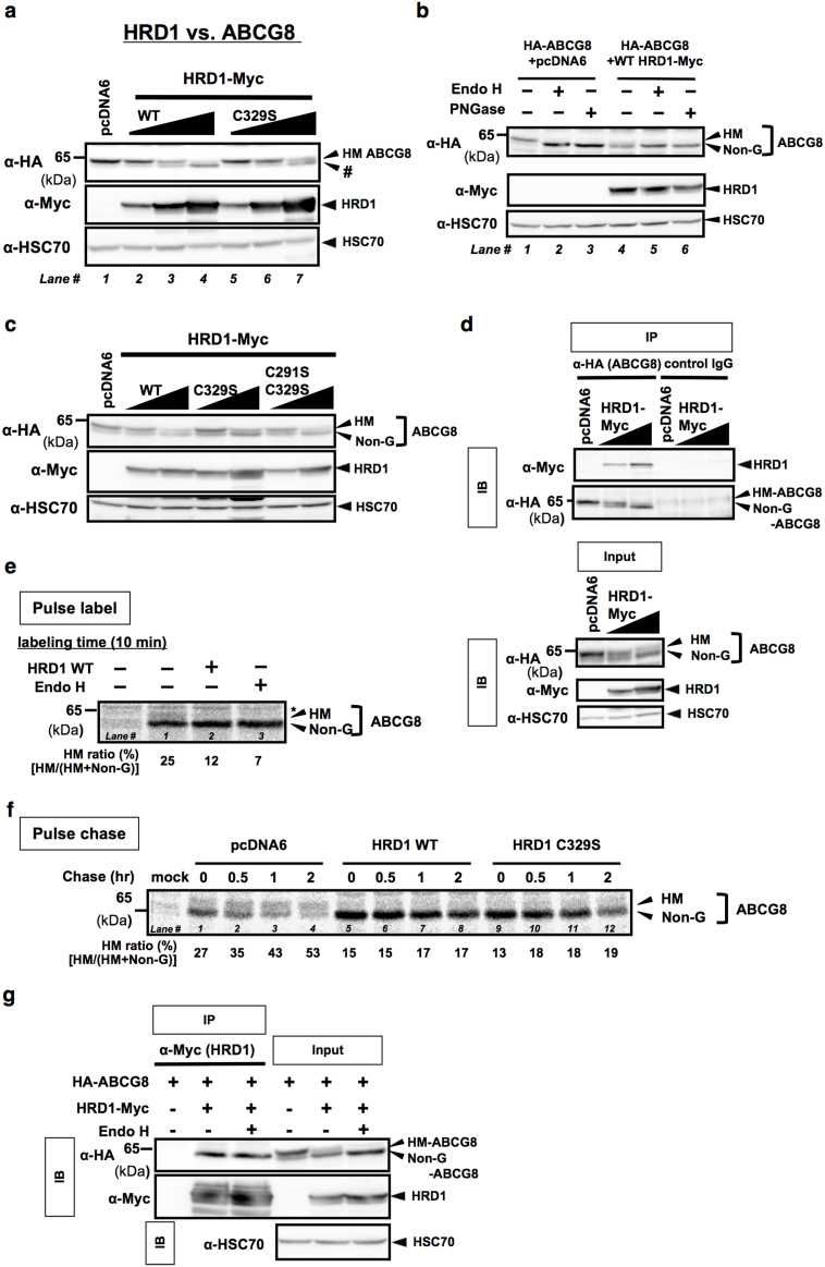
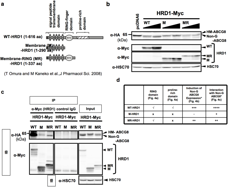
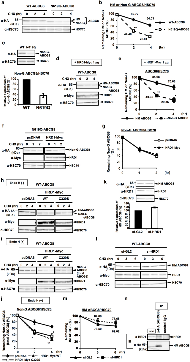
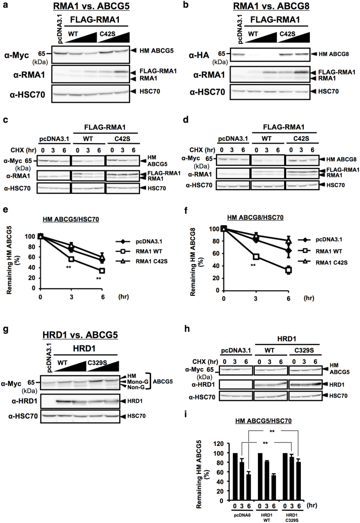
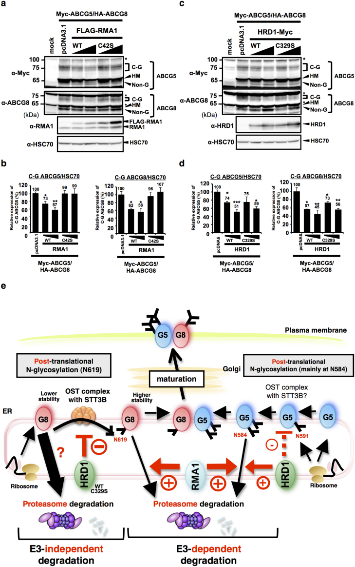
N-glycosylation of proteins in endoplasmic reticulum is critical for protein quality control. We showed here a post-translational N-glycosylation affected by the HRD1 E3 ubiquitin ligase. Both WT- and E3-defective C329S-HRD1 decreased the level of high mannose form of ABCG8, a protein that heterodimerizes with ABCG5 to control sterol balance. Meanwhile, HRD1 increased the non-glycosylated ABCG8 regardless of its E3 activity, thereby suppressing full maturation of ABCG5/8 transporter. Pulse chase and mutational analysis indicated that HRD1 inhibits STT3B-dependent post-translational N-glycosylation of ABCG8. Whereas, HRD1 had only slight effect on the N-glycosylation status of ABCG5; rather it accelerated ABCG5 degradation in an E3 activity-dependent manner. Finally, RMA1, another E3 ubiquitin ligase, accelerated the degradation of both ABCG5 and ABCG8 via E3 activity-dependent manner. HRD1 and RMA1 may therefore be negative regulators of disease-associated transporter ABCG5/ABCG8. The findings also highlight the unexpected E3 activity-independent role of HRD1 in the regulation of N-glycosylation.


Figures

Figure 1
Figure 1. Determination of N-linked glycosylation sites in human ABCG5 and ABCG8.
(a) Putative asparagine residues to be N-glycosylated in human ABCG5 protein. (b) Steady-state expression of monomeric Myc-tagged WT-, N584Q-, N591Q- and N584Q-/N591Q-ABCG5 was analyzed by immunoblotting. (c) Endo H (500 U) and PNGase F (500 U) sensitivity of Myc-tagged WT-, N584Q-, N591Q- and N584Q-/N591Q-ABCG5 proteins shown in (b). (d) Putative asparagine residue to be N-glycosylated in human ABCG8 protein. (e) Endo H and PNGase F sensitivity of HA-tagged WT- or N619Q-ABCG8 proteins. (f) Schematic flow of intracellular trafficking pathway of ABCG5 and ABCG8 proteins. Complex-glycosylated, high-mannose, di-glycosylated, mono-glycosylated and non-glycosylated forms of ABCG5 and ABCG8 proteins are represented as C-G, HM, Di-G, Mono-G and Non-G, respectively. Gels have been cropped for clarity; full-length gels of Figure 1b, 1c and 1e are presented in Supplementary Figure S4.
Figure 2
Figure 2. Co- and post-transcriptional N-glycosylation in human ABCG5 and ABCG8.
(a), (b) STT3 siRNAs (100 nM)-transfected HEK293 cells were co-transfected with Myc-ABCG5 (a) or HA-ABCG8 (b). Cell lysates were analyzed by immunoblotting. (c) Knockdown efficiencies of STT3A and STT3B genes were analyzed by quantitative RT-PCR (Q-RT-PCR). Human 18s ribosmal RNA (18srRNA) was used as an internal control. *p < 0.05, ***p < 0.001, versus con siRNA-transfected cells; Dunnett's test (n = 3). (d), (e) Myc-ABCG5 or HA-ABCG8-transfected HEK293 cells were pulse labeled for indicated time (5, 15, 30 min) and cell lysates were harvested immediately and digested with or without EndoH. Samples were immunoprecipitated with anti-Myc or anti-HA antibody, followed by autoradiography (d). Relative quantity of each glycosylated form of ABCG5 or ABCG8 within all ABCG5 or ABCG8 bands in a short pulse-labeling period (5 min) was quantified (e). (f–i) Pulse chase (pulse labeled for 10 min) of HEK293 cells expressing Myc-tagged WT-, N584Q- and N591Q-ABCG5 (f). Relative quantity of each glycosylated form of WT-ABCG5 (g), N591Q-ABCG5 (h) and N584Q-ABCG5 (i) within all ABCG5 was quantified. (j), (k) Pulse chase of HEK293 cells expressing HA-tagged WT-ABCG8 (j) and relative quantity of each glycosylated form of WT-ABCG8 within all ABCG8 (k). Gels have been cropped for clarity; the bands were confirmed by the comparison with full-length gel images (Supplementary Figure S4) and molecular weight.
Figure 3
Figure 3. HRD1 accumulates non-glycosylated human ABCG8 by inhibiting STT3B-dependent post-translational N-glycosylation.
(a–c) Cell lysates from HA-ABCG8-expressing HEK293 cells transfected with empty vector or serial amounts of expression vectors that encode Myc-tagged WT or C329S HRD1 (a), (c) or C291S/C329S HRD1 (c) were analyzed by immunoblotting. # indicates the band of LMW. Cell lysates from WT HRD1-transfected HEK293 cells were digested with EndoH or PNGase F, and analyzed by immunoblotting (b). (d) HEK293 cells were transiently co-transfected with HA-ABCG8 and Myc-HRD1. ABCG8 protein was immunoprecipitated by using anti-HA antibody or non-immunized IgG (control IgG) and immunoprecipitants were analyzed by Western blotting (upper panels). Input protein, cell lystes without immunoprecipitation of antibodies, was used as a positive control (lower panels). (e) HA-tagged ABCG8-expressing HEK293 cells were transfected with WT-HRD-1, and pulse labeled for 10 min. Cell lysates were harvested immediately and were digested with or without EndoH, and immunoprecipitated with anti-HA antibody, followed by autoradiography. HM ratio (%) was calculated based on the band intensities of HM and Non-G ABCG8 proteins [HM/(HM + Non-G)]. (f) Pulse chase analysis of HA-ABCG8-expressing HEK293 cells transfected with empty vector, Myc-tagged WT HRD-1 or C329S HRD1. After the indicated chase period, cell lysates were immunoprecipitated with anti-Myc antibody, followed by analysis with autoradiography. HM ratio (%) was calculated as explained in (e). (g) Samples in (d) were digested with or without EndoH and immunoprecipitated with HRD1 protein using anti-Myc antibody or control IgG and immunoprecipitants were analyzed by Western blotting. Input protein (cell lystes without immunoprecipitation of antibodies) was used as positive control. Gels have been cropped for clarity; the bands were confirmed by the comparison with full-length gel images (Supplementary Figure S4) and molecular weight.
Figure 4
Figure 4. RING finger domain is critical for HRD1-induced accumulation of non-glycosylated human ABCG8.
(a) Schematic representation of the HRD1 constructs. The panel diagrammatically represents WT-HRD1 and a variety of deletion mutants-HRD1 (M- and MR-HRD1). Numbers in parentheses indicate the corresponding amino acid residues of HRD1. (b) HEK293 cells were co-transfected with HA-ABCG8 and WT-, M- or MR-HRD1. Cell lysates were recovered 48 hr after transfection, followed by immunoblotting. HSC70 was used as internal control. (c) HEK293 cells were co-transfected with HA-ABCG8 plasmid and expression vectors that encode Myc-tagged WT-, M- or MR-HRD1. HRD1 protein was immunoprecipitated by using anti-Myc antibody or control IgG, and immunoprecipitants were analyzed by immunoblotting. Input protein was used as positive control. (d) Summary of the effects of WT-, M- and MR-HRD1 on the induction of non-G ABCG8 expression based on the analysis as shown in (b), (c). A tick indicates the presence of the domains whereas a cross indicates the absence of the domains in each protein structure. The levels of Non-G ABCG8 induction and the strength of interaction with Non-G ABCG8 were indicated as a different number of plus signs (+) in each column. Gels have been cropped for clarity; the bands were confirmed by the comparison with full-length gel images (Supplementary Figure S4) and molecular weight.
Figure 5
Figure 5. HRD1 accelerates ABCG8 degradation by reducing its protein stability through the inhibition of ABCG8 N-glycosylation.
(a), (b) Stability of WT- or N619Q-ABCG8 in HEK293 cells was determined by CHX (50 μM) chase experiment (a). HM or Non-G forms of ABCG8 were quantified and normalized by HSC70 expression. Data are presented as the percentage of the amount detected at 0 hr (b). **p < 0.01, ***p < 0.001, versus WT ABCG8-transfected cells; Student's t test (n = 3). (c) Steady-state expression of HEK293 cells transfected with WT- or N619Q-ABCG8. Cell lysates were subjected to immunoblotting (upper panel). Relative expression of Non-G form of ABCG8 were calculated (n = 3) (lower panel). (d), (e) Stabilities of HM and Non-G forms of WT-ABCG8 protein derived from Myc-HRD1 (1 μg)-transfected HEK293 cells were determined by CHX chase experiment (d). HM or Non-G forms of WT-ABCG8 were quantified (e). *p < 0.05, **p < 0.01, versus HM ABCG8 bands; Student's t test (n = 3). (f), (g) Stability of Non-G N619Q-ABCG8 protein in HEK293 cells transfected with Myc-HRD1 (f). Non-G N619Q-ABCG8 protein was quantified (n = 3) (g). (h–j) WT-ABCG8 and Myc-tagged WT- or C329S-HRD1 were co-transfected and cells are subjected to CHX chase experiment. After recovery, cell lysates were digested with (i) or without (h) EndoH, and were analyzed by immunoblotting. Stability of total ABCG8 protein after transfection with HRD1 constructs was quantified using Endo H (+) shown in (i) and normalized by HSC70 expression (j). *p < 0.05, **p < 0.01, #p < 0.05, versus pcDNA6-transfected cells; Student's t test (n = 3). Student's t test (n = 3). (k) Steady-state expression of HEK293 cells transfected with si-GL2 (control si-RNA) or si-HRD1. Cell lysates were subjected to immunoblotting (upper panel). Relative expression of HM ABCG8 was calculated (n = 3) (lower panel). (l), (m) Stabilities of HM form of WT-ABCG8 protein derived from si-GL2 or si-HRD1-transfected HEK293 cells (l). HM form of WT-ABCG8 was quantified (m) (n = 6 for si-GL2, n = 3 for si-HRD1). (n) ABCG8 protein transfected in HEK293 cells was immunoprecipitated and immunoprecipitants were analyzed by Western blotting (IB). Input protein, cell lystes without immunoprecipitation of antibodies, was used as a positive control (input). Gels have been cropped for clarity; the bands were confirmed by the comparison with full-length gel images (Supplementary Figure S4) and molecular weight.
Figure 6
Figure 6. RMA1 and/or HRD1 facilitates ERAD of HM forms of human ABCG5 and/or ABCG8 proteins.
(a), (b) Steady-state expression of ABCG5 (a) and ABCG8 (b) proteins in HEK293 cells transfected with empty vector or serial amounts of expression vectors that encode Flag-tagged WT or C42S RMA1 were analyzed by immunoblotting. (c–f) Stability of ABCG5 (c) or ABCG8 (d) proteins in HEK293 cells transfected with empty vector, WT or C42S RMA1 was determined by CHX (50 μM) chase experiments. HM forms of ABCG5 (e) or ABCG8 (f) were quantified and normalized by HSC70 expression. Data are presented as the percentage of the amount detected at 0 hr. **p < 0.01, versus pcDNA3.1-transfected cells; Student's t test (n = 3–6). (g) Steady-state expression of ABCG5 protein in HEK293 cells transfected with empty vector or serial amounts of expression vectors that encode WT or C329S HRD1 were analyzed by immunoblotting. (h), (i) Stability of ABCG5 protein in HEK293 cells transfected with empty vector, WT or C329S HRD1 was determined by CHX (50 μM) chase experiments (h). HM form of ABCG5 was quantified and normalized by HSC70 expression. Data are presented as the percentage of the amount detected at 0 hr (i). **p < 0.01, versus pcDNA6-transfected cells; Student's t test (n = 6). (j) Schematic flow of E3 activity-dependent posttranslational regulation of ABCG5 and ABCG8 by RMA1 and HRD1. RMA1 accelerates the degradation of both ABCG5 and ABCG8 proteins, and HRD1 also seems to accelerate the degradation of ABCG5 protein, in an E3 activity-dependent manner. Gels have been cropped for clarity; the bands were confirmed by the comparison with full-length gel images (Supplementary Figure S4) and molecular weight.
Figure 7
Figure 7. RMA1 and HRD1 are negative regulators of ABCG5/G8 maturation.
(a–d) Steady-state expression of C-G forms of ABCG5 and ABCG8 proteins in HEK293 cells transfected with empty vector or serial amounts of expression vectors that encode Flag-tagged WT or C42S RMA1 (a) and Myc-tagged WT or C329S HRD1 (c) was analyzed by immunoblotting. * indicates non-specific band. Mature C-G forms of ABCG5 and ABCG8 protein bands after transfection with RMA1 (b) and HRD1 (d) constructs were calculated and data are presented as the percentage of the amount detected in empty vectors-transfected control. *p < 0.05, **p < 0.01, ***p < 0.001, versus empty vector-transfected cells; Dunnett's test (n = 3). Gels have been cropped for clarity; the bands were confirmed by the comparison with full-length gel images (Supplementary Figure S2 and S3) and molecular weight. (e) Schematic flow of RMA1- and HRD1-dependent post-translational regulation of ABCG5 and ABCG8 proteins. RMA1 accelerates the degradation of both ABCG5 and ABCG8 proteins, and HRD1 mainly accelerates the degradation of ABCG5 protein, in an E3 activity-dependent manner, while HRD1 accelerates ABCG8 degradation by diminishing the STT3B-dependent post-translational N-glycosylation of ABCG8 in an E3 activity-independent manner. Since ABCG5 N-glycosylation is also mediated by post-translational mechanism (mainly at N584 and partially at N591), HRD1 may inhibit ABCG5 N-glycosylation although the possibility seems to be minor based on the data. Hence, both RMA1 and HRD1 are negative regulators of ABCG5/ABCG8 maturation.

Similar articles

Cited by

References

    1. Braakman I. & Bulleid N. J. Protein folding and modification in the mammalian endoplasmic reticulum. Annu. Rev. Biochem. 80, 71–99 (2011). - PubMed
    1. Helenius A. & Aebi M. Roles of N-linked glycans in the endoplasmic reticulum. Annu. Rev. Biochem. 73, 1019–1049 (2004). - PubMed
    1. Welply J. K., Shenbagamurthi P., Lennarz W. J. & Naider F. Substrate recognition by oligosaccharyltransferase. Studies on glycosylation of modified Asn-X-Thr/Ser tripeptides. J. Biol. Chem. 258, 11856–11863 (1983). - PubMed
    1. Whitley P., Nilsson I. M. & von Heijne G. A nascent secretory protein may traverse the ribosome/endoplasmic reticulum translocase complex as an extended chain. J. Biol. Chem. 271, 6241–6244 (1996). - PubMed
    1. Ruiz-Canada C., Kelleher D. J. & Gilmore R. Cotranslational and posttranslational N-glycosylation of polypeptides by distinct mammalian OST isoforms. Cell 136, 272–283 (2009). - PMC - PubMed

Publication types

MeSH terms

Substances

LinkOut - more resources