. 2014 Apr 18;289(16):11059-11067.
doi: 10.1074/jbc.M114.552281. Epub 2014 Feb 28.

The GTPase-deficient Rab27A(Q78L) mutant inhibits melanosome transport in melanocytes through trapping of Rab27A effector protein Slac2-a/melanophilin in their cytosol: development of a novel melanosome-targetinG tag

Affiliations

The GTPase-deficient Rab27A(Q78L) mutant inhibits melanosome transport in melanocytes through trapping of Rab27A effector protein Slac2-a/melanophilin in their cytosol: development of a novel melanosome-targetinG tag

Morié Ishida et al. J Biol Chem. .

Abstract

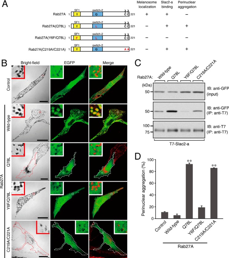
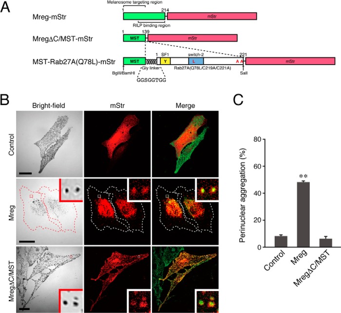
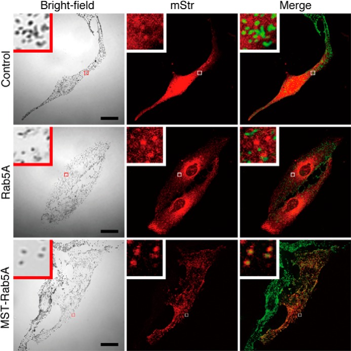
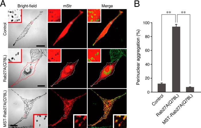
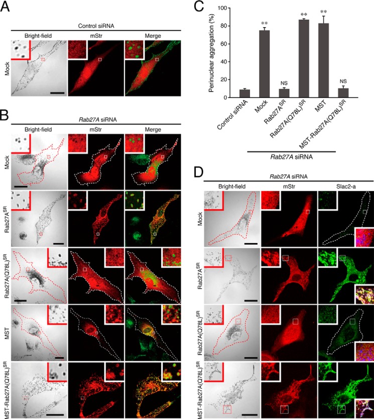
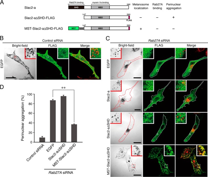
The small GTPase Rab27A is a crucial regulator of actin-based melanosome transport in melanocytes, and functionally defective Rab27A causes human Griscelli syndrome type 2, which is characterized by silvery hair. A GTPase-deficient, constitutively active Rab27A(Q78L) mutant has been shown to act as an inhibitor of melanosome transport and to induce perinuclear aggregation of melanosomes, but the molecular mechanism by which Rab27A(Q78L) inhibits melanosome transport remained to be determined. In this study, we attempted to identify the primary cause of the perinuclear melanosome aggregation induced by Rab27A(Q78L). The results showed that Rab27A(Q78L) is unable to localize on mature melanosomes and that its inhibitory activity on melanosome transport is completely dependent on its binding to the Rab27A effector Slac2-a/melanophilin. When we forcibly expressed Rab27A(Q78L) on mature melanosomes by using a novel melanosome-targeting tag that we developed in this study and named the MST tag, the MST-Rab27A(Q78L) fusion protein behaved in the same manner as wild-type Rab27A. It localized on mature melanosomes without inducing melanosome aggregation and restored normal peripheral melanosome distribution in Rab27A-deficient cells. These findings indicate that the GTPase activity of Rab27A is required for its melanosome localization but is not required for melanosome transport.

Keywords: Low Molecular Weight G Proteins; Melanogenesis; Membrane Trafficking; Protein Targeting; Rab; Small GTPases; Subcellular Organelles.


Figures

FIGURE 1.
FIGURE 1.
Effect of expression of Rab27A mutants on melanosome distribution in melanocytes. A, schematic of the Rab27A mutants used in this study. The melanosome localization activity, Slac2-a binding activity, and perinuclear aggregation activity of each mutant are shown in the right panel. The SF1 region is one of the conserved regions of a certain Rab subfamily (28), and the switch-2 region is thought to be sensitive to the nucleotide-binding state of Rab proteins. B, typical images of melanocytes expressing EGFP-tagged Rab27A mutants and EGFP alone (EGFP fluorescence images and their corresponding bright-field images). Cells exhibiting perinuclear melanosome aggregation are outlined with a dashed line. The melanosomes in the merged images (right column) are pseudocolored in red. The insets show magnified views of the boxed areas. Note that wild-type Rab27A alone, and not any of the Rab27A mutants, is present on mature melanosomes (second row, yellow signals in the inset). Scale bars = 20 μm. C, Slac2-a binding activity of the Rab27A mutants. Testing for associations between T7-tagged Slac2-a and EGFP-tagged Rab27A (wild-type and mutants) was performed by coimmunoprecipitation assays with anti-T7 tag antibody-conjugated agarose beads as described previously (23, 27). Coimmunoprecipitated EGFP-Rab27A (center panel) and immunoprecipitated T7-Slac2-a (IP) (bottom panel) were detected with the antibodies indicated. Input means 1/80 volume of the reaction mixture used for immunoprecipitation (top panel). The positions of the molecular mass markers (in kilodaltons) are shown at the left. IB, immunoblot. D, the number of melanocytes showing perinuclear melanosome aggregation is expressed as a percentage of the number of transfected melanocytes shown in B. **, p < 0.01, unpaired Student's t test.
FIGURE 2.
FIGURE 2.
The N-terminal portion of Mreg is necessary and sufficient for melanosome targeting in melanocytes. A, schematic of the Mreg mutants and MST tag used in this study. The N-terminal portion of Mreg is required for its melanosome localization (19, 40), whereas its C-terminal portion binds Rab-interacting lysosomal protein, which forms a retrograde melanosome transport complex (19). MST-Rab27A(Q78L)-mStr was produced by insertion of Gly-linker+Rab27A(Q78L/C219A/C221A) into the site between MST and mStr. BamHI, BglII, and SalI are restriction enzyme sites that were used for the construction of MST-Rab27A(Q78L)-mStr (see “Experimental Procedures” for details). RILP, Rab-interacting lysosomal protein. B, typical images of melanocytes expressing mStr-tagged Mreg mutants and mStr alone (mStr fluorescence images and their corresponding bright-field images). Cells exhibiting perinuclear melanosome aggregation are outlined with a dashed line. The melanosomes in the merged images (right column) are pseudocolored green. The insets show magnified views of the boxed areas. Note that the full-length Mreg with mStr-tag was clearly targeted to mature melanosomes and that Mreg-mStr-expressing cells often exhibited perinuclear melanosome aggregation (center row). MregΔC/MST-mStr, on the other hand, was able to target mature melanosomes without altering peripheral melanosome distribution (bottom row). Scale bars = 20 μm. C, the number of melanocytes exhibiting perinuclear melanosome aggregation expressed as a percentage of the number of transfected melanocytes shown in B. **, p < 0.01; unpaired Student's t test.
FIGURE 3.
FIGURE 3.
Forcible targeting of Rab5A, an early endosome-resident Rab, to mature melanosomes in melanocytes by the MST tag. Typical images of melanocytes expressing mStr-tagged Rab5A and MST-Rab5A (mStr fluorescence images and their corresponding bright-field images). The melanosomes in the merged images (right column) are pseudocolored green. The insets show magnified views of the boxed areas. Note that MST-Rab5A was clearly targeted to mature melanosomes without altering peripheral melanosome distribution (bottom row). Scale bars = 20 μm.
FIGURE 4.
FIGURE 4.
Forcible targeting of Rab27A(Q78L) to mature melanosomes in melanocytes by the MST tag. A, typical images of melanocytes expressing mStr-tagged Rab27A(Q78L) and MST-Rab27A(Q78L) (mStr fluorescence images and their corresponding bright-field images). Cells exhibiting perinuclear melanosome aggregation are outlined with a dashed line. The melanosomes in the merged images (right column) are pseudocolored green. The insets show magnified views of the boxed areas. In contrast to Rab27A(Q78L), MST-Rab27A(Q78L) was clearly targeted to mature melanosomes without inducing perinuclear melanosome aggregation (bottom row). Scale bars = 20 μm. B, the number of melanocytes exhibiting perinuclear melanosome aggregation expressed as a percentage of the number of transfected melanocytes shown in A. **, p < 0.01, unpaired Student's t test.
FIGURE 5.
FIGURE 5.
MST-Rab27A(Q78L)-mStr can compensate for the function of endogenous Rab27A in melanosome transport in melanocytes. A and B, typical images of melanocytes expressing mStr together with control siRNA (A) and mStr, mStr-Rab27ASR, mStr-Rab27A(Q78L)SR, MST-mStr, or MST-Rab27A(Q78L)SR-mStr together with Rab27A siRNA (B) (mStr fluorescence images and their corresponding bright-field images). Cells exhibiting perinuclear melanosome aggregation are outlined with a dashed line. The melanosomes in the merged images (right column) are pseudocolored green. The insets show magnified views of the boxed areas. Note that MST-Rab27A(Q78L)SR-mStr was able to restore peripheral melanosome distribution in Rab27A-deficient cells (B, fifth row), the same as wild-type Rab27ASR (B, second row). Scale bars = 20 μm. C, the number of melanocytes exhibiting perinuclear melanosome aggregation expressed as a percentage of the number of transfected melanocytes shown in B. **, p < 0.01, unpaired Student's t test. NS, not significant. D, subcellular localization of endogenous Slac2-a molecules in melanocytes expressing mStr, mStr-Rab27ASR, mStr-Rab27A(Q78L)SR, or MST-Rab27A(Q78L)SR-mStr together with Rab27A siRNA. Cells exhibiting perinuclear melanosome aggregation are outlined with a dashed line. The insets show magnified views of the boxed areas. Note that both wild-type mStr-Rab27ASR and MST-Rab27A(Q78L)SR-mStr recruited Slac2-a to mature melanosomes in Rab27A-deficient cells (second and fourth rows), whereas mStr-Rab27A(Q78L)SR did not (third row). The lower insets in the right column are merged images of mStr fluorescence, Slac2-a, and melanosomes (pseudocolored in blue). Scale bars = 20 μm.
FIGURE 6.
FIGURE 6.
Expression of MST-Slac2-aΔSHD in Rab27A-deficient melanocytes restores peripheral melanosome distribution. A, schematic of the Slac2-a mutants used in this study. Slac2-a contains a Rab27A-binding Slp homology domain (SHD) at the N terminus and a myosin Va-binding domain (MBD) in the middle region (12–14, 16). Slac2-aΔSHD lacks the N-terminal SHD and, as a result, is unable to support peripheral melanosome distribution (26). MST-Slac2-aΔSHD contains the MST tag at the N terminus instead of at the SHD. The melanosome localization activity, Rab27A binding activity, and perinuclear aggregation phenotype of Rab27A-deficient cells expressing each mutant are shown in the right panel. B and C, typical images of melanocytes expressing FLAG-EGFP together with control siRNA (B) and FLAG-EGFP, FLAG-Slac2-a, Slac2-aΔSHD-FLAG, or MST-Slac2-aΔSHD-FLAG together with Rab27A siRNA (C) (fluorescence images and their corresponding bright-field images). Cells exhibiting perinuclear melanosome aggregation are outlined with a dashed line. The melanosomes in the merged images (right column) are pseudocolored red. The insets show magnified views of the boxed areas. Note that MST-Slac2-aΔSHD-FLAG was clearly targeted to mature melanosomes even in the absence of Rab27A and that it was able to restore peripheral melanosome distribution in Rab27A-deficient cells (C, fourth row). Scale bars = 20 μm. D, the number of melanocytes exhibiting perinuclear melanosome aggregation expressed as a percentage of the number of transfected melanocytes shown in B and C. **, p < 0.01, unpaired Student's t test.

Similar articles

Cited by

References

    1. Fukuda M. (2008) Regulation of secretory vesicle traffic by Rab small GTPases. Cell. Mol. Life Sci. 65, 2801–2813 - PMC - PubMed
    1. Stenmark H. (2009) Rab GTPases as coordinators of vesicle traffic. Nat. Rev. Mol. Cell Biol. 10, 513–525 - PubMed
    1. Barr F., Lambright D. G. (2010) Rab GEFs and GAPs. Curr. Opin. Cell Biol. 22, 461–470 - PMC - PubMed
    1. Fukuda M. (2011) TBC proteins. GAPs for mammalian small GTPase Rab? Biosci. Rep. 31, 159–168 - PubMed
    1. Pfeffer S. R. (2013) Rab GTPase regulation of membrane identity. Curr. Opin. Cell Biol. 25, 414–419 - PMC - PubMed

Publication types

MeSH terms

Supplementary concepts

LinkOut - more resources