Review
. 2014 Mar;52(3):243-58.
doi: 10.1007/s12275-014-4087-z. Epub 2014 Mar 1.

Phage lysis: three steps, three choices, one outcome

Affiliations
Review

Phage lysis: three steps, three choices, one outcome

Ryland Young. J Microbiol. 2014 Mar.

Abstract

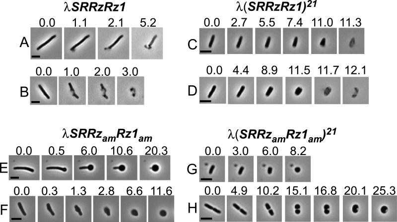
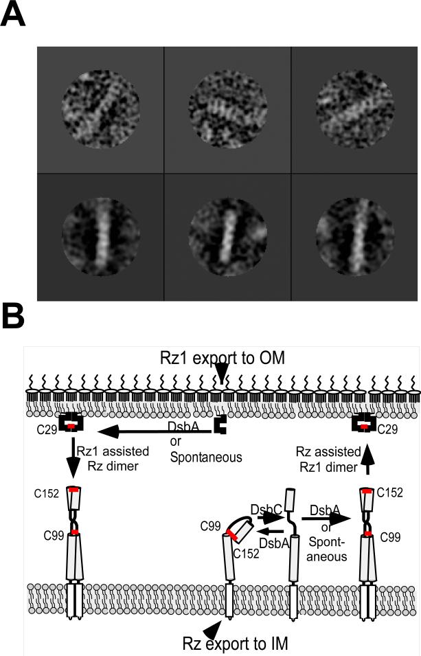
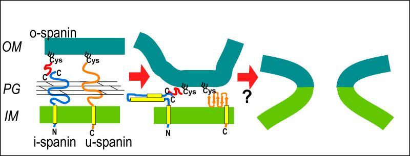
The lysis of bacterial hosts by double-strand DNA bacteriophages, once thought to reflect merely the accumulation of sufficient lysozyme activity during the infection cycle, has been revealed to recently been revealed to be a carefully regulated and temporally scheduled process. For phages of Gramnegative hosts, there are three steps, corresponding to subversion of each of the three layers of the cell envelope: inner membrane, peptidoglycan, and outer membrane. The pathway is controlled at the level of the cytoplasmic membrane. In canonical lysis, a phage encoded protein, the holin, accumulates harmlessly in the cytoplasmic membrane until triggering at an allele-specific time to form micron-scale holes. This allows the soluble endolysin to escape from the cytoplasm to degrade the peptidoglycan. Recently a parallel pathway has been elucidated in which a different type of holin, the pinholin, which, instead of triggering to form large holes, triggers to form small, heptameric channels that serve to depolarize the membrane. Pinholins are associated with SAR endolysins, which accumulate in the periplasm as inactive, membrane-tethered enzymes. Pinholin triggering collapses the proton motive force, allowing the SAR endolysins to refold to an active form and attack the peptidoglycan. Surprisingly, a third step, the disruption of the outer membrane is also required. This is usually achieved by a spanin complex, consisting of a small outer membrane lipoprotein and an integral cytoplasmic membrane protein, designated as o-spanin and i-spanin, respectively. Without spanin function, lysis is blocked and progeny virions are trapped in dead spherical cells, suggesting that the outer membrane has considerable tensile strength. In addition to two-component spanins, there are some single-component spanins, or u-spanins, that have an N-terminal outer-membrane lipoprotein signal and a C-terminal transmembrane domain. A possible mechanism for spanin function to disrupt the outer membrane is to catalyze fusion of the inner and outer membranes.


Figures

Figure 1
Figure 1. The genes and proteins of lysis
A. The lysis cassettes of phages lambda and lambdoid phage 21 are shown. In both of these lambdoid phages, the lysis genes are proximal to the late promoter, pR’. Lengths of primary translation products are shown above each gene; upper and lower numbers for S and S21 are for antiholin and holin, respectively. Gene color codes: light blue = canonical holin, red = pinholin; black = canonical endolysin (R transglycosylase); dark blue = SAR endolysin; brown = i-spanin; green = o-spanin. B. Dual start motifs for lambda holin and phage 21 pinholin genes. Start codons for each product indicated by star; red = antiholin, green = holin (S105) or pinholin (S2168). Red and green rectangles indicate Shine-Dalgarno sequences. Inverted arrows indicate RNA stem-loops that control choice of start codons. C. Topological dynamics of lysis proteins. In the energized membrane (PMF = ~180 mV), TMD1 of antiholin forms (S107 and S2171) are inhibited from entering (S107) or exiting (S2171) the bilayer, whereas TMD1 of the pinholin (S2168) exits spontaneously during pinhole pathway. N-terminal SAR domain of R21 exits the membrane at a low rate spontaneously, or quantitatively upon membrane depolarization. The R transglycosylase is shown as fully active muralytic enzyme (oval with open “active site” cleft) in the periplasm, whereas the R21 SAR endolysin is shown in its inactive, membrane tethered form. The Rz-Rz1 complex is shown with the Rz i-spanin embedded in the IM with its N-terminal TMD and its periplasmic domain (elongated oval) disposed in the periplasm (dark grey = alpha-helical, coiled-coil domains; light gray = predicted hinge region). The Rz1 o-spanin is shown as a small oval attached to the inner leaflet of the OM by the three fatty acid chains of the lipoprotein motif. Intermolecular disulfide bonds are shown in approximate position. Finally, gp11 is shown with its N-terminal OM lipoprotein motif already sorted to the OM by the Lol system, and its C-terminal TMD embedded in the IM. Modified from Young (2013) , with permission.
Figure 2
Figure 2. Two pathways to murein degradation
Shown are cartoon views of the (A) canonical holin-endolysin and (B) pinholin-SAR endolysin pathways to murein degradation. Only the IM (grey rectangle) and PG (grid) are shown. The cartoon series begins early in late gene expression (morphogenesis period) and progresses downwards. Holins (blue ovals in A), pinholins (blue ovals in B), soluble endolysins (green ovals with open “active site cleft”, and SAR endolysins (green ovals with N-terminal SAR domains depicted either in TMD conformation (green rectangle in top two panels under B) or extracted, refolded conformation (jack-knifed green rectangles in bottom panel), attached to the green globular (enzymatic and PG binding) domain. Holins accumulate in the IM (top two panels of A and B). Note that the prototype holin, S105, and pinholin S2168, accumulate as homodimers or heterodimers with their cognate antiholins (Fig. 1; see text); however, holins are represented as single ovals here, for simplicity. The bottom panels represent the triggered cells, in which the canonical holins form a large (“micron-scale”) hole (A) or the pinholins form many small heptameric pinholes (B) in the IM. Pmf of IM is indicated for each stage.
Figure 3
Figure 3. Lysis morphologies
Shown are phase-contrast images of representative individual cells after induction of lambda lysogens with the indicated lysis genotype. Cultures were thermally induced and then, at just before the pre-determined time for triggering, a 10 μl sample was placed on a glass slide, covered with a coverslip, and imaged; all manipulations were done at 37°C. Numbers represent elapsed time in sec. For details see Berry et al.(2012), from which this was modified, with permission.
Figure 4
Figure 4. Lysis gene clusters in paradigm phages
Genes are color-coded as follows: green = canonical holin; yellow = pinholin; grey = canonical endolysin; light blue = SAR endolysin; red = i-spanin; black = o-spanin; red/black hatch = u-spanin. Other genes are unrelated to lysis function and are marked by dots, except P2 lysA, thought to encode an antiholin (purple). No significant sequence homology exists between any genes except ~96% identity between the i-spanin/o-spanin genes of lambda and 21 (Bonovich & Young, 1991) and 57% identity between the endolysins R of lambda and K of P2 (Ziermann et al., 1994).
Figure 5
Figure 5. Spanin sequences
Primary structures of i-spanin/o-spanin pairs from prototype embedded (lambda Rz-Rz1), overlapped (P2 lysB/lysC) and separated (T4 pseT.3/pseT.2) gene architectures, as well as the prototype u-spanin, gp11. The primary structure for the embedded and overlapped o-spanins are shown aligned to the i-spanin sequence as they are in the gene structure. Predicted secondary structure elements: green stripes = alpha-helix; orange hatch = beta (extended) strand. Pro residues are highlighted in red. The Cys residue subjected to signal peptidase II processing and lipoylation is highlighted light blue. Signal peptidase II signal peptides and TMDs are colored grey. Charges predicted for individual residues are shown above or below the sequence. Modified from Summer et al. (2007) with permission.
Figure 6
Figure 6. Spanin complex structure and formation
A. Single particle analysis of sRz-sRz1 complexes imaged by transmission electron microscopy with negative stain. Upper 3 panels are representative single particles and lower 3 panels are class averages. For details, see Fig. 6 of Berry et al. (2010), from which this was modified with permission. B. Model for intermolecular disulfide bond formation in the spanin complex. Red bars indicate disulfide bonds. Normally, DsbA catalyzes the formation of a non-productive intramolecular disulfide between the two Cys residues, Cys99 and Cys152, of Rz. DsbC reduces this bond, allowing DsbA to catalyze the formation of intermolecular disulfide bonds in the covalent Rz homodimer. In the presence of Rz1, the disulfide linkages of Rz–Rz covalent homodimer can form spontaneously. For Rz1, DsbA catalyzes the covalent dimerization of Rz1 using its single Cys, Cys29. In the absence of DsbA, the homodimer disulfide linkage in each subunit can form as long as the cognate subunit is present, presumably as a template. For details see Berry et al. (2013), from which this panel was derived, with permission.
Figure 7
Figure 7. Model for spanin-mediated disruption of the OM by membrane fusion
Both two-component spanin complexes and u-spanins accumulate in the envelope during the morphogenesis period, trapped within lacuna of the PG network. For simplicity, the two-component spanin complex is shown as monomer-monomer complexes interacting by C-terminal domains, although in fact, both the i-spanin and the o-spanin are covalent homodimers with intermolecular disulfide linkages in the lambda complex. (See cartoon in Fig. 1C.) After removal of the PG by the endolysin or SAR endolysin, the spanins undergo conformational changes that bring the two membranes together. Again, a simplified model is shown where the two coiled-coil domains of the Rz i-spanin interact in the collapsed conformation of the complex. Not shown is the two-dimensional oligomerization of these complexes (see Fig. 6) (Berry et al., 2010). With the u-spanin, which lacks predicted alpha-helical or coiled-coil structures, the cartoon depicts formation of a beta-sheet from the predicted beta-strand elements. Whether the u-spanin also oligomerizes after removal of the PG layer is not known. In both cases, some feature of the collapsed conformation (middle cartoon) acts to destabilize either the inner leaflet of the OM or the outer leaflet of the IM, or both. The Rz1 periplasmic domain may be a good candidate for this activity (Bryl et al., 2000). From Young (2013), with permission.

Similar articles

Cited by

References

    1. Adhya S, Sen A, Mitra S. In: The role of gene S In: The Bacteriophage Lambda. Hershey AD, editor. Cold Spring Harbor Laboratory; Cold Spring Harbor, NY: 1971. pp. 743–746.
    1. Altman E, Altman RK, Garrett JM, Grimaila RJ, Young R. S gene product: identification and membrane localization of a lysis control protein. J.Bacteriology. 1983;155:1130–1137. - PMC - PubMed
    1. Altman E, Young K, Garrett J, Altman R, Young R. Subcellular localization of lethal lysis proteins of bacteriophages λ and øX174. J. Virol. 1985;53:1008–1011. - PMC - PubMed
    1. Atkins JF, Steitz JA, Anderson CW, Model P. Binding of mammalian ribosomes to MS2 phage RNA reveals an overlapping gene encoding a lysis function. Cell. 1979;18:247–256. - PubMed
    1. Barenboim M, Chang CY, dib Hajj F, Young R. Characterization of the dual start motif of a class II holin gene. Mol.Microbiol. 1999;32:715–727. - PubMed

Publication types

MeSH terms