

Use of synthetic serum-free medium for culture of human dermal fibroblasts to establish an experimental system similar to living dermis
- PMID: 24585098
- PMCID: PMC4371565
- DOI: 10.1007/s10616-014-9709-0
Use of synthetic serum-free medium for culture of human dermal fibroblasts to establish an experimental system similar to living dermis
Abstract
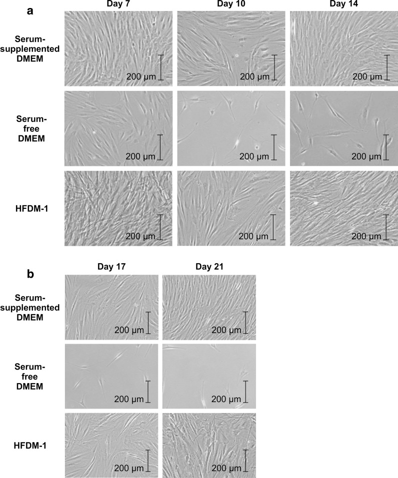
In this study, we sought to establish a defined experimental system for fibroblast growth similar to that of the living dermis. To this end, we evaluated the growth and biochemical characteristics of fibroblasts cultured with serum-free HFDM-1, a finely tuned synthetic medium for human fibroblast culture. Three culture conditions were used to grow fibroblasts obtained from primary culture: (1) culture with Dulbecco's modified Eagle medium (DMEM) plus 10 % fetal bovine serum (serum-supplemented DMEM), (2) culture with DMEM (serum-free DMEM), and (3) culture with HFDM-1 (HFDM-1), and fibroblast morphology, growth, collagen type I production, and lipid composition were analyzed. Fibroblasts grown in HFDM-1 maintained cell numbers at nearly 100 % from days 14 to 21 and produced more collagen type I than cells grown in serum-supplemented and serum-free DMEM. Arachidonic acid (20:4) and total polyunsaturated fatty acids were lower in cells grown in serum-free DMEM and HFDM-1 than in serum-supplemented DMEM. These results suggested that HFDM-1 recapitulated growth conditions in the dermis better than traditional, serum-supplemented DMEM. In addition, the controlled chemical composition of HFDM-1 eliminated a potential source of variability in cell culture conditions.
Figures


Similar articles
-
The utilization of animal product-free media and autologous serum in an autologous dermal substitute culture.J Surg Res. 2011 Nov;171(1):339-46. doi: 10.1016/j.jss.2009.11.724. Epub 2010 Feb 6. J Surg Res. 2011. PMID: 20189600
-
Fabrication of cultured oral gingiva by tissue engineering techniques without materials of animal origin.J Periodontol. 2006 Apr;77(4):672-7. doi: 10.1902/jop.2006.050223. J Periodontol. 2006. PMID: 16584349
-
Bovine colostrum supports the serum-free proliferation of epithelial cells but not of fibroblasts in long-term culture.J Cell Biol. 1980 Mar;84(3):808-14. doi: 10.1083/jcb.84.3.808. J Cell Biol. 1980. PMID: 7358799 Free PMC article.
-
Conditions for the culture of bovine embryonic myogenic cells.Tissue Cell. 1997 Apr;29(2):207-15. doi: 10.1016/s0040-8166(97)80020-1. Tissue Cell. 1997. PMID: 9149443
-
Growth factor profile of irradiated human dermal fibroblasts using a serum-free method.Plast Reconstr Surg. 2003 May;111(6):1960-8. doi: 10.1097/01.PRS.0000055065.41599.75. Plast Reconstr Surg. 2003. PMID: 12711958
Cited by
-
Development of a tissue-engineered skin substitute on a base of human amniotic membrane.J Tissue Eng. 2019 Feb 2;10:2041731418825378. doi: 10.1177/2041731418825378. eCollection 2019 Jan-Dec. J Tissue Eng. 2019. PMID: 30746119 Free PMC article.
-
Dermal fibroblasts cultured from donors with type 2 diabetes mellitus retain an epigenetic memory associated with poor wound healing responses.Sci Rep. 2021 Jan 14;11(1):1474. doi: 10.1038/s41598-020-80072-z. Sci Rep. 2021. PMID: 33446687 Free PMC article.
-
Diabetic Foot: The Role of Fasciae, a Narrative Review.Biology (Basel). 2021 Aug 7;10(8):759. doi: 10.3390/biology10080759. Biology (Basel). 2021. PMID: 34439991 Free PMC article. Review.
-
Functional responses of dermal fibroblasts to low nutrition and pro-inflammatory stimuli mimicking a wound environment in vitro.In Vitro Cell Dev Biol Anim. 2022 Sep;58(8):643-657. doi: 10.1007/s11626-022-00713-7. Epub 2022 Aug 10. In Vitro Cell Dev Biol Anim. 2022. PMID: 35948856
-
Collagen-Derived Di-Peptide, Prolylhydroxyproline (Pro-Hyp): A New Low Molecular Weight Growth-Initiating Factor for Specific Fibroblasts Associated With Wound Healing.Front Cell Dev Biol. 2020 Nov 27;8:548975. doi: 10.3389/fcell.2020.548975. eCollection 2020. Front Cell Dev Biol. 2020. PMID: 33330443 Free PMC article. Review.
References
-
- Balazs L, Okolicany J, Ferrebee M, Tolley B, Tigyi G. Topical application of the phospholipids growth factor lysophosphatidic acid promotes wound healing in vivo. Am J Physiol Regul Integr Comp Physiol. 2001;280:R466–R472. - PubMed
-
- Bettger WJ, Boyce ST, Walthall BJ, Ham RG. Rapid clonal growth and serial passage of human diploid fibroblasts in a lipid-enriched synthetic medium supplemented with epidermal growth factor, insulin, and dexamethasone. Proc Natl Acad Sci USA. 1981;78:5588–5592. doi: 10.1073/pnas.78.9.5588. - DOI - PMC - PubMed
-
- Folch J, Lees M, Stanley GHS (1957) A simple method for the isolation and purification of total lipides from animal tissues. J Biol Chem 226:497–509 - PubMed
LinkOut - more resources
Full Text Sources
Other Literature Sources
