. 2014 Apr;7(2):132-143.
doi: 10.1161/CIRCGENETICS.113.000324. Epub 2014 Feb 28.

Desensitization of myofilaments to Ca2+ as a therapeutic target for hypertrophic cardiomyopathy with mutations in thin filament proteins

Affiliations

Desensitization of myofilaments to Ca2+ as a therapeutic target for hypertrophic cardiomyopathy with mutations in thin filament proteins

Marco L Alves et al. Circ Cardiovasc Genet. 2014 Apr.

Abstract

Background: Hypertrophic cardiomyopathy (HCM) is a common genetic disorder caused mainly by mutations in sarcomeric proteins and is characterized by maladaptive myocardial hypertrophy, diastolic heart failure, increased myofilament Ca(2+) sensitivity, and high susceptibility to sudden death. We tested the following hypothesis: correction of the increased myofilament sensitivity can delay or prevent the development of the HCM phenotype.

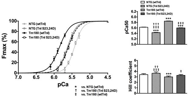
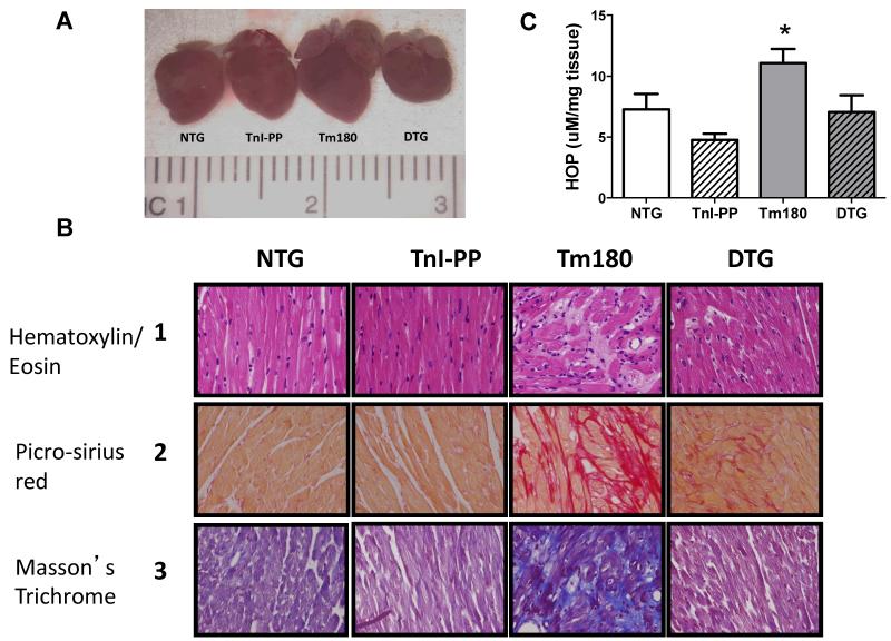
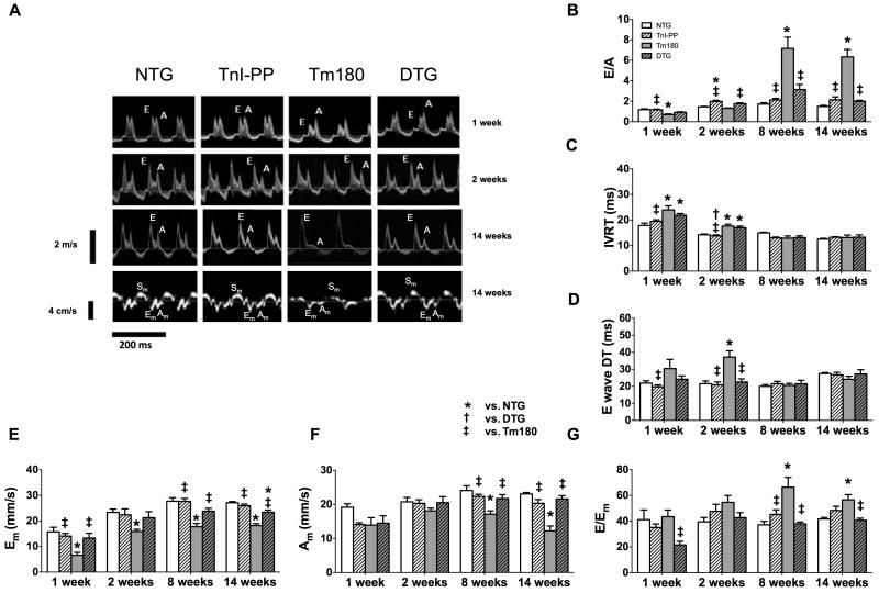
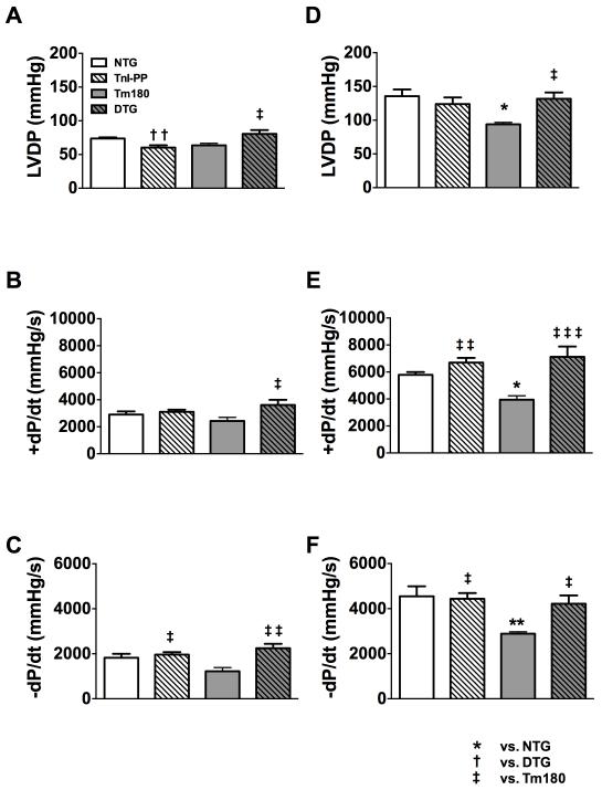
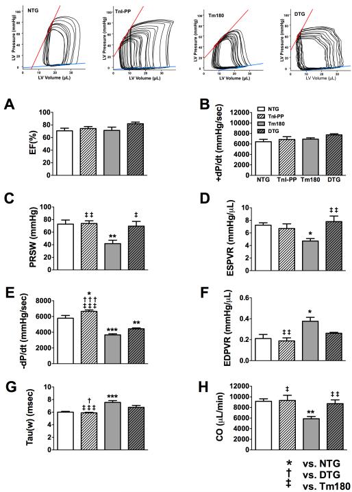
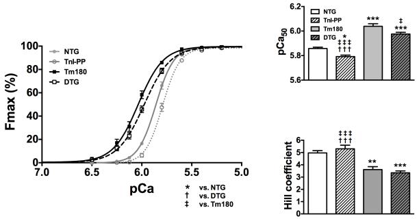
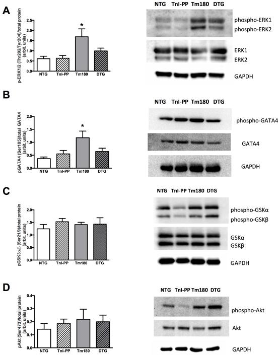
Methods and results: We used an HCM mouse model with an E180G mutation in α-tropomyosin (Tm180) that demonstrates increased myofilament Ca(2+) sensitivity, severe hypertrophy, and diastolic dysfunction. To test our hypothesis, we reduced myofilament Ca(2+) sensitivity in Tm180 mice by generating a double transgenic mouse line. We crossed Tm180 mice with mice expressing a pseudophosphorylated cardiac troponin I (S23D and S24D; TnI-PP). TnI-PP mice demonstrated a reduced myofilament Ca(2+) sensitivity compared with wild-type mice. The development of pathological hypertrophy did not occur in mice expressing both Tm180 and TnI-PP. Left ventricle performance was improved in double transgenic compared with their Tm180 littermates, which express wild-type cardiac troponin I. Hearts of double transgenic mice demonstrated no changes in expression of phospholamban and sarcoplasmic reticulum Ca(2+) ATPase, increased levels of phospholamban and troponin T phosphorylation, and reduced phosphorylation of TnI compared with Tm180 mice. Moreover, expression of TnI-PP in Tm180 hearts inhibited modifications in the activity of extracellular signal-regulated kinase and zinc finger-containing transcription factor GATA in Tm180 hearts.

Conclusions: Our data strongly indicate that reduction of myofilament sensitivity to Ca(2+) and associated correction of abnormal relaxation can delay or prevent development of HCM and should be considered as a therapeutic target for HCM.

Keywords: cardiac remodeling; cardiomyopathies; cardiomyopathy, hypertrophic; therapeutics.


Figures

Figure 1
Figure 1
Effects of TnI-S23/24 pseudo-phosphorylation (TnI-S23,24D) on Ca2+-activated tension in skinned fiber preparations. pCa-force relations in NTG and Tm180 skinned fiber bundles exchanged with Tn complex containing either wtTnI or TnI -S23,24D. Exchange with TnI-S23,24D in both NTG and Tm180 fiber bundles resulted in a significant rightward shift (decrease in pCa50) and increase in cooperativity (Hill coefficient) compared to fibers exchanged with Tn complex containing wtTnI. Data are presented as mean±SE. *significantly different vs. NTG(wtTnI); †significantly different vs. Tm180(TnI-S23,24D); ‡significantly different vs. Tm180(wtTnI). ***, ††† or ‡‡‡ p<0.001; †† p<0.01; ‡ p<0.05 based on post-hoc multiple comparison analysis (Tukey’s test). n=8 per group.
Figure 2
Figure 2
Evaluation of cardiac morphology by two-dimentional (2-D) and M-mode of high resolution echocardiography. Panel A. 2-D short axis views of NTG, TnI-PP, Tm180 and DTG 8 week-old mouse hearts. Left atrium (LA) size is increased only in Tm180 mouse (white arrow). No morphological abnormalities were detected in TnI-PP or DTG hearts. Panels B-E. Time dependent changes in morphological parameters in NTG, TnI-PP, Tm180 and DTG mice: LA anterio-posterior internal dimension (B), LV internal diastolic dimension (LVDd) (C), septal wall thickness (SWT) (D), LV calculated mass (E). Panel F. HW/TL ratio in 14 week-old mice. Data are presented as mean±SE. *Significantly different from NTG, †significantly different from DTG, ‡significantly different from Tm180 based on post-hoc multiple comparison analysis (Tukey’s test). Numerical data and p values for significance are presented in Table 1 in the Supplemental Material. n=4-8 per group.
Figure 3
Figure 3
Morphology and histology of 14-week old mouse heart. Panel A. Comparative gross morphology. Panel B. 40x magnification of heart sections (septal wall) stained with Hematoxylin and Eosin (HE, line 1), Picro-sirius red (PSR, line 2) and Masson’s Trichrome (line 3). The intense myocyte disarray is depicted in HE-stained sections and the extensive fibrosis is observed in PSR-(red stain) and Masson’s Trichrome-stained sections (blue stain) in Tm180 hearts. DTG hearts show less disarray and fibrosis than Tm180. Panel C. Hydroxyproline content in NTG, TnI-PP, Tm180 and DTG mice. Data are presented as mean±SE. *Significantly different from all other groups based on post-hoc multiple comparison analysis (Fisher’s test; p<0.05). n=4-5 per group.
Figure 4
Figure 4
Serial evaluation of diastolic function by echocardiography. Panel A. Representative images of pulsed Doppler of mitral inflow (top 3 panels) and TDI recordings of the mitral annulus (bottom panel) in NTG, TnI-PP, Tm180 and DTG mice at 1, 2 and 14 weeks of age. Panels B-D. Diastolic function parameters measured by pulsed Doppler: peak velocity of mitral blood inflow in early diastole (E) to peak velocity of mitral blood inflow in late diastole (A) (E/A ratio) (B), isovolumic relaxation time (IVRT) (C), E wave deceleration time (DT) (D) in NTG, TnI-PP, Tm180 and DTG mice. Tm180 hearts show progression from a mild form of diastolic dysfunction (1 week) to a severe form (8 weeks). Panels E-G. Diastolic function parameters measured by TDI: peak myocardial velocity in early diastole (Em) (E), peak myocardial velocity in late diastole (Am) (F) and E to Em ratio (E/Em) (G). Tm180 hearts showed decreased Em in all ages, decreased Am and increased E/Em ratio after 2 weeks, suggesting early impaired relaxation and progressive decrease in LV compliance. DTG hearts showed mitral inflow and TDI patterns similar to NTG controls. Data are presented as mean±SE. *Significantly different from NTG, †significantly different from DTG, ‡significantly different from Tm180 based on post-hoc multiple comparison analysis (Tukey’s test). Numerical data and p values for significance are presented in Table 2 in the Supplemental Material. n=4-8 per group.
Figure 5
Figure 5
Inotropic and lusitropic responses to β-adrenergic stimulation in isolated 6 weeks old Langendorff perfused NTG, TnI-PP, Tm180 and DTG hearts. A. LV developed pressure (LVDP), B. the maximal rate of contraction (+dP/dt) and C. the maximal rate of relaxation (-dP/dt) at baseline. D. LVDP, E. +dP/dt and F. -dP/dt during perfusion with 25 nmol/L isoproterenol. Data are presented as mean±SE. *Significantly different from NTG, †significantly different from DTG, ‡significantly different from Tm180 based on post-hoc multiple comparison analysis (Tukey’s test). ‡‡‡ p<0.001; ** or †† p<0.01; ‡ or * p<0.05. Numerical data are presented in Table 3 in the Supplemental Material. n=4-5 per group.
Figure 6
Figure 6
In situ cardiac function in 14-week old NTG, TnI-PP, Tm180 and DTG mice. A. Ejection fraction (EF), B. the maximal rate of contraction (+dP/dt), C. Preload recruited stoke work (PRSW), D. end systolic pressure-volume relation slope (ESPVR), E. the maximal rate of relaxation (-dP/dt), F. end diastolic pressure-volume relation (EDPVR), G. relaxation time constant calculated by Weiss method (tau), H. cardiac output (CO). Data are presented as mean±SE. *Significantly different from NTG, †significantly different from DTG, ‡significantly different from Tm180 based on post-hoc multiple comparison analysis (Tukey’s test). *** or ‡‡‡ p<0.001; ** or ‡‡ p<0.01; ‡, * or † p<0.05. Numerical data are presented in Table 4 in the Supplemental Material. n=6-8 per group.
Figure 7
Figure 7
pCa-force relations in skinned fiber bundles prepared from papillary muscles of NTG, Tm180, TnI-PP and DTG mice. Expression of TnI-PP in the presence of Tm180 mutations (DTG) caused a decrease in the myofilament Ca2+ (pCa50) sensitivity and cooperativity of activation (Hill coefficient). Data are presented as mean±SE; *Significantly different from NTG, †significantly different from DTG, ‡significantly different from Tm180 based on post-hoc multiple comparison analysis (Tukey’s test). ***, ††† or ‡‡‡ p<0.001; ** p<0.01; * or ‡ p<0.05. n=6-8 per group.
Figure 8
Figure 8
Phosphorylation of ERK1/2, GATA4, GSK3α/β and Akt. A. phosphorylation of ERK1/2, B. phosphorylation of GATA4, C. phosphorylation of GSK3α/β and D. phosphorylation of Akt. Data are presented as mean±SE. Phosphorylated protein bands were normalized to total protein. No significant differences in total protein expression were observed. *significantly different from all other groups based on post-hoc multiple comparison analysis (Fisher’s test; p<0.05). n=6-7 per group.

References

    1. Gersh BJ, Maron BJ, Bonow RO, Dearani JA, Fifer MA, Link MS, et al. 2011 ACCF/AHA guideline for the diagnosis and treatment of hypertrophic cardiomyopathy: A report of the american college of cardiology foundation/american heart association task force on practice guidelines. Circulation. 2011;124:e783–831. - PubMed
    1. Maron BJ, Towbin JA, Thiene G, Antzelevitch C, Corrado D, Arnett D, et al. Contemporary definitions and classification of the cardiomyopathies: An american heart association scientific statement from the council on clinical cardiology, heart failure and transplantation committee; quality of care and outcomes research and functional genomics and translational biology interdisciplinary working groups; and council on epidemiology and prevention. Circulation. 2006;113:1807–1816. - PubMed
    1. Hughes SE. The pathology of hypertrophic cardiomyopathy. Histopathology. 2004;44:412–427. - PubMed
    1. Elliott P, McKenna WJ. Hypertrophic cardiomyopathy. Lancet. 2004;363:1881–1891. - PubMed
    1. Bos JM, Towbin JA, Ackerman MJ. Diagnostic, prognostic, and therapeutic implications of genetic testing for hypertrophic cardiomyopathy. J Am Coll Cardiol. 2009;54:201–211. - PubMed

Publication types