. 2014 Feb 27;10(2):e1003945.
doi: 10.1371/journal.ppat.1003945. eCollection 2014 Feb.

The post-transcriptional regulator rsmA/csrA activates T3SS by stabilizing the 5' UTR of hrpG, the master regulator of hrp/hrc genes, in Xanthomonas

Affiliations

The post-transcriptional regulator rsmA/csrA activates T3SS by stabilizing the 5' UTR of hrpG, the master regulator of hrp/hrc genes, in Xanthomonas

Maxuel O Andrade et al. PLoS Pathog. .

Abstract

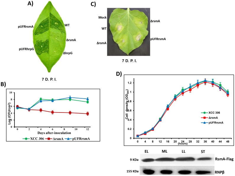
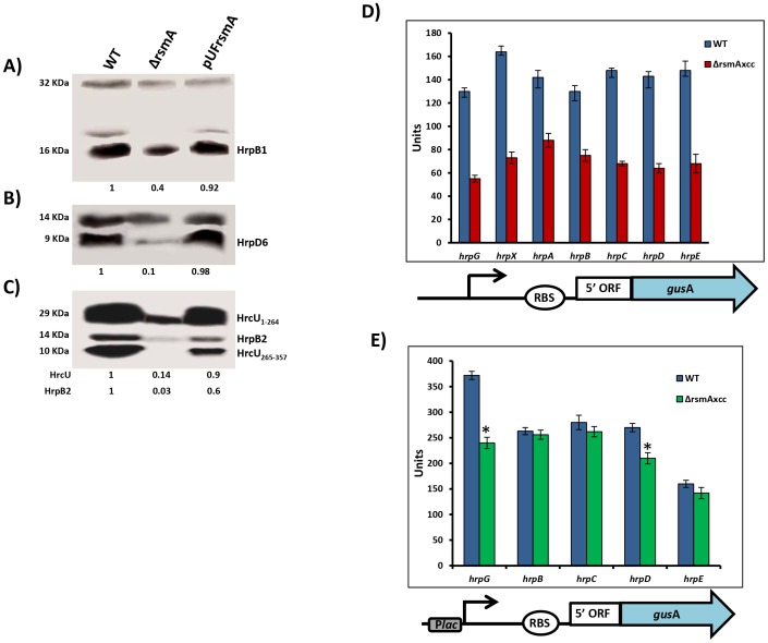
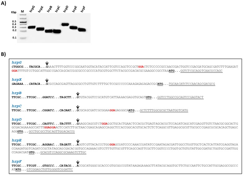
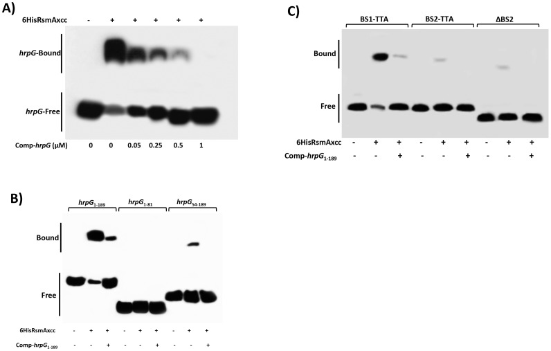
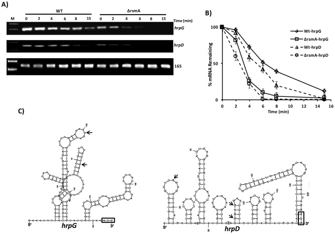
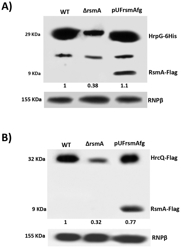
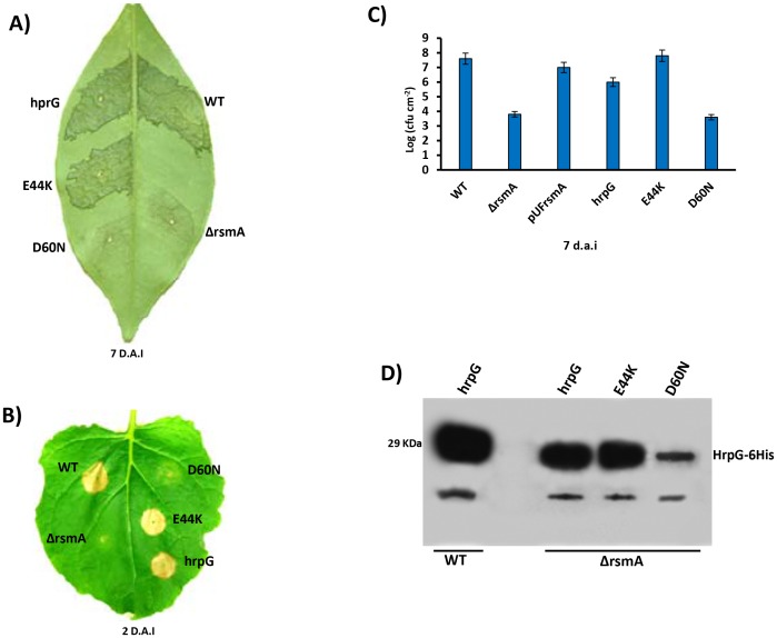
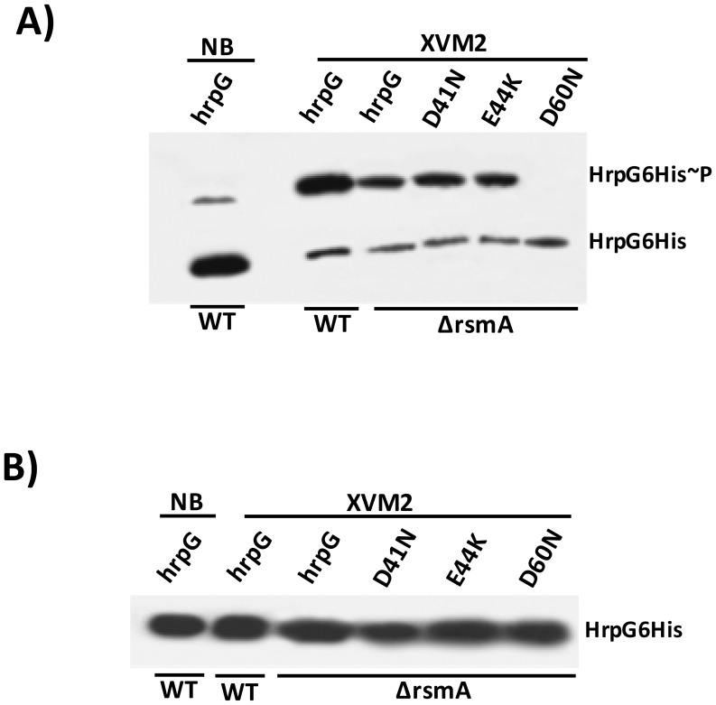
The RsmA/CsrA family of the post-transcriptional regulators of bacteria is involved in the regulation of many cellular processes, including pathogenesis. In this study, we demonstrated that rsmA not only is required for the full virulence of the phytopathogenic bacterium Xanthomonas citri subsp. citri (XCC) but also contributes to triggering the hypersensitive response (HR) in non-host plants. Deletion of rsmA resulted in significantly reduced virulence in the host plant sweet orange and a delayed and weakened HR in the non-host plant Nicotiana benthamiana. Microarray, quantitative reverse-transcription PCR, western-blotting, and GUS assays indicated that RsmA regulates the expression of the type 3 secretion system (T3SS) at both transcriptional and post-transcriptional levels. The regulation of T3SS by RsmA is a universal phenomenon in T3SS-containing bacteria, but the specific mechanism seems to depend on the interaction between a particular bacterium and its hosts. For Xanthomonads, the mechanism by which RsmA activates T3SS remains unknown. Here, we show that RsmA activates the expression of T3SS-encoding hrp/hrc genes by directly binding to the 5' untranslated region (UTR) of hrpG, the master regulator of the hrp/hrc genes in XCC. RsmA stabilizes hrpG mRNA, leading to increased accumulation of HrpG proteins and subsequently, the activation of hrp/hrc genes. The activation of the hrp/hrc genes by RsmA via HrpG was further supported by the observation that ectopic overexpression of hrpG in an rsmA mutant restored its ability to cause disease in host plants and trigger HR in non-host plants. RsmA also stabilizes the transcripts of another T3SS-associated hrpD operon by directly binding to the 5' UTR region. Taken together, these data revealed that RsmA primarily activates T3SS by acting as a positive regulator of hrpG and that this regulation is critical to the pathogenicity of XCC.


Conflict of interest statement

The authors have declared that no competing interests exist.

Figures

Figure 1
Figure 1. rsmA is required for the pathogenicity of Xanthomonas citri subsp. citri in the host plant sweet orange and contributes to the hypersensitive response (HR) in tobacco leaves (Nicotiana benthamiana).
A) Disease symptoms on host sweet orange (Citrus sinensis) leaves 7 days post inoculation (D.P.I.) of bacterial cells at a concentration of 106 CFU/ml. B) Growth assay in planta. Bacterial cells were inoculated into sweet orange leaves at a concentration of 106 CFU/ml and recovered at different time points. The values represent the means of three replicates. The experiment was repeated three times with similar results. Means ± standard deviations are plotted. C) Macroscopic symptoms induced 7 D.A.I of tobacco leaves by infiltrating bacterial cells at a concentration of 106 CFU/ml. D) Growth curve in the minimal medium XVM2 and Western-blotting assay using protein extracts of rsmA mutant cells harboring the pUFR047-rsmA-Flag construct. Cells were collected in different growth stages: EL, early log; ML, medium log); LL, late log; and ST, stationary phase. Wt = X. citri subsp. citri strain 306, ΔrsmA = mutant with a deletion of XAC1743 (rsmA) harboring the empty plasmid pUFR047, pUFRrsmA = complementation of ΔrsmA with rsmA cloned in pUFR047, ΔhrpG = hprG mutant , pUFRhrpG = complementation of ΔhrpG with hrpG cloned into pUFR047, and Mock, 10 mM MgCl2. RNPβ: antibody to the β-subunit of RNA polymerase.
Figure 2
Figure 2. RsmA regulates protein levels of T3SS in X. citri subsp. citri.
Immunoblotting analyses of the total protein extracts of the wild-type (Wt), the rsmA mutant (ΔrsmA) harboring the empty plasmid pUFR047 and the complemented strain (pUFRrsmA) are shown. Bacterial cells were grown in the XVM2 medium and collected at OD600 nm = 0.5. Proteins were separated by SDS-PAGE and transferred to a nitrocellulose membrane. The blots were probed with A) HrpB1, B) HrpD6 or C) HrcU and HrpB2 polyclonal antibodies, respectively. Protein-A conjugated with horseradish peroxidase was used to detect the blots. Beneath the panels are presented the values of the relative levels of detected proteins in rsmA mutant and complemented strains which were estimated according to wild-type results. The estimated values were normalized with the values obtained to unspecific protein bands also recognized by the antibodies. D) and E) GUS assays using translational fusion constructs. The different constructs used in this assay are represented by diagrams bellow of the graphics. D) Wild-type and rsmA mutant cells harboring plasmid-borne promoterless gusA in-frame fused to the native promoters and the first codons of the hrp genes. E) Wild-type and rsmA mutant cells transformed with translational fusions driven by the constitutive Plac promoter. Values presented are means ± standard deviations of three independent experiments. * represents the significant difference between the wild-type and ΔrsmA values by using ANOVA. The GUS assay was repeated twice with similar results.
Figure 3
Figure 3. Determination of hrp/hrc transcriptional start sites of X. citri subsp. citri.
The transcriptional start sites for the genes hrpG, hrpX and hrpF, and the operons hrpB, hrpC, hrpD and hrpE of XCC were determined by 5′RACE. A) Specific PCR products were detected after the amplification of reverse-transcribed cDNA with the gene-specific primers for hrp/hrc genes together with an adapter primer (Roche), respectively. B) Sequencing of the PCR products identified the nucleotides indicated by arrow as the transcription start sites of hrpG, hrpX, hrpB, hrpC, hrpD, hrpE and hrpF of XCC. Analysis of the 5′ leader sequences of the hrp/hrc transcripts suggests potential RsmA binding sites (highlighted) in hrpG, hrpC, hrpD and hrpE. However, the 5′ leader regions of the hrpB, hrpF and hrpX do not contain the GGA motifs. The +1 nucleotide is indicated with an arrow, putative RsmA binding sites (GGA) in the leader sequences are highlighted in red color, and the PIP-box motif within each promoter are in bold. The −35 and −10 sequences are shown in bold and italics. ATG or GTG are indicated in bold and underlined. The specific primers used to amplify the fragments are underlined.
Figure 4
Figure 4. RNA mobility shift assays with purified 6HisRsmA of X. citri subsp. citri
A) 6HisRsmAxcc (65 nM) binds to the high affinity RNA target R9-43. Biotin 3′-end-labeled R9-43 (6.25 nM) was incubated with 6HisRsmAxcc (65 nM) for 30 minutes at room temperature, followed by analysis on a 5% native polyacrylamide gel. A competitive assay in which unlabeled R9-43 RNA (6.25 nM) was added to the reaction reduced the signal resulting from the biotinylated nucleotide. B) 6HisRsmAxcc directly interacts with the 5′ UTRs of hrpG and hrpD. The leader sequences of hrpD, hrpE and hrpG cloned were transcribed in vitro and biotinylated with RNA Labeling kit (Roche). Biotinylated RNA probes were incubated with 6HisRsmAxcc and resolved in a 5% native polyacrylamide gel. The addition of unlabeled competitor R9-43 to the reactions reduced the intensity of the shifted band, which confirmed the specificity of the RsmAxcc-hrpG and RsmAxcc-hrpD interactions. C) 3′-end-biotin-labeled RNA probes encoding the leader sequences of hrpB, hrpC, hrpF and hrpX were tested for interactions with 6HisRsmAxcc (Table S4). In addition, 3′-end-biotin–labeled RNA probes hrpG1 and hrpG2, which bear the GGA motifs encoded by the 5′ leader sequence of hrpG, were used to map the interaction RsmAxcc-hrpG. Only the GGA motif between nucleotides 80 and 120 in the hrpG leader sequence (hrpG2 probe) interacted with 6HisRsmAxcc. D) To determinate the apparent equilibrium binding constant (Kd), 3′ end-labeled hrpG2 RNA (6.25 nM) was incubated with increasing concentrations of 6HisRsmAxcc as noted at the bottom of each lane. The binding curve for the 6HisRsmAxcc-hrpG2 interaction was determined as a function of 6HisRsmAxcc concentration and shifted band intensity. The average pixel value of each shifted band was calculated with ImageJ software , , . The apparent equilibrium binding constant (Kd) for this reaction was 0.18±0.2 µM 6HisRsmAxcc. Samples were loaded and resolved onto a 5% native polyacrylamide gel. All probes were transferred and cross-linked to a nylon membrane, incubated with streptavidin conjugated with horseradish peroxidase, and detected according to manufacturer's instructions (LightShiftChemiluminescent RNA EMSA Kit, Thermo Scientific). Signals + and − correspond to the presence and absence in the reaction, respectively. Positions of bound and free probes are shown.
Figure 5
Figure 5. Gel mobility shift analysis for mapping the RsmA binding sites within the hrpG transcript.
A) For a competition assay, interactions between 6HisRsmAxcc (0.5 µM) and biotinylated hprG 1-189 transcript were tested in the presence of the competitor hprG 1-189 non-biotinylated (Comp-hrpG). The specific concentrations of the unlabeled Comp-hprG 1-189 are indicated at below of each lane. B) Biotinylated hrpG 1-189 transcripts carrying mutations in the potential RsmA-binding sites 1 and 2 (BS1-TTA and BS2-TTA) or a deletion of the GGA motif plus 20 nucleotides in the BS2 (ΔBS2) were incubated with 6HisRsmA (0.5 µM) and competitor hprG 1-189 non-biotinylated (Comp-hprG 1-189). C) The interaction with 6HisRsmA (0.5 µM) was tested using the full length hrpG leader sequence (hrpG1-189) and two fragments hrpG 1-81 and hrpG 54-189 in vitro transcribed and biotinylated. Signals + and − correspond to the presence and absence in the reaction, respectively. Positions of bound and free hrpG transcripts are shown.
Figure 6
Figure 6. Analysis of hrpG and hrpD mRNA stability in the wild-type and rsmA mutant strains by RT-PCR.
A) Cells of the XCC wild-type and ΔrsmA strains were grown in XVM2 medium to OD600 nm = 0.6, treated with 10 µg/µL ciprofloxacin and harvested at several time points after treatment. Total RNA was isolated, and 2 µg of RNA was used for One Step RT-PCR (Qiagen) in 25 µL reactions. Reactions were subjected to PCR amplification for 26 cycles. Ten microliters of each reaction were resolved on a 1.5% agarose gel. The stability of the hrpD transcript was evaluated using primers annealing within the first orf hrpQ. The 16S RNA was analyzed as a control for normalizing the hrpG and hrpD amplification products. B) The relative values of hrpG and hrpD mRNA half-lives were estimated by determinating the average pixel value of each amplified product and subtracting the background using ImageJ software , . The mean values were normalized to the corresponding 16S amplification product. Mean values derived from two independent experiments are shown. C) Model for the predicted secondary structure of the hrpG and hrpD leader sequences obtained with MFold software . The positions of GGA motifs in the structures are indicated with arrows. AUG is shown in an open box.
Figure 7
Figure 7. Immunoblotting experiments showed reduction of HrpG-6His and HrcQ-Flag protein levels in the rsmA mutant cells of Xanthomonas citri subsp. citri.
The wild-type, ΔrsmA and complemented strains of XCC carrying the chromosomally inserted recombinant constructs pPM7-hrpG-6His and pPM7-hrcQ-Flag were grown in XVM2 medium to OD600 nm = 0.6. Total cell extracts were analyzed by SDS-PAGE and immunoblotting using specific antibodies. A) For detection of HrpG-6His, after transferred to membrane protein extracts were incubated with anti-6HisTag antibodies (Medical and Biology Lab, MBL). Also, expression of RsmA-Flag was verified only in protein extracts of complemented strain (pUFrsmAfg) probed with anti-FlagTag antibodies (Sigma). B) For detection of HrcQ-Flag, protein extracts were incubated with anti-FlagTag antibodies (Sigma).
Figure 8
Figure 8. Ectopic expression of hrpG under control of a constitutive promoter restores full pathogenicity and HR of the rsmA mutant of Xanthomonas citri subsp. citri.
A) XCC strains 306 (WT) and the rsmA mutant carrying the empty plasmid pBBR5 (ΔrsmA) or pBBR5 with hrpG wild-type (hrpG) or hrpG alleles with E44K and D60N mutations were inoculated into A) sweet orange (Citrus sinensis) leaves or B) tobacco leaves by infiltrating bacterial cells at a concentration of 106 CFU/ml. Sweet orange leaves inoculated with WT and rsmA mutant strains harboring both hrpG and hrpG-E44K alleles showed canker symptoms 7 days after inoculation, while a strong HR was observed in tobacco leaves only 2 days after infiltration. However, constitutive expression of the hrpGD60N allele was not able to recover the pathogenicity and HR in the rsmA mutant. C) In plant growth curve experiments confirmed that the rsmA mutant transformed with both empty pBBR5 (ΔrsmA) and pBBR5Lac-hrpGD60N-6His (D60N) have impaired growth in host plants. Error bars represent standard deviations. D) Western-blotting analysis of HrpG-His protein levels in the wild-type and rsmA mutant strains carrying the hrpG (hrpG) or mutated hrpG alleles (E44K and D60N) under control of a constitutive promoter. Equal amounts of total cell extracts were analyzed by immunoblotting with anti-6HisTag antibodies (MBL, USA).
Figure 9
Figure 9. In vivo phosphorylation of HrpG Asp60 residue is critical to restore the virulence in the ΔrsmA mutant of XCC.
A) Western-blotting analysis of XCC lysates on a 25 µM Phos-tag acrylamide gel (Wako, USA). Bacteria cells were grown in nutrient broth or XVM2 medium (T3SS inducible medium) to OD600 nm = 0.6, and equal amounts of total cell extracts were analyzed by immunoblotting with anti-6HisTag antibodies (MBL, USA). B) Western-blotting analysis to determine HrpG-His protein levels in XCC cell extracts resolved in a 12% acrylamide gel system without Manganese(II)-Phos-tag. Strains analyzed: WT harboring the wild-type hrpG-6His allele; ΔrsmA carrying the wild-type hrpG-6His allele and the mutated hrpG-6His alleles D41N, E44K and D60N. All hrpG-6His constructs were cloned into the pBBR5 plasmid and placed under the control of a constitutive promoter.

References

    1. Buttner D, Bonas U (2010) Regulation and secretion of Xanthomonas virulence factors. FEMS microbiology reviews 34: 107–133. - PubMed
    1. Kay S, Bonas U (2009) How Xanthomonas type III effectors manipulate the host plant. Current opinion in microbiology 12: 37–43. - PubMed
    1. Laia ML, Moreira LM, Dezajacomo J, Brigati JB, Ferreira CB, et al. (2009) New genes of Xanthomonas citri subsp. citri involved in pathogenesis and adaptation revealed by a transposon-based mutant library. BMC microbiology 9: 12. - PMC - PubMed
    1. Koebnik R, Kruger A, Thieme F, Urban A, Bonas U (2006) Specific binding of the Xanthomonas campestris pv. vesicatoria AraC-type transcriptional activator HrpX to plant-inducible promoter boxes. Journal of bacteriology 188: 7652–7660. - PMC - PubMed
    1. Boch J, Bonas U (2010) Xanthomonas AvrBs3 family-type III effectors: discovery and function. Annual review of phytopathology 48: 419–436. - PubMed

Publication types

MeSH terms

Substances