. 2014 Feb 27;10(2):e1003981.
doi: 10.1371/journal.ppat.1003981. eCollection 2014 Feb.

Human genome-wide RNAi screen identifies an essential role for inositol pyrophosphates in Type-I interferon response

Affiliations

Human genome-wide RNAi screen identifies an essential role for inositol pyrophosphates in Type-I interferon response

Niyas Kudukkil Pulloor et al. PLoS Pathog. .

Erratum in

  • PLoS Pathog. 2014 Mar;10(3):e1004095
  • PLoS Pathog. 2014 Oct;10(10):e1004519. McCaffrey, Kathleen [added]

Abstract

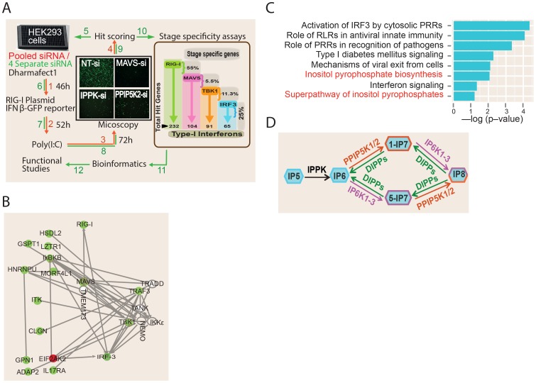
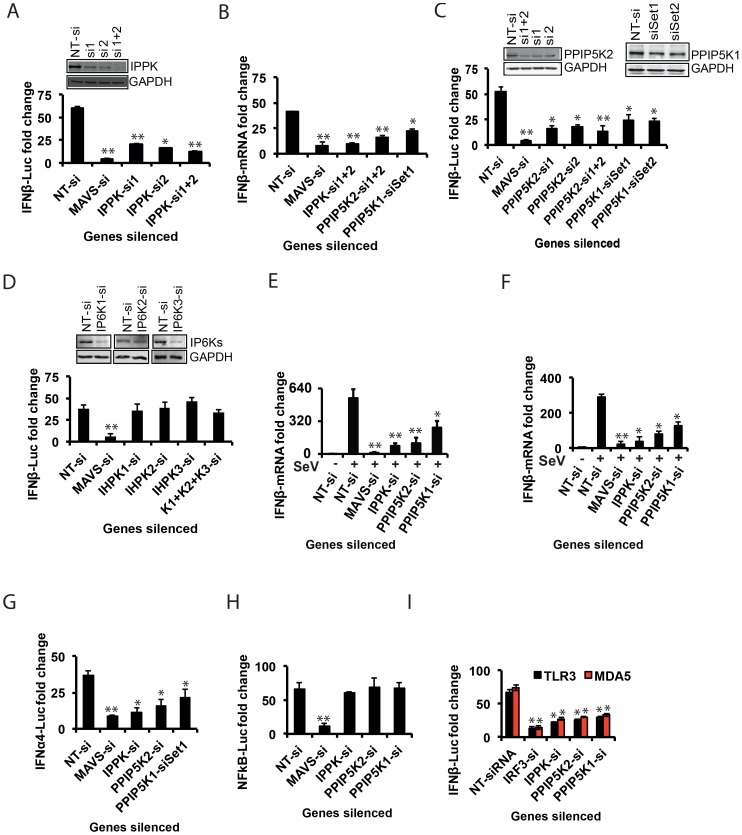
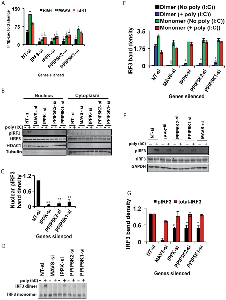
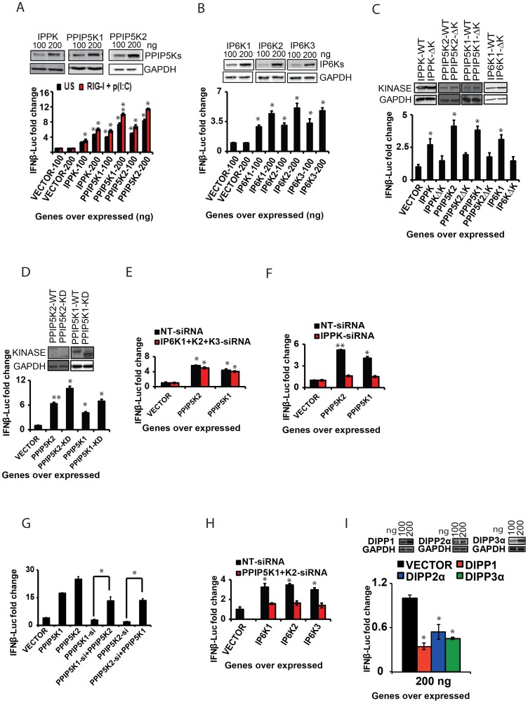
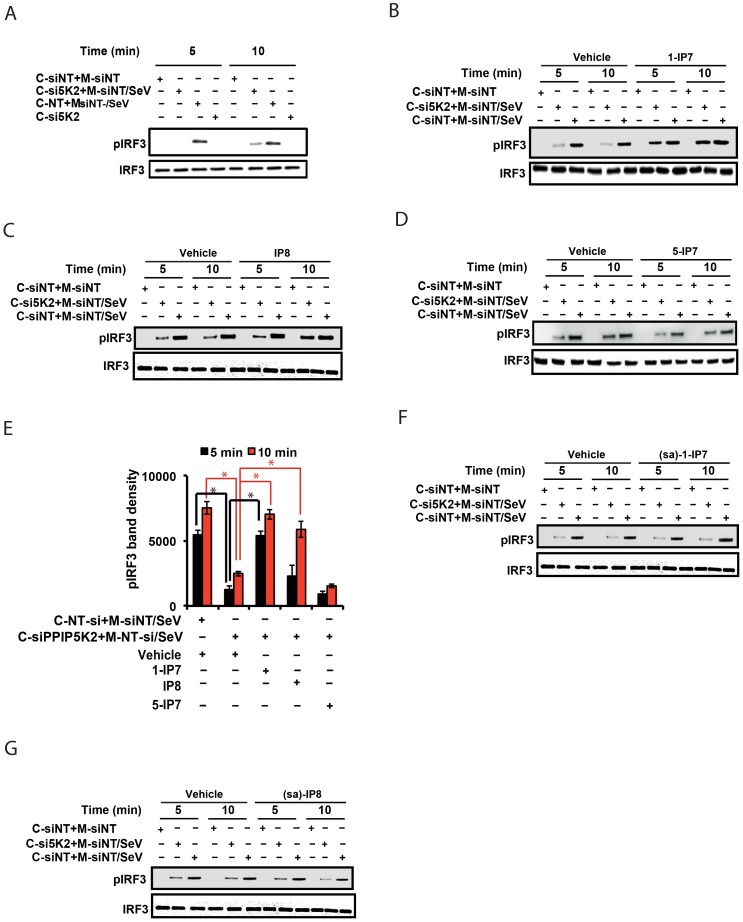
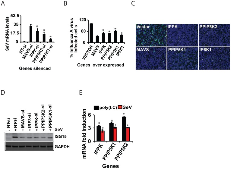
The pattern recognition receptor RIG-I is critical for Type-I interferon production. However, the global regulation of RIG-I signaling is only partially understood. Using a human genome-wide RNAi-screen, we identified 226 novel regulatory proteins of RIG-I mediated interferon-β production. Furthermore, the screen identified a metabolic pathway that synthesizes the inositol pyrophosphate 1-IP7 as a previously unrecognized positive regulator of interferon production. Detailed genetic and biochemical experiments demonstrated that the kinase activities of IPPK, PPIP5K1 and PPIP5K2 (which convert IP5 to1-IP7) were critical for both interferon induction, and the control of cellular infection by Sendai and influenza A viruses. Conversely, ectopically expressed inositol pyrophosphate-hydrolases DIPPs attenuated interferon transcription. Mechanistic experiments in intact cells revealed that the expression of IPPK, PPIP5K1 and PPIP5K2 was needed for the phosphorylation and activation of IRF3, a transcription factor for interferon. The addition of purified individual inositol pyrophosphates to a cell free reconstituted RIG-I signaling assay further identified 1-IP7 as an essential component required for IRF3 activation. The inositol pyrophosphate may act by β-phosphoryl transfer, since its action was not recapitulated by a synthetic phosphonoacetate analogue of 1-IP7. This study thus identified several novel regulators of RIG-I, and a new role for inositol pyrophosphates in augmenting innate immune responses to viral infection that may have therapeutic applications.


Conflict of interest statement

The authors have declared that no competing interests exist.

Figures

Figure 1
Figure 1. RNAi screening and bioinformatics analysis.
(A) Screen methodology. See methods for detailed description of screening methodology. The results of stage specific assays are shown in the brown box on the right side of panel A. Numbers and arrows shown in red and green colour indicate steps of the primary and secondary screens, respectively. (B) Network analysis involving all ‘hits’ in the siRNA screen that have been experimentally validated in the literature to interact with one or more components of the RIG-I pathway. Putative positive regulators of RIG-I are indicated in green, and putative negative regulators in red. Empty circles are previously known genes of the RIG-I pathway that were not defined as hits in our RNAi screen. (C) Gene ontology canonical pathway enrichment analysis among the novel RIG-I regulators identified in this screen. Most enriched categories are shown. RLR, RIG-I Like Receptors. (D) Schematic showing inositol pyrophosphate synthesis pathway. IPPK, inositol 1,3,4,5,6-pentakisphosphate 2-kinase; PPIP5K1/2, diphosphoinositol pentakisphosphate kinase 1/2; IP6K1-3, inositol hexakisphosphate kinase 1–3; IP5, inositol pentakisphosphate; IP6, inositol hexakisphosphate; IP7, diphosphoinositol-pentakisphosphate; IP8, bisdiphosphoinositol-tetrakisphosphate.
Figure 2
Figure 2. Inositol pyrophosphates pathway is needed for RIG-I mediated IFNβ response.
(A, C, D) Effect of silencing of IPPK, PPIP5K1, PPIP5K2, and IP6K1-3 on p(I:C) stimulated RIG-I mediated IFNβ-promoter driven luciferase reporter activity in HEK293 cells is shown. (B) Effect of silencing of IPPK, PPIP5K1 and PPIP5K2 on poly (I:C) stimulated RIG-I mediated IFNβ transcription (detected using q-RTPCR) in HEK293 cells. (E) Effect of silencing of IPPK, PPIP5K1 and PPIP5K2 on IFNβ transcription (detected using q-RTPCR) during Sendai virus infection of HEK293 cells. (F) Effect of silencing of IPPK, PPIP5K1 and PPIP5K2 in human primary macrophages on Sendai virus induced IFNβ transcription, measured by q-RTPCR. (G, H) Effect of silencing of IPPK, PPIP5K1 and PPIP5K2 on RIG-I mediated IFNα4 (G) and NFκB (H) driven luciferase reporter activities. (I) Effect of silencing of IPPK, PPIP5K1 and PPIP5K2 on IFNβ promoter reporter activity driven by activation of TLR3 (black bar) and MDA5 (red bar). The IFNβ or NFκB luciferase reporter values were normalized with Renilla luciferase reporter values, and expressed as fold change from uninduced NT-si treated samples. The mRNA level data was quantified by q-RTPCR and expressed as fold-change, determined using the comparative Ct value based approach, using the formula 2 (Ct of kinase gene - Ct of β-actin), with untreated value as 1. The statistical significance was determined by comparing the values for each gene silencing with that of corresponding stimulated NT-si treated samples. The Western blot based silencing confirmation experiments for PPIP5K1 and PPIP5K2 were performed using ectopically expressed proteins. The values are mean ± SD of one representative experiment performed in triplicates. si, siRNA; NT-si, non-targeting negative control siRNA; US, unstimulated. GAPDH, Glyceraldehyde 3-Phosphate Dehydrogenase, cytoplasmic marker.
Figure 3
Figure 3. Inositol pyrophosphate synthesis pathway is needed for IRF3 activation.
(A) Effect of silencing of IPPK, PPIP5K1 and PPIP5K2 on IFNβ promoter driven luciferase reporter activity induced by ectopic expression of RIG-I, MAVS and TBK1, in HEK293 cells. (B, C) Effect of silencing of IPPK, PPIP5K1 and PPIP5K2 on nuclear translocation of IRF3, shown by Western blot and densitometry, respectively. (D, E) Effect of silencing of IPPK, PPIP5K1 and PPIP5K2 on dimerization of IRF3, shown by Western blot and densitometry, respectively. (F, G) Effect of silencing of IPPK, PPIP5K1 and PPIP5K2 on p(I:C) induced phosphorylation of IRF3, shown by Western blot and densitometry, respectively. A representative Western blot for each experiment is shown. The IFNβ-luciferase values were normalized with Renilla luciferase reporter values, and expressed as fold change from uninduced NT-si samples. Densitometry values represent measured intensities of the indicated bands from three different Western blots, shown as mean ± SD. The significance of densitometry data was calculated by comparing the values obtained from gene silenced conditions with that of stimulated NT-si controls. For panel A, the significance was determined by comparing the values of IFNβ-luciferase activity obtained upon over expression of RIG-I/MAVS/TBK1 in IPPK, PPIP5K1 and PPIP5K2 gene silenced samples with that of corresponding NT-si samples. The values are mean ± SD of one representative experiment performed in triplicates. si, siRNA; NT-si, non-targeting negative control siRNA. GAPDH, Glyceraldehyde 3-Phosphate Dehydrogenase, cytoplasmic marker.
Figure 4
Figure 4. The catalytic activity of inositol pyrophosphate synthesis pathway kinases regulates IFNβ response.
(A, B) Ectopic expression of inositol pyrophosphate-synthesis-pathway kinases enhanced RIG-I mediated IFNβ-promoter driven luciferase reporter activity. HEK293 cells transfected with the indicated concentrations of empty vector, or expression plasmids of (A) IPPK, PPIP5K1, and PPIP5K2, and (B) IP6K1-3, with (red bars) or without (black bars) p(I:C)stimulation. (C) Effect of ablation of the catalytic activity of IPPK, PPIP5K1, PPIP5K2 and IP6K1 on IFNβ response. IFNβ promoter driven luciferase activity was measured in HEK293 cells ectopically expressing wild type and catalytically inactive mutants of the indicated proteins. (D) Interferon stimulating effect of the ectopic expression of the isolated kinase domains of PPIP5K1 (PPIP5K1-KD) and PPIP5K2 (PPIP5K2-KD) in HEK293 cells and measured by IFNβ promoter driven luciferase reporter. (E) Effect of silencing of IP6K1-3 on the ability of ectopically expressed PPIP5K1 and PPIP5K2 to enhance IFNβ promoter driven luciferase activity in HEK293 cells. (F) Effect of silencing of IPPK on the ability of ectopically expressed PPIP5K1 and PPIP5K2 to enhance IFNβ promoter driven luciferase activity in HEK293 cells. (G) Effect of overexpression of PPIP5K1 or PPIP5K2 on IFNβ promoter driven luciferase activity in HEK293 cells silenced for PPIP5K2 and PPIP5K1 respectively. (H) Effect of silencing of PPIP5K1 and PPIP5K2 on the ability of ectopically expressed IP6K1-3 to enhance IFNβ promoter driven luciferase activity in HEK293 cells. (I) Effect of ectopic expression of DIPP1, and DIPP2a on RIG-I mediated IFNβ promoter driven luciferase activity in HEK293 cells. RIG-I pathway activated HEK293 cells were transfected with the indicated concentrations of the genes or empty vector, and IFNβ promoter driven luciferase activity was measured. The IFNβ-luciferase values were normalized with Renilla luciferase reporter values, and expressed as fold change from empty vector transfected samples. The significance was determined by comparing the values for each gene with that of corresponding empty vector samples. For panel G, the significance of the recovery of IFNβ-luciferase signal upon gene over expression was determined by comparing to that from kinase silenced cells. The values are mean ± SD of one representative experiment performed in triplicates. NT-si, non-targeting negative control siRNA; WT, wild type; ΔK, kinase catalytically activity defective mutant; KD, isolated functional kinase domain only. GAPDH, Glyceraldehyde 3-Phosphate Dehydrogenase, cytoplasmic marker.
Figure 5
Figure 5. 1-IP7 is needed for IRF3 phosphorylation.
(A) In vitro reconstituted RIG-I signaling assay reveals defect in IRF3 phosphorylation upon the silencing of PPIP5K2. Western blot was performed at 5 and 10 minutes after mixing the uninfected cytoplasm with mitochondrial fraction from Sendai virus (SeV) infected cells. (B–E) Effect of addition of various physiological inositol pyrophosphates on the attenuated IRF3 phosphorylation in PPIP5K2 silenced cell extracts, determined by the in vitro reconstituted RIG-I signaling assay. Reactions were quenched at 5 or 10 min after the addition of inositol pyrophosphates as indicated. A representative Western blot showing the effect of addition of 0.5 uM of 1-IP7 (B), IP8 (C), and 5-IP7 (D) is given. (E) Graph showing densitometry analysis of the intensity of the pIRF3 bands of the experiments shown in “B–D” at both 5 and 10 minute time points. Densitometry values represent measured average intensities of the indicated bands from three different Western blots, expressed as mean ± SD. The significance of densitometry data was calculated by comparing the values obtained from gene silenced conditions with that of indicated negative control NT-si controls with vehicle. (F, G) Effect of addition (0.5 uM) of the synthetic phosphonoacetate analogues 1-IP7 (F) and IP8 (G) on the attenuated IRF3 phosphorylation in PPIP5K2 silenced cell extracts, determined by the in vitro reconstituted RIG-I signaling assay. Reactions were quenched at 5 or 10 min after the addition of inositol pyrophosphates as indicated. (sa)-1-IP7, synthetic analogue of 1-IP7; (sa)-IP8, synthetic analogue of IP8; si, siRNA; siNT, non-targeting negative control siRNA; si5K2, siRNA targeting PPIP5K2. C-siNT+M-siNT, cytoplasm of negative control NT siRNA treated cells, mixed with mitochondria of uninfected negative control NT siRNA treated cells; C-si5K2+M-siNT/SeV, cytoplasm of PPIP5K2 siRNA treated cells, mixed with mitochondria of Sendai virus (SeV) infected negative control NT siRNA treated cells; C-siNT+M-siNT/SeV, cytoplasm of negative control NT siRNA treated cells, mixed with mitochondria of Sendai virus (SeV) infected negative control NT siRNA treated cells. C, cytoplasm; M, mitochondria. GAPDH, Glyceraldehyde 3-Phosphate Dehydrogenase, cytoplasmic marker.
Figure 6
Figure 6. Inositol pyrophosphates synthesis pathway is required for cellular antiviral immunity.
(A) Data showing increased Sendai virus RNA load in IPPK, PPIP5K1 and PPIP5K2 silenced HEK293 cells, measured by q-RTPCR. (B) Effect of ectopic expression of IPPK, PPIP5K1, PPIP5K2 and IP6K1 on the infectivity of GFP-tagged influenza A virus (12 h infection assay) on HEK293 cells. Results are expressed as percentage of cells expressing Influenza-GFP virus (from 15 images per condition), with empty vector transfected value taken as 100%. (C) Microscopic images showing effect of ectopic expression of IPPK, PPIP5K1, PPIP5K2 and IP6K1 in HEK293 cells on influenza-GFP virus infection. A representative image is shown. (D) Effect of silencing of IPPK, PPIP5K1 and PPIP5K2 on Sendai virus mediated induction of ISG15 protein, shown as a representative Western blot. (E) Effect of infection with Sendai virus (SeV) or treatment with p(I:C) on the transcription of the indicated genes, measured by q-RTPCR. The mRNA level data are expressed as fold-change, determined using the comparative Ct value based approach, using the formula 2 (Ct of kinase gene - Ct of β-actin), with untreated value as 1. The significance is determined by comparing the values for each gene with that of corresponding NT-siRNA or vector controls. The values are mean ± SD of one representative experiment performed in triplicates. si, siRNA; NT, non-targeting negative control siRNA. GAPDH, Glyceraldehyde 3-Phosphate Dehydrogenase, cytoplasmic marker.

Similar articles

Cited by

References

    1. Kumar H, Kawai T, Akira S (2009) Pathogen recognition in the innate immune response. Biochem J 420: 1–16. - PubMed
    1. Liu SY, Sanchez DJ, Cheng G (2011) New developments in the induction and antiviral effectors of type I interferon. Curr Opin Immunol 23: 57–64. - PMC - PubMed
    1. Yoneyama M, Fujita T (2010) Recognition of viral nucleic acids in innate immunity. Rev Med Virol 20: 4–22. - PubMed
    1. Belgnaoui SM, Paz S, Hiscott J (2011) Orchestrating the interferon antiviral response through the mitochondrial antiviral signaling (MAVS) adapter. Curr Opin Immunol 23: 564–572. - PubMed
    1. Ramos HJ, Gale M Jr (2011) RIG-I like receptors and their signaling crosstalk in the regulation of antiviral immunity. Curr Opin Virol 1: 167–176. - PMC - PubMed

Publication types

MeSH terms

LinkOut - more resources