. 2014 Feb 27;10(2):e1004166.
doi: 10.1371/journal.pgen.1004166. eCollection 2014 Feb.

DAAM is required for thin filament formation and Sarcomerogenesis during muscle development in Drosophila

Affiliations

DAAM is required for thin filament formation and Sarcomerogenesis during muscle development in Drosophila

Imre Molnár et al. PLoS Genet. .

Abstract

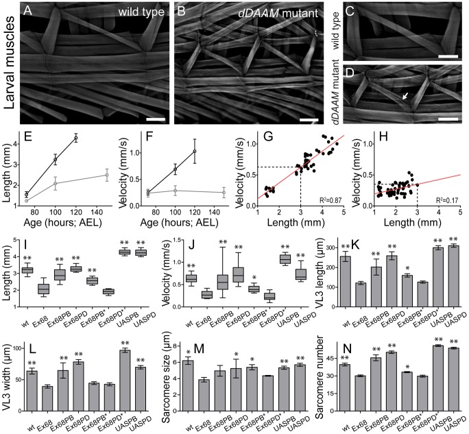
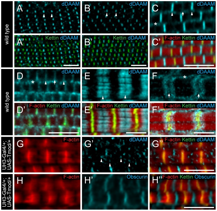
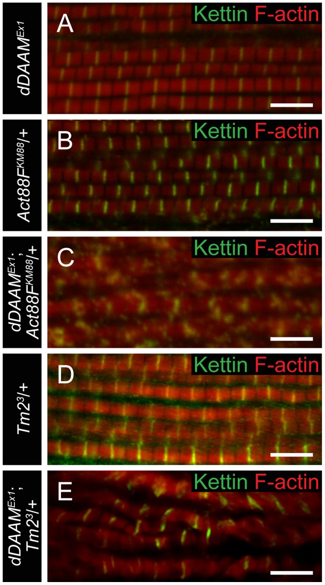
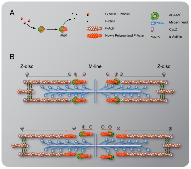
During muscle development, myosin and actin containing filaments assemble into the highly organized sarcomeric structure critical for muscle function. Although sarcomerogenesis clearly involves the de novo formation of actin filaments, this process remained poorly understood. Here we show that mouse and Drosophila members of the DAAM formin family are sarcomere-associated actin assembly factors enriched at the Z-disc and M-band. Analysis of dDAAM mutants revealed a pivotal role in myofibrillogenesis of larval somatic muscles, indirect flight muscles and the heart. We found that loss of dDAAM function results in multiple defects in sarcomere development including thin and thick filament disorganization, Z-disc and M-band formation, and a near complete absence of the myofibrillar lattice. Collectively, our data suggest that dDAAM is required for the initial assembly of thin filaments, and subsequently it promotes filament elongation by assembling short actin polymers that anneal to the pointed end of the growing filaments, and by antagonizing the capping protein Tropomodulin.


Conflict of interest statement

The authors have declared that no competing interests exist.

Figures

Figure 1
Figure 1. dDAAM impairs IFM structure.
(A) Quantification of the flight ability of wild type and dDAAM mutant flies with the genotypes indicated. Bars display mean±SEM. (B) Western blot shows that wild type IFM expresses two dDAAM protein isoforms, of 130 kD and 163 kD. The larger isoform is more highly expressed of the two. The dDAAMEx1 allele reduces the level of the 130 kD isoform, whereas RNAi silencing results in a strong reduction of the level of the 163 kD isoform. Lower panel shows the loading control (α-glycogen-phosphorylase). (C–C″) Myofibrils of a wild type IFM display a regular sarcomere organization. (D–E″) Myofibrils from two different IFMs of the dDAAMEx1, UDT mutant combination. Note the complex sarcomeric defects (D–E″) including the reduced F-actin level (in red, C′–E′), the irregularities in fiber width, the disorganized Z-discs stained with anti-Kettin (in green, C–E″) and the sarcomere length shortening in E–E″. Bars, 5 µm.
Figure 2
Figure 2. EM analysis of IFM morphology in dDAAM mutants.
Electronmicrographs of IFM from wild type (A, C, E, G, I) and dDAAMEx1; UDT mutants (B, D, F, H, J). Longitudinal sections of adult IFM (A–D) show that, as compared to the wild type, highly ordered and tightly packed sarcomeres (A, C), the dDAAM mutant myofibrils (B, D) display Z-disc and M-band defects, and shortened sarcomeres with loosely organized thin and thick filaments. Transverse sections of wild type (E, G) muscles reveal the hexagonal lattice organization of thin and thick filaments, which is almost entirely lost in dDAAM mutant myofibrils (F, H). Instead, the mutant fibrils are irregularly shaped, consisting of clusters of thick filaments, and individual thin filaments are hardly detectable. Note: wild type thick filaments are hollow (G), while those of the dDAAM mutant are very dark, irregularly shaped and almost never hollow (H). Longitudinal sections of pupal IFM (48 hours APF) (I, J) show that, as compared to wild type (I), mutants (J) have strong Z-disc and M-line defects, shorter sarcomeres and irregular filament organisation. Arrows mark the Z-discs, asterisks mark the M-bands, m labels the mitochondria. Bars, 500 nm.
Figure 3
Figure 3. Structural and functional analysis of the larval body wall muscles.
Wild type (A, C) and dDAAMEx68 null mutant (B, D) larval body wall muscles stained with phalloidin. Mutant muscles are smaller, some myofibers are split (arrow on D) and the overall muscle pattern is looser than in wild type. The relationship of larval age and length (E), and of larval age and velocity (F) in wt (wild type; black line) and dDAAMEx68 (grey line) larvae. The relationship of larval length and velocity of wt (G) and dDAAMEx68 mutant (H) larvae. Quantification of larval length (I), crawling velocity (J), VL3 muscle length (K), width (L), mean sarcomere length (M) and serial sarcomere number (N) in larvae 100 hours AEL with the following genotypes: wt (wild type), Ex68 (dDAAMEx68), Ex68PB (dDAAMEx68; DMef2-Gal4; UAS-dDAAM-PB), Ex68PD (dDAAMEx68; DMef2-Gal4; UAS-dDAAM-PD), Ex68PB* (dDAAMEx68; DMef2-Gal4; UAS-dDAAM-PBI732A), Ex68PD* (dDAAMEx68; DMef2-Gal4; UAS-dDAAM-PDI732A), UASPB (DMef2-Gal4; UAS-dDAAM-PB) and UASPD (DMef2-Gal4; UAS-dDAAM-PD). Bars represent mean values with respective SDs in I–N. Statistical significance: * 0.05>p<0.001; ** p≤0.001. Wild type and rescue data were compared to dDAAMEx68 data, unless otherwise indicated in the text. Bars, 100 µm (A–D).
Figure 4
Figure 4. Sarcomeric localization of the dDAAM protein in the IFM and the larval body wall muscles.
dDAAM staining of the IFM myofibrils of wild type pupae 48 hours (A, A′) and 72 hours APF (B, B′), freshly eclosed adult (C, C′) and 4 day-old adult (D, D′). dDAAM accumulates at the M-line (arrowheads), at the Z-disc (arrow) and in the sarcoplasm (asterisk). Note: accumulation at Z-disc is weak in pupae and young adults (A–C), but in 4 day-old adults staining is equally strong at the M-line and the Z-disc (D). In developing larval body wall muscles (72 hours AEL) dDAAM staining resolves into two bands along the M-line (E, E′). In fully matured larval body wall muscles dDAAM staining relocates to a region flanking the Z-disc (F, F′). Arrowheads mark the M-line in E; arrows mark the Z-disc, asterisk marks the sarcoplasm in F. (G–H″) Excess Tmod in UH3-Gal4/+; UAS-Tmod/+ flies leads to shorter thin filaments that are not in perfect register and vary in length as judged by F-actin staining (G, H). In these IFMs dDAAM protein displays a punctate distribution (arrowheads in G′) most of which colocalizes with the pointed end region of the thin filaments (G″). The M-line in these mutant muscles remains nearly intact as judged by Obscurin staining (H′). Phalloidin staining is in red (C′–H″), Kettin (C′–H″) and sls-GFP (A′, B′) as Z-disc markers are in green, anti-dDAAM (A–F′, G′, G″) and anti-Obscurin (H, H′) are in cyan. Bars, 5 µm.
Figure 5
Figure 5. Sarcomeric localization of the mDaam1 protein.
(A–B″) mDaam1 staining (in cyan) of mouse muscle sections (the Z-disc marker α-actinin is in red). In m. tibialis anterior sarcomeres mDaam1 accumulates in two bands either side of the M-line (A–A″), whereas in m. vastus lateralis it is mostly detected along the Z-discs (B–B″). In C2C12 cells differentiated for 96 hours mDaam1 (cyan) accumulates in two broad bands at the middle of the sarcomere that does not significantly overlap with titin staining (yellow; 9D10 antibody) (C–C″) or myomesin (green), an M-line marker (D–D″). (E–F″) Distribution of mDaam1 (cyan) and α-actinin (red) in C2C12 cells induced to differentiate for 24 (E–E″) or 96 hours (F–F″). Bars: 5 µm (A–D″); 15 µm (E–F″).
Figure 6
Figure 6. dDAAM interacts with thin filament mutants.
IFM myofibrils from (A) dDAAMEx1, (B) Act88FKM88/+, (C) dDAAMEx1; Act88FKM88/+, (D) Tm23/+ and (E) dDAAMEx1; Tm23/+ flies (actin in red, Kettin in green in all panels). Note: sarcomere organization in dDAAMEx1 (A) is nearly wild type; likewise Act88F (B) and Tm2 (D) heterozygotes display a largely regular myofibril and Z-disc organization. Myofibrils of the dDAAMEx1; Act88FKM88/+ (C) and dDAAMEx1; Tm23/+ (E) genotypes are extremely disorganized compared to the controls. Bars, 5 µm.
Figure 7
Figure 7. The interaction of dDAAM and tmod.
Upon silencing of tmod myofibrils get severely disrupted (A–A″), though ∼10% of them show a milder effect with regular Z-disc arrangement but missing H-zones (B–B″). In dDAAMEx1, UH3-Gal4; UAS-tmodRNAi muscles most myofibrils have a nearly wild type sarcomeric organization with regularly spaced Z-discs and M-lines, and almost normal sarcomere length (C–C″). (D–D″) In UH3-Gal4; UAS-Tmod IFMs the sarcomeric thin filaments often appear to be shorter than wild type as judged by phalloidin staining, whereas myofibrils of UH3-Gal4; UAS-FLDAAM muscle look wild type (E–E″). Simultaneous overexpression of FLDAAM and Tmod results in the same effect as the expression of Tmod alone (F–F″; compare to D–D″). Kettin in green, actin in red in A–F″. (G) An end-to-end actin annealing assay, dark grey: 0 minute control, average filament length in the presence of 1 µM F-actin (F-actin), light gray: average filament length after 60 minutes incubation, in the presence of 1 µM F-actin (F-actin), 1 µM F-actin+ 10 nM capping protein (F-actin+CP), 1 µM F-actin+100 nM DAAM-FH1-FH2 (F-actin+DAAM), 1 µM F-actin+1 µM skeletal tropomyosin (F-actin+TM), 1 µM F-actin+100 nM DAAM-FH1-FH2+1 µM skeletal tropomyosin (F-actin+DAAM+TM). Bars represent mean values with respective SEMs. (H) Electronmicrograph of a tmodRNAi IFM. Black arrowheads mark the borders of the mid-sarcomeric region where the M-line structures are not evident but thin filaments appear to cross this area. White arrows on the inset, corresponding to the dashed area, mark thin filaments that fail to terminate in the H-zone. Bars: 5 µm (A–F″); 500 nm (H) 100 nm (H, inset).
Figure 8
Figure 8. A model of DAAM mediated ‘pointed end elongation’.
(A) Nucleation and elongation of short actin filaments by the barbed (+) end binding formin DAAM. (B) A possible mechanism of thin filament elongation from the pointed end (−) is the end-to-end annealing of DAAM assembled short actin filaments (in orange) to the Z-disc anchored growing “mother filament” (in brown).

Similar articles

Cited by

References

    1. Luther PK (2009) The vertebrate muscle Z-disc: sarcomere anchor for structure and signalling. J Muscle Res Cell Motil 30: 171–185. - PMC - PubMed
    1. Sparrow JC, Schock F (2009) The initial steps of myofibril assembly: integrins pave the way. Nat Rev Mol Cell Biol 10: 293–298. - PubMed
    1. Pollard TD, Blanchoin L, Mullins RD (2000) Molecular mechanisms controlling actin filament dynamics in nonmuscle cells. Annu Rev Biophys Biomol Struct 29: 545–576. - PubMed
    1. Campellone KG, Welch MD (2010) A nucleator arms race: cellular control of actin assembly. Nat Rev Mol Cell Biol 11: 237–251. - PMC - PubMed
    1. Chesarone M, Gould CJ, Moseley JB, Goode BL (2009) Displacement of formins from growing barbed ends by bud14 is critical for actin cable architecture and function. Dev Cell 16: 292–302. - PMC - PubMed

Publication types

MeSH terms

LinkOut - more resources