. 2014 Feb 19;9(2):e88712.
doi: 10.1371/journal.pone.0088712. eCollection 2014.

DNA aptamers against exon v10 of CD44 inhibit breast cancer cell migration

Affiliations

DNA aptamers against exon v10 of CD44 inhibit breast cancer cell migration

Joji Iida et al. PLoS One. .

Abstract

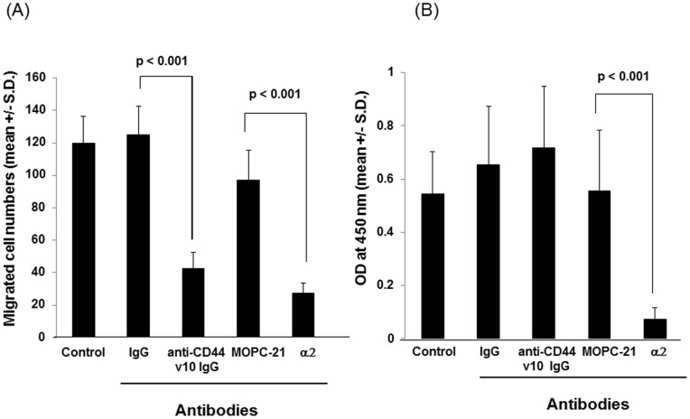
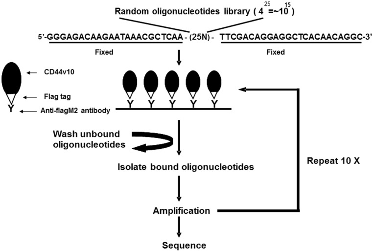
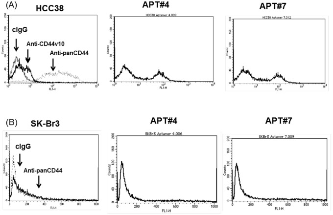
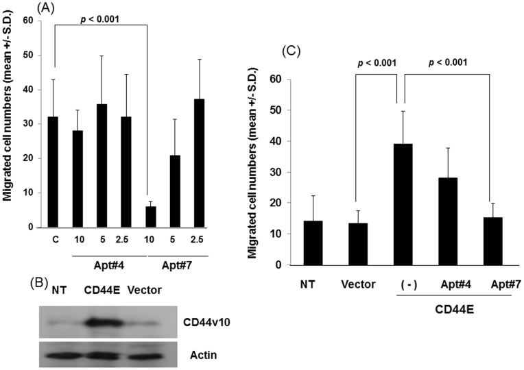
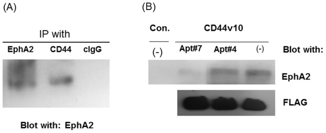
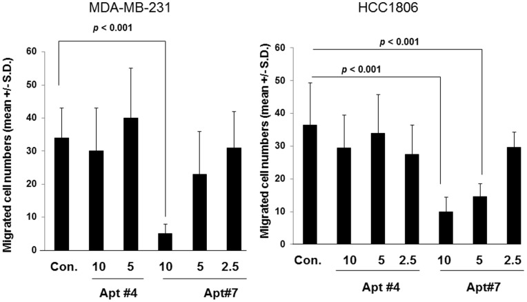
CD44 adhesion molecules are expressed in many breast cancer cells and have been demonstrated to play a key role in regulating malignant phenotypes such as growth, migration, and invasion. CD44 is an integral transmembrane protein encoded by a single 20-exon gene. The diversity of the biological functions of CD44 is the result of the various splicing variants of these exons. Previous studies suggest that exon v10 of CD44 plays a key role in promoting cancer invasion and metastasis, however, the molecular mechanisms are not clear. Given the fact that exon v10 is in the ectodomain of CD44, we hypothesized that CD44 forms a molecular complex with other cell surface molecules through exon v10 in order to promote migration of breast cancer cells. In order to test this hypothesis, we selected DNA aptamers that specifically bound to CD44 exon v10 using Systematic Evolution of Ligands by Exponential Enrichment (SELEX). We selected aptamers that inhibited migration of breast cancer cells. Co-immunoprecipitation studies demonstrated that EphA2 was co-precipitated with CD44. Pull-down studies demonstrated that recombinant CD44 exon v10 bound to EphA2 and more importantly aptamers that inhibited migration also prevented the binding of EphA2 to exon v10. These results suggest that CD44 forms a molecular complex with EphA2 on the breast cancer cell surface and this complex plays a key role in enhancing breast cancer migration. These results provide insight not only for characterizing mechanisms of breast cancer migration but also for developing target-specific therapy for breast cancers and possibly other cancer types expressing CD44 exon v10.


Conflict of interest statement

Competing Interests: Dr. Richard Somiari is employed by ITSI-Biosciences, which is a commercial company. This does not alter the authors’ adherence to all the PLOS ONE policies on sharing data and materials.

Figures

Figure 1
Figure 1. Inhibition of migration of HCC38 cells with anti-CD44 exon v10 antibody.
(A) Cells were harvested, washed, and resuspended in RPMI1640-serum free media. Migration assays were performed using type I collagen (10 µg/ml) as an adhesive substrate in the lower compartment of Transwell by incubating at 37°C for 4 hours. The antibodies were added in both upper and lower chambers at a concentration of 5 µg/ml. Experiments were performed by triplicates three times. Statistical significance was calculated by Student’s two-tailed paired t-test. (B) Cells were harvested, washed, resuspended in RPMI1640 containing 1 mg/ml BSA (adhesion buffer) at a concentration of 105 cell/ml. Cells (100 µl/well) were incubated on plates coated with type I collagen (5 µg/ml) for 1 hour in the presence or absence of antibodies (5 µg/ml). Plates were washed, fixed followed by staining with crystal violet. After extensive washing to remove excess dye, cells were lysed with 100 µl of PBS containing 2% SDS and then the absorbance was measured at 550 nm. Experiments were performed in quadruplicates and repeated three times. The representative data were shown as mean +/− S.D. of absorbance values.
Figure 2
Figure 2. Protocol for isolating CD44 exon v10-specific DNA aptamers by Systematic Evolution of Ligands by Exponential Enrichment (SELEX).
The CD44 exon v10 peptide was expressed as a fusion protein with FLAG tag. The purified recombinant peptide was immobilized on plates coated with anti-FLAG (M2) antibody. Library of DNA aptamers were incubated on the plates and extensively washed to remove non-bound aptamers. The isolated aptamers were then amplified and this selection process was repeated 10 times. Aptamers isolated from the last selection were amplified and ligated to pCR2.1-TOPOTA vector for sequencing.
Figure 3
Figure 3. FACS analysis of aptamers against CD44 exon v10.
HCC38 or SK-Br3 cells were harvested and resuspended in RPMI1640 containing 1 mg/ml BSA and 0.025% NaN3 (FACS buffer). Cells were incubated with aptamers of which 5′-end was conjugated to biotin (5 µg/ml), anti-panCD44 (clone 156-3C11) (5 µg/ml), or anti-CD44 exon v10 antibody (AB2082) (5 µg/ml) for 1 hour at 4°C. After washing with the same buffer, cells were incubated with Streptavidin-FITC, FITC-goat anti-mouse IgG, or FITC-goat anti-rabbit IgG for 1 hour at 4°C. After extensive washing with FACS buffer, cells were resuspended in PBS containing 1% paraformaldehyde. Expression was measured on FACS Calibur (Becton-Dickinson, NJ USA) by collecting 10,000 events and analyzed by CellQuest.
Figure 4
Figure 4. Inhibition of cell migration by aptamers against CD44 exon v10.
(A) HCC38 cells were harvested, washed and resuspended in RPMI1640-serum free media. Migration assays were performed using type I collagen (10 µg/ml) as an adhesive substrate in the lower compartment of Transwell by incubating at 37°C for 4 hours. The aptamers were added in both upper and lower chambers at the indicated concentrations in the text. Experiments were performed in triplicate. Statistical significance was calculated by Student’s two-tailed paired t-test. (B) MCF-7 cells were stably transfected with CD44E according to the methods described in Materials and Methods. MCF-7 cells transfected control vector, [MCF-7(pIRES2)], MCF-7 (CD44E), and parental MCF-7 cells (NT) were lysed and subjected to western blotting for the expression of CD44E using anti-CD44 exon v10 antibody followed by HRP-conjugated goat-anti-rabbit IgG. The signal was detected by ECL reactions. Actin was used as a loading control. (C) The migration of these cells was evaluated as described above in the presence or absence of aptamers against CD44 exon v10 at a concentration of 10 nM as described above. Experiments were performed in triplicate. Statistical significance was calculated by Student’s two-tailed paired t-test.
Figure 5
Figure 5. CD44 associated with EphA2 on breast cancer cells.
(A) HCC38 cells were lysed with 100 mM Tris-HCl (pH 7.5) containing 1% Brij35, 0.14 M NaCl, 1 mM CaCl2, 1 mM MnCl2, and a protease inhibitor cocktail by scraping and pipetting. The cell lysates were precleared and immunoprecipitated with anti-CD44 antibody clone (clone 156-3C11)-, anti-EphA2- or control IgG-protein G beads for 4 hours at 4°C. The bound proteins were released by boiling at 95°C in SDS-sample buffer under reducing conditions and separated on SDS-PAGE followed by western blotting with anti-EphA2 antibody followed by HRP-secondary antibody. The proteins are visualized with enhanced chemiluminescence (ECL) reaction. (B) Cells were harvested using 5 mM EDTA in PBS and were lysed in 100 mM Tris-HCL (pH 7.5) containing 1% Brij35, 0.14 M NaCl, 1 mM CaCl2, 1 mM MnCl2, and a protease inhibitor cocktail. The lysates were precleared with anti-FLAG (M2) agarose beads for 4 hours by shaking at 4°C. The cleared lysates were incubated with recombinant protein CD44v10 P-FLAG-anti-FLAG (M2) antibody-agarose beads (CD44v10) or anti-FLAG (M2) antibody-agarose beads (Con.) in the presence or absence of aptamers (Apt#4 and Apt#7) by shaking at 4°C for 4 hours. The beads were washed extensively with the lysis buffer. The bound proteins were released by boiling at 95°C in SDS-sample buffer under reducing conditions and separated on SDS-PAGE followed by western blotting with anti-EphA2 followed by HRP-secondary antibody or HRP-conjugated anti-FLAG (M2) antibody. The proteins are visualized with enhanced chemiluminescence (ECL) reaction. CD44 exon v10 peptide was used as a loading control for ensuring the same amount of proteins was loaded in each lane.
Figure 6
Figure 6. Inhibition of migration of MDA-MB-231 and HCC1806.
MDA-MB-231 (A) or HCC1806 (B) cells were harvested, washed and resuspended in RPMI1640-serum free media. Migration assays were performed using type I collagen (10 µg/ml) as an adhesive substrate in the lower compartment of Transwell by incubating at 37°C for 4 hours. The aptamers were added in both upper and lower chambers at the indicated concentrations. Experiments were performed in triplicate. Statistical significance was calculated by Student’s two-tailed paired t-test.

Similar articles

Cited by

References

    1. Ferreira CS, Matthews CS, Missailidis S (2006) DNA aptamers that bind to MUC1 tumour marker: design and characterization of MUC1-binding single-stranded DNA aptamers. Tumour Biol 27: 289–301. - PubMed
    1. Fitzwater T, Polisky B (1996) A SELEX primer. Methods Enzymol 267: 275–301. - PubMed
    1. Lupold SE, Hicke BJ, Lin Y, Coffey DS (2002) Identification and characterization of nuclease-stabilized RNA molecules that bind human prostate cancer cells via the prostate-specific membrane antigen. Cancer Res 62: 4029–4033. - PubMed
    1. Kimoto M, Shirouzu M, Mizutani S, Koide H, Kaziro Y, et al. (2002) Anti-(Raf-1) RNA aptamers that inhibit Ras-induced Raf-1 activation. Eur J Biochem 269: 697–704. - PubMed
    1. Daniels DA, Chen H, Hicke BJ, Swiderek KM, Gold L (2003) A tenascin-C aptamer identified by tumor cell SELEX: systematic evolution of ligands by exponential enrichment. Proc Natl Acad Sci U S A 100: 15416–15421. - PMC - PubMed

Publication types