. 2014 Feb 20;9(2):e88828.
doi: 10.1371/journal.pone.0088828. eCollection 2014.

C. elegans CEP-1/p53 and BEC-1 are involved in DNA repair

Affiliations

C. elegans CEP-1/p53 and BEC-1 are involved in DNA repair

Sandy Hoffman et al. PLoS One. .

Abstract

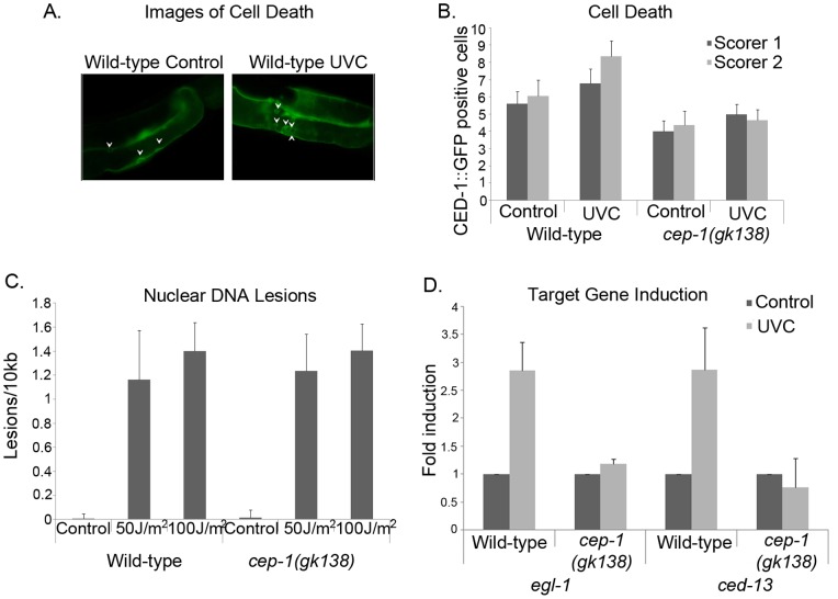
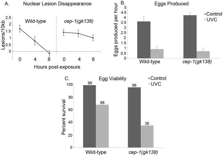
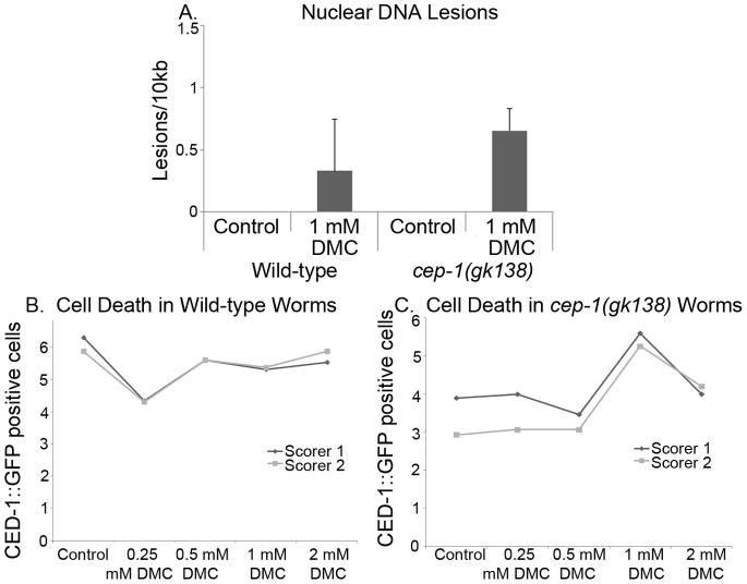
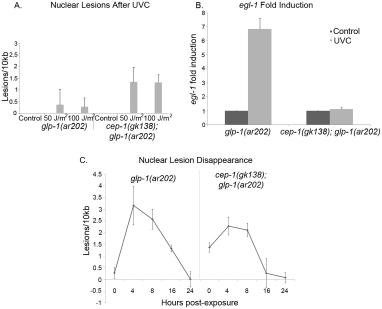
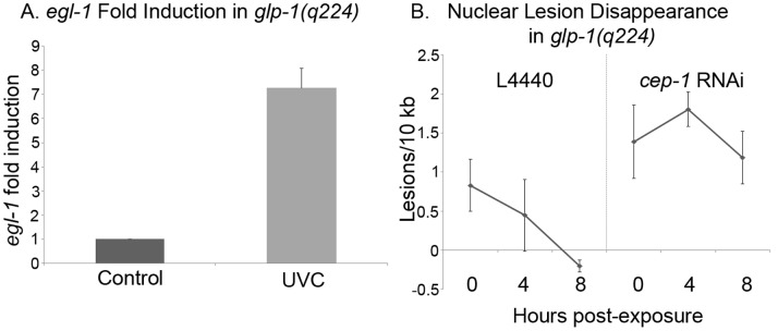
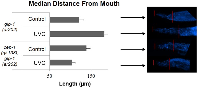
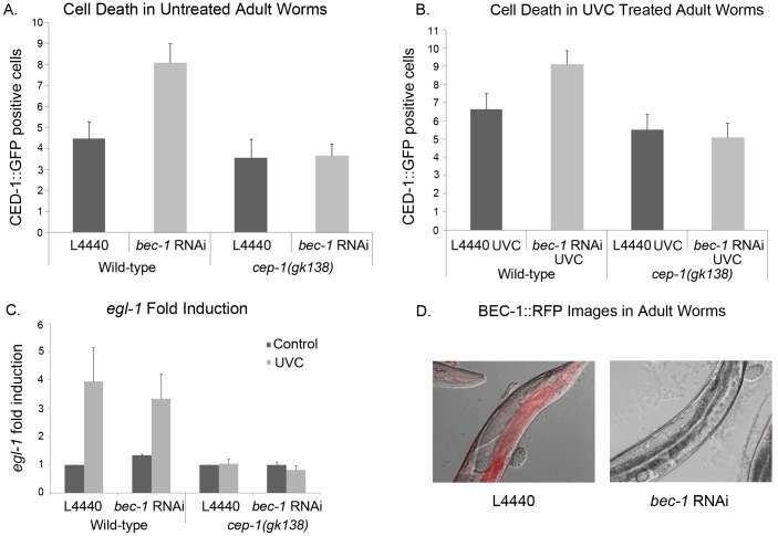
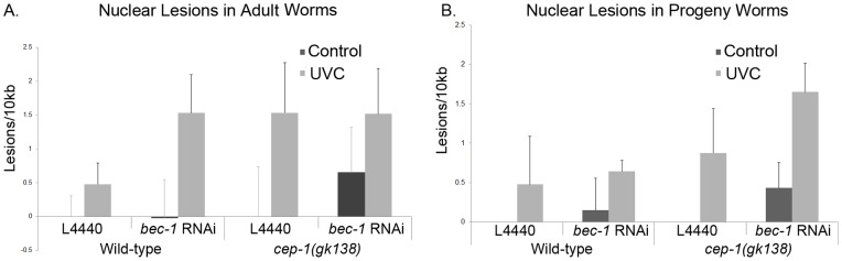
p53 is a transcription factor that regulates the response to cellular stress. Mammalian p53 functions as a tumor suppressor. The C. elegans p53, cep-1, regulates DNA-damage induced germline cell death by activating the transcription of egl-1 and ced-13. We used the C. elegans model to investigate how, in the whole animal, different forms of DNA damage can induce p53-dependent versus p53-independent cell death and DNA repair. DNA damage was induced by ultraviolet type C (UVC) radiation, or 10-decarbamoyl mitomycin C (DMC, an agent known to induce mammalian p53-independent cell death). Wild-type or cep-1 loss-of-function mutant animals were assayed for germline cell death and DNA lesions. Wild-type animals displayed greater removal of UVC-lesions over time, whereas cep-1 mutant animals displayed increased UVC-lesion retention. The cep-1 mutation increased UVC-lesion retention directly correlated with a reduction of progeny viability. Consistent with DMC inducing p53-independent cell death in mammalian cells DMC induced a C. elegans p53-independent germline cell death pathway. To examine the influence of wild-type CEP-1 and DNA damage on C. elegans tumors we used glp-1(ar202gf)/Notch germline tumor mutants. UVC treatment of glp-1 mutant animals activated the CEP-1 target gene egl-1 and reduced tumor size. In cep-1(gk138);glp-1(ar202gf) animals, UVC treatment resulted in increased susceptibility to lesions and larger tumorous germlines. Interestingly, the partial knockdown of bec-1 in adults resulted in a CEP-1-dependent increase in germline cell death and an increase in DNA damage. These results strongly support cross-talk between BEC-1 and CEP-1 to protect the C. elegans genome.


Conflict of interest statement

Competing Interests: The authors have declared that no competing interests exist.

Figures

Figure 1
Figure 1. Wild-type and cep-1(gk138) mutant worms had similar amounts of nuclear DNA damage after UVC exposure.
A) Images of CED-1::GFP positive cells of wild-type worms either untreated or 24 hours after 100 J/m2 UVC. Arrows point to CED-1::GFP positive cells. Magnification is 400x. B) Worms were treated 24 hours post L4 and imaged 24 hours later. The number of CED-1::GFP positive cells in the germ line was scored blindly by two independent people. Error bars indicate standard error and the number of worms scored was 20. The difference between wild-type untreated and UVC treated animals had a P value were all equal to or less than 0.002. C) Analysis of nuclear DNA lesions on 24 hour post L4 wild-type or cep-1(gk138) mutant animals, after exposure to 50 or 100 J/m2 of UVC. Average of three representative experiments is shown. Error bars indicate standard error. The wild-type P values were 0.0001 and 0.003 for a comparison to untreated at 50 or 100 J/m2 respectively. The cep-1(gk138) mutant P values were 0.012 and 0.02 for a comparison to untreated at 50 or 100 J/m2 respectively. D) Q-RTPCR of egl-1 and ced-13 mRNA levels four hours post UVC treatment of 100 J/m2. Fold induction compared to cDNAs amplified from untreated worms. Average of three representative experiments is shown. Error bars indicate standard error. Normalized to tbg-1. The P values for wild-type animals were less than or equal to 0.02 for both egl-1 and ced-13. No significant change was detected in cep-1(gk138) mutant animals.
Figure 2
Figure 2. cep-1(gk138) mutants had less repair and decreased egg viability than wild-type worms after UVC treatment.
A) Analysis of nuclear DNA lesions at 0, 4 h or 8 h time points of post L4 wild-type or cep-1(gk138) mutant animals after exposure to 100 J/m2 of UVC. Average of three representative experiments is shown. Error bars indicate standard error. Significant lesions were observed in wild-type worms with a P value of 0.0002 at the zero hours and 0.03 after four hours of repair when compared to untreated wild-type worms. The P values for cep-1(gk138) mutant animals were equal or less than 0.03 for each time point when compared to untreated animals. B) Analysis of the rate of egg laying of animals treated with UVC as L4 and allowed to recover for 24 hours. Egg laying values represent number of eggs laid per hour per adult hermaphrodite. Average of 18 worms are shown. Error bars indicate standard error. P values were less than or equal to 0.003 for both strains when compared to untreated animals. C) Percent survival of eggs from wild-type or cep-1(gk138) mutant animals after no treatment or exposure to UVC as L4s and allowed to recover for 24 hours. Survival of eggs was determined as the number of eggs that hatched after 1 day over the total number of eggs laid after 24 h. P values were less than or equal to 0.001 for both strains when compared to untreated animals.
Figure 3
Figure 3. DMC caused an increase in lesions and increased germline cell death in cep-1(gk138) mutant worms.
A) Lesions of nuclear DNA were measured on 24 hour old adult wild-type or cep-1(gk138) mutant animals, after five hours of treatment with 1 mM DMC or 30% methanol for 5 hours (as L4 larvae). The lesions detected were not statistically significant. B) Average of three representative experiments is shown. Error bars indicate standard error. The cell death compared to the control was not statistically significant at 0.5, 1 and 2 mM. The number of dead CED-1::GFP positive cells in the germ line was determined in wild-type adults or in C) cep-1(gk138) mutant animals after feeding with 30% methanol or increasing concentrations of DMC (0.25 mM, 0.5 mM, 1.0 mM or 2.0 mM DMC), overnight. Only 1 mM DMC treatment demonstrated statistically significant cell death with a P value of 0.02. The number of CED-1::GFP positive cells was scored blindly by two independent people. The number of worms scored per point was 20.
Figure 4
Figure 4. CEP-1 was activated in glp-1(ar202gf) tumor mutant worms.
A) Nuclear DNA lesions were measured in glp-1(ar202gf) or cep-1(gk138); glp-1(ar202gf) double mutants, immediately following UVC exposure. Animals were exposed to 50 or 100 J/m2 of UVC, as adults (24 h post L4 stage). Average of three representative experiments is shown. Error bars indicate standard error. Only cep-1(gk138); glp-1(ar202gf) double mutants had a statistically significant increase in lesions with P value of 0.003 after 100 J/m2. B) Expression of egl-1 mRNA determined by Q-RTPCR in glp-1(ar202gf) or cep-1(gk138); glp-1(ar202gf) double mutant animals, 4 hours post UVC treatment of 100 J/m2. Data are reported as fold induction compared to expression in untreated worms and normalized to tbg-1. Average of six representative experiments is shown. Error bars indicate standard error. The egl-1 fold change had a P value in glp-1(ar202gf) of 0.008 and the P value in cep-1(gk138); glp-1(ar202gf) double mutants was 0.03. C) Nuclear lesions were quantified immediately, four, eight, sixteen and twenty-four hours after UV exposure in glp-1(ar202gf) and cep-1(gk138); glp-1(ar202gf) worms. Average of three representative experiments is shown. Error bars indicate standard error. In glp-1(ar2020gf) mutant worms significant lesions were detected with the P values equal or less than 0.02 for time points four, eight and sixteen hours after UVC treatment when compared to untreated animals. In cep-1(gk138); glp-1(ar202gf) mutant worms significant lesions were detected with the P values equal or less than 0.004 for time points zero, four and eight hours after UVC treatment when compared to untreated animals.
Figure 5
Figure 5. CEP-1 acts to remove UVC induced lesions in somatic cells.
A) egl-1 expression measure by Q-RTPCR in glp-1(q224lf) animals that lack a germline, 4 hours post UVC treatment of 100 J/m2, or control animals without treatment. Data are reported as fold induction compared to expression in untreated worms and normalized to tbg-1. Average of two representative experiments is shown. Error bars indicate standard error, P value of 0.008. B) Nuclear DNA lesions were measured immediately, four and eight hours after 100 J/m2 of UVC in glp-1(q224lf) worms grown on either L4440 or cep-1 RNAi plates at 25°C. Average of three representative experiments is shown. Error bars indicate standard error. Wild-type animals at 0 hours had significant lesions with a P value of 0.03 and no significant lesions at 4 and 8 hours. cep-1 depleted animals had significant lesions at 0, 4, and 8 hours after UVC treatment with P values of equal to or less than 0.05.
Figure 6
Figure 6. glp-1(ar202gf) tumor mutants displayed an increase in tumor size in the absence of cep-1, four days after UVC treatment.
The size of the germ line tumors was determined in glp-1(ar202gf) or cep-1(gk138); glp-1(ar202gf) double mutant animals grown at the restrictive temperature after treatment with 100 J/m2 of UVC, and allowed to recover for four days. The size of the tumor is inversely proportional to the distance between the mouth and the tumor. Median distances of 18 worms are shown. glp-1(ar202gf) or cep-1(gk138); glp-1(ar202gf) double mutant animals had P values of 0.04 and 0.009 respectively for their change in tumor size versus untreated controls.
Figure 7
Figure 7. In treated adult worms, increased death from bec-1 RNAi depletion required CEP-1.
A) Animals carrying the CED-1::GFP reporter were fed control (empty vector) or bec-1 RNAi expressing bacteria and scored for germline cell death in the absence or B) presence of 100 J/m2 of UVC. The number of CED-1::GFP positive dead cells was scored blindly by two independent people. Error bars indicate standard error and the number of worms scored per bar was 20. In wild-type worms, the knockdown of bec-1 caused a significant increase of CED-1::GFP positive cells with P values of equal or less than 0.003 when compared to untreated animals. C) Expression of egl-1 was detected by Q-RTPCR, 4 hours post 100 J/m2 of UVC treatment. Data are reported as fold induction compared to expression in untreated worms and normalized to tbg-1. The average of three representative experiments is shown. Error bars indicate standard error. L4440 UVC induction was significant with a P value of 0.03 as was the P value for animal fed bec-1 RNAi with a P value of 0.0004 when compared to untreated L4440 animals. No significant change was detected in cep-1(gk138) mutant animals. D) Images of BEC-1::RFP worms 48 hours after L4 worms were placed on L4440 or bec-1 RNAi plates. The Nomarski and red channels were placed on top of each other. Magnification is 400x.
Figure 8
Figure 8. UVC treatment after bec-1 depletion increased the number of DNA lesions in the treated adult animals.
Wild-type and cep-1(gk138) mutant animals were exposed to 100 J/m2 of UVC. A) The number of nuclear DNA lesions were measured in adult worms fed control (empty vector) RNAi or bec-1 RNAi expressing bacteria, during adulthood. All comparisons were made to L4440 untreated animals. For UVC treated wild-type animals fed L4440, the lesions were significantly increased with a P value of 0.04. For UVC treated wild-type animals fed bec-1 RNAi, the lesions were significantly increased with a P value of 0.02. For UVC treated cep-1(gk138) mutant worms animals fed L4440, the lesions were significantly increased with a P value of 0.001. For UVC treated cep-1(gk138) mutant worms animals fed bec-1 RNAi, the lesions were significantly increased with a P value of 0.004. B) RNAi depletion was also achieved throughout development. Average of three representative experiments is shown. Error bars indicate standard error. All comparisons were made to L4440 untreated animals. For UVC treated wild-type animals fed L4440, the lesions were significantly increased with a P value of 0.05. For bec-1 knockdown without UV treatment, there was a significant increase with a P value of 0.01. For UVC treated wild-type animals fed bec-1 RNAi, the lesions were significantly increased with a P value of 0.0007. For UVC treated cep-1(gk138) mutant worms animals fed L4440, the lesions were significantly increased with a P value of 0.05. For UVC treated cep-1(gk138) mutant worms animals fed bec-1 RNAi, the lesions were significantly increased with a P value of 0.003.

Similar articles

Cited by

References

    1. Riley T, Sontag E, Chen P, Levine A (2008) Transcriptional control of human p53-regulated genes. Nat Rev Mol Cell Biol 9: 402–412. - PubMed
    1. Schumacher B, Hofmann K, Boulton S, Gartner A (2001) The C. elegans homolog of the p53 tumor suppressor is required for DNA damage-induced apoptosis. Curr Biol 11: 1722–1727. - PubMed
    1. Derry WB, Putzke AP, Rothman JH (2001) Caenorhabditis elegans p53: role in apoptosis, meiosis, and stress resistance. Science 294: 591–595. - PubMed
    1. Lu WJ, Abrams JM (2006) Lessons from p53 in non-mammalian models. Cell Death Differ 13: 909–912. - PubMed
    1. Derry WB, Bierings R, van Iersel M, Satkunendran T, Reinke V, et al. (2007) Regulation of developmental rate and germ cell proliferation in Caenorhabditis elegans by the p53 gene network. Cell Death Differ 14: 662–670. - PubMed

Publication types

MeSH terms