. 2014 Feb 20;9(2):e89006.
doi: 10.1371/journal.pone.0089006. eCollection 2014.

Stromal cells from human decidua exert a strong inhibitory effect on NK cell function and dendritic cell differentiation

Affiliations

Stromal cells from human decidua exert a strong inhibitory effect on NK cell function and dendritic cell differentiation

Daniele Croxatto et al. PLoS One. .

Abstract

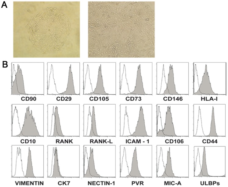
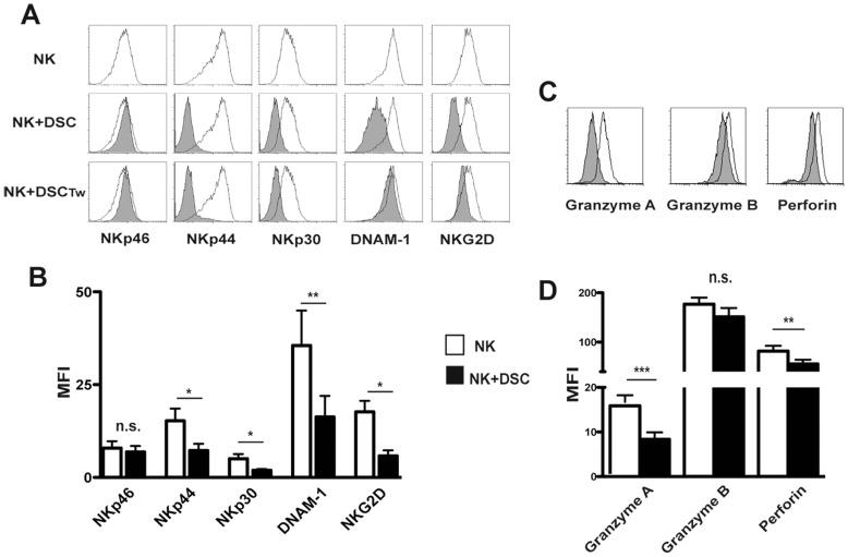
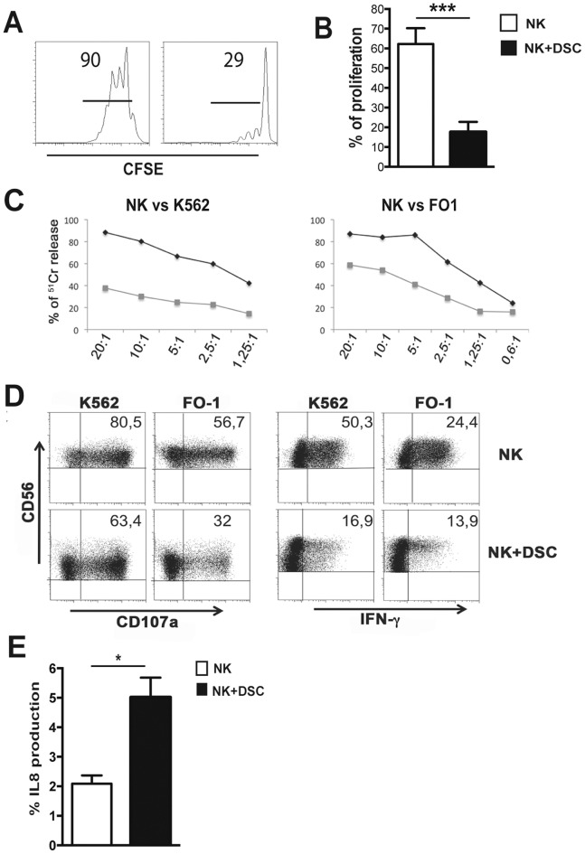
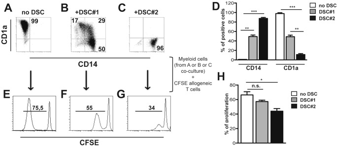
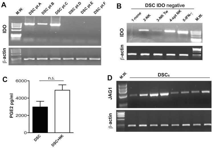
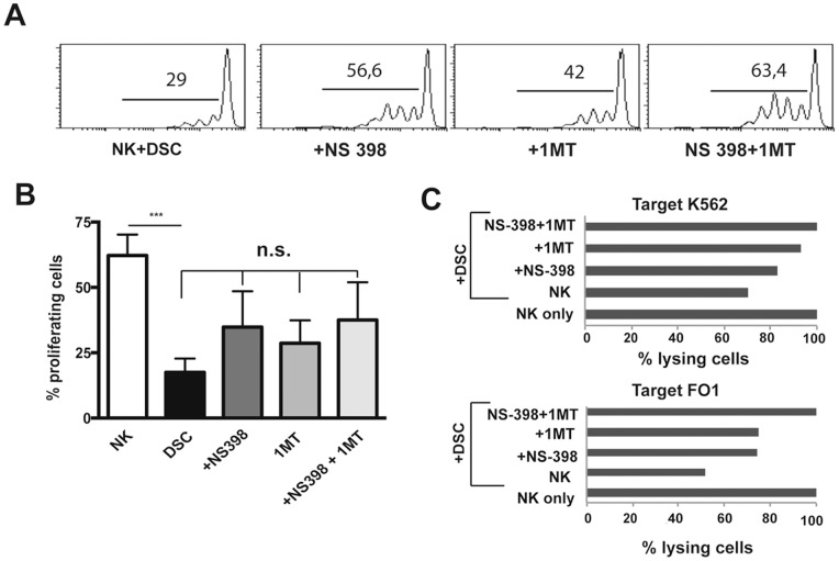
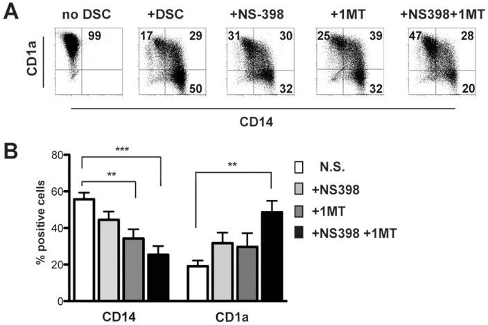
Stromal cells (SC) are an important component of decidual tissues where they are in strict proximity with both NK and CD14(+) myelomonocytic cells that play a role in the maintenance of pregnancy. In this study we analyzed whether decidual SC (DSC) could exert a regulatory role on NK and CD14(+) cells that migrate from peripheral blood (PB) to decidua during pregnancy. We show that DSCs inhibit the IL15-mediated up-regulation of major activating NK receptors in PB-derived NK cells. In addition, the IL15-induced NK cell proliferation, cytolytic activity and IFN-γ production were severely impaired. DSCs sharply inhibited dendritic cells differentiation and their ability to induce allogeneic T cell proliferation. Indoleamine 2,3-dioxygenase (IDO) and prostaglandin E2 (PGE2) mediated the inhibitory effect of DSCs. Our results strongly suggest an important role of DSCs in preventing potentially dangerous immune response, thus contributing to maintenance of pregnancy.


Conflict of interest statement

Competing Interests: The authors have declared that no competing interests exist.

Figures

Figure 1
Figure 1. Informative surface markers expressed by DSCs.
Characterization of DSCs. (A) Morphology of adherent cells isolated from decidua tissues (magnification 20X and 40X). (B) Phenotypic analysis of DSC by flow cytometry. The histograms depict the expression of different molecules (filled gray profiles) compare to negative controls (white profiles). One representative experiment out of eight performed.
Figure 2
Figure 2. DSCs inhibit the IL15-induced expression of activating receptors, perforin and granzymes in PB-derived NK cells.
(A) Expression of NKp46, NKp30, NKp44, DNAM-1 and NKG2D on IL15-activated PB NK cells, at day 5 of culture, in the absence (white profiles) or in the presence of DSCs (grey profiles) by cell-to-cell contact or by transwell (Tw) conditions. Cells were analyzed by gating on CD56+CD3 cells. One representative experiment out of 10 performed. (B) Statistical analysis of MFI of activating NK receptors on IL15-activated PB NK cells in the absence (white bar) or in presence (black bar) of DSCs. Data is shown as a MFI±SEM of ten experiments. (C) Expression of granzyme A, granzyme B and perforin in IL15-activated PB NK cells in the absence (white profiles) or in the presence (grey profiles) of DSCs. One representative experiment out of 6 performed. (D) Statistical analysis of MFI±SEM of six experiments.
Figure 3
Figure 3. DSCs inhibit the NK cell functions.
IL15-activated NK cells were cultured in the presence of in the absence of DSCs. (A) Proliferation of CFSE-labeled NK cells was evaluated at day 5 of culture by flow cytometry. One representative experiment out of 9 performed. (B) Statistical analysis of proliferating CFSE-labeled NK cells. (C) Cytolytic activity of IL15-activated NK cells in the presence (grey line) or in the absence (black line) of DSCs was evaluated by 51Cr release against K562 and FO1 target cell lines. One representative experiment out of 6 performed. (D) CD107a expression and IFN-γ production, after 4 h of co-culture with K562 and FO1 target cell lines by IL15-activated NK cells in the presence or in the absence of DSCs for 5 days. Cells were analyzed by gating on CD56+CD3 cells. One representative experiment out of 7 performed. (E) Percentages of IL-8 positive cells, after 4 h of co-culture with PMA/ionomycin in IL15-activated NK cells cultured in the absence (white bars) or in the presence (black bars) of DSCs.
Figure 4
Figure 4. DSCs inhibit DC differentiation from peripheral blood monocytes.
PB-CD14+ cells were cultured with GM-CSF and IL4 for 5 days in the presence or in the absence of DSCs. (A-C) Phenotypic analysis of CD14 and CD1a markers. One representative experiment out of 35 performed. (D) Statistical analysis of CD14 and CD1a expression ± SEM. (E-G) Myeloid cells were purified at day 5 from co-culture with DSCs and used as stimulator for allogeneic CFSE-labeled T cells. Proliferation of allogeneic CFSE-labeled T cells was analyzed at day 7. One representative experiment out of 8 performed. (H) Statistical analysis of proliferating CFSE-labeled T cells ± SEM.
Figure 5
Figure 5. DSC express IDO and produce PGE2.
(A) Analysis of IDO mRNA expression in DSCs isolated from different patients (pt). (B) IDO mRNA expression was analyzed in IDO DSCs cultured alone (first line) or with IL15-activated NK cells in cell-to-cell contact or Tw conditions or after ON incubation with supernatant (spt) of IL15-activated NK cells or 100 UI of IFN-γ. RT–PCR was performed with primers specific for IDO and for β-actin as positive control. PCR products were run on a 0.8% agarose gel and visualized by ethidium bromide staining. (C) PGE2 production was analyzed in supernatants by DSCs culture alone (black bar) or with IL15-activated NK cells. (D) Analysis of Jagged-1 mRNA expression in DSCs isolated from different patients.
Figure 6
Figure 6. Role of IDO and PGE2 in the DSC-mediated inhibition of NK-cell functions.
PB IL15-activated NK cells were cultured with DSCs in the presence of in the absence of IDO and PGE2 inhibitors. (A) After 7 days of cultured, proliferation of CFSE-labeled NK cells was analyzed. One representative experiment out of 3 performed. (B) Statistical analysis of proliferating CFSE-labeled NK cells ± SEM. (C) Cytolytic activity of IL15-activated NK cells cultured with DSC in the presence or in the absence of IDO and/or PGE2 inhibitors. 51Cr release against K562 and FO1 target cell lines was evaluated. One representative experiment out of 3 performed. Since we analyzed the inhibitory potential of different DSC, we considered, in each experiment, the cytotoxicity of IL15-activated NK cells cultured alone as 100%. The percentages of cytotoxicity of NK cells cultured in the presence of DSC ± inhibitors were normalized respect to IL15-activated NK cells cultured alone (set to 100%).
Figure 7
Figure 7. Role of IDO and PGE2 in the DC differentiation.
PB-CD14+ cells were cultured with DSC, GM-CSF and IL4 for 5 days in the presence or in the absence of IDO and/or PGE2 inhibitors. (A) Analysis of CD1a and CD14 surface expression. One representative experiment out of 7 performed. (B) Statistical analysis of CD14 and CD1a markers. Data indicate the percentages of positive cells ± SEM of 7 independent experiments.

Similar articles

Cited by

References

    1. Caligiuri MA (2008) Human natural killer cells. Blood 112: 461–469. - PMC - PubMed
    1. Cerwenka A, Lanier LL (2001) Natural killer cells, viruses and cancer. Nat Rev Immunol 1: 41–49. - PubMed
    1. Fauriat C, Long EO, Ljunggren HG, Bryceson YT (2010) Regulation of human NK-cell cytokine and chemokine production by target cell recognition. Blood 115: 2167–2176. - PMC - PubMed
    1. Le Bouteiller P, Tabiasco J (2006) Killers become builders during pregnancy. Nat Med 12: 991–992. - PubMed
    1. Moretta A (2002) Natural killer cells and dendritic cells: rendezvous in abused tissues. Nat Rev Immunol 2: 957–964. - PubMed

Publication types