. 2014 Feb 21;9(2):e89027.
doi: 10.1371/journal.pone.0089027. eCollection 2014.

Distinct structural features of the peroxide response regulator from group A Streptococcus drive DNA binding

Affiliations

Distinct structural features of the peroxide response regulator from group A Streptococcus drive DNA binding

Chang Sheng-Huei Lin et al. PLoS One. .

Abstract

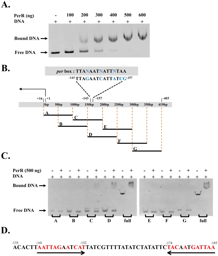
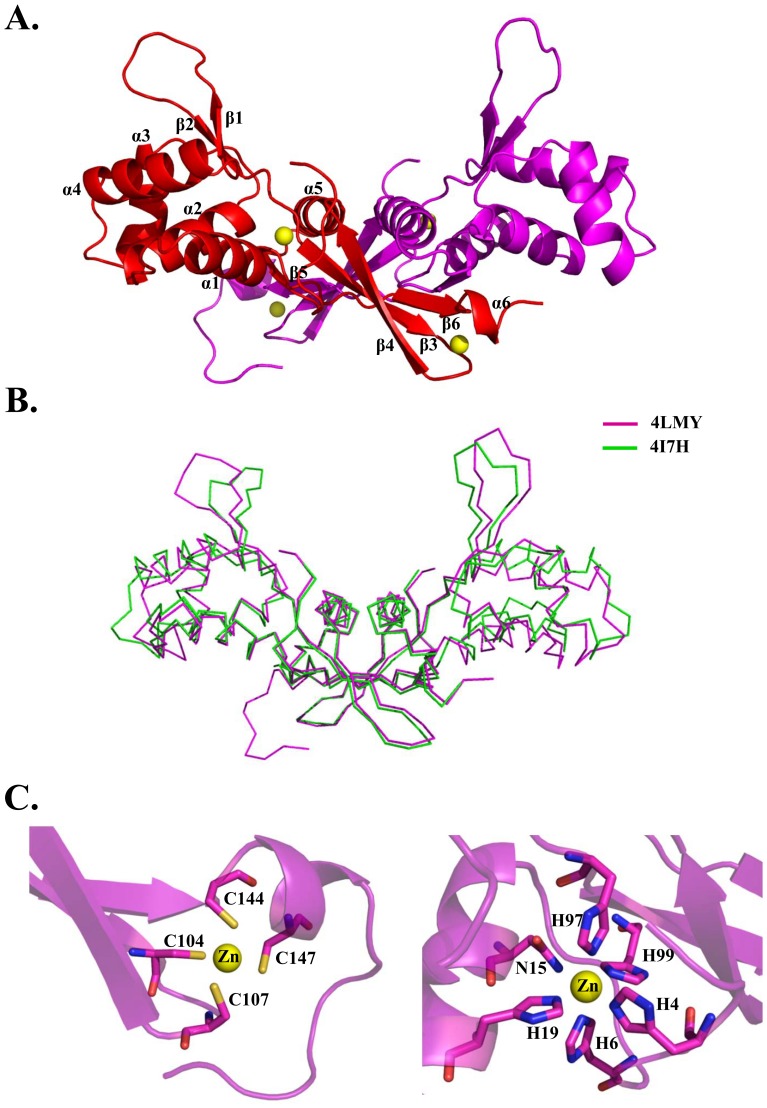
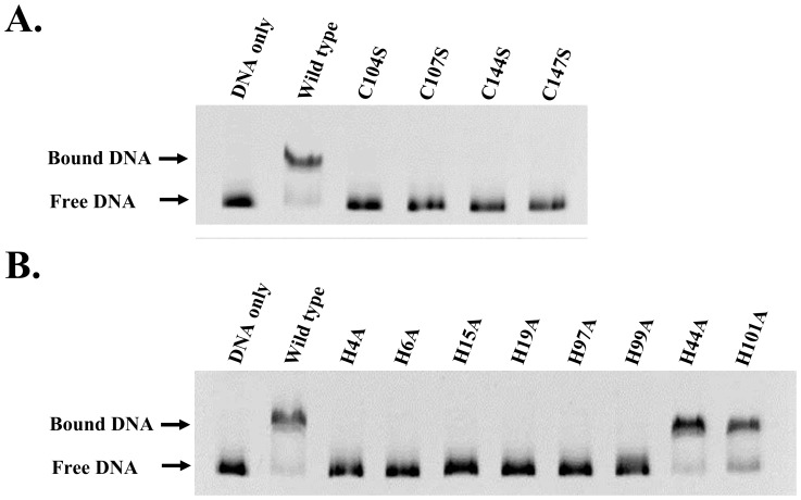
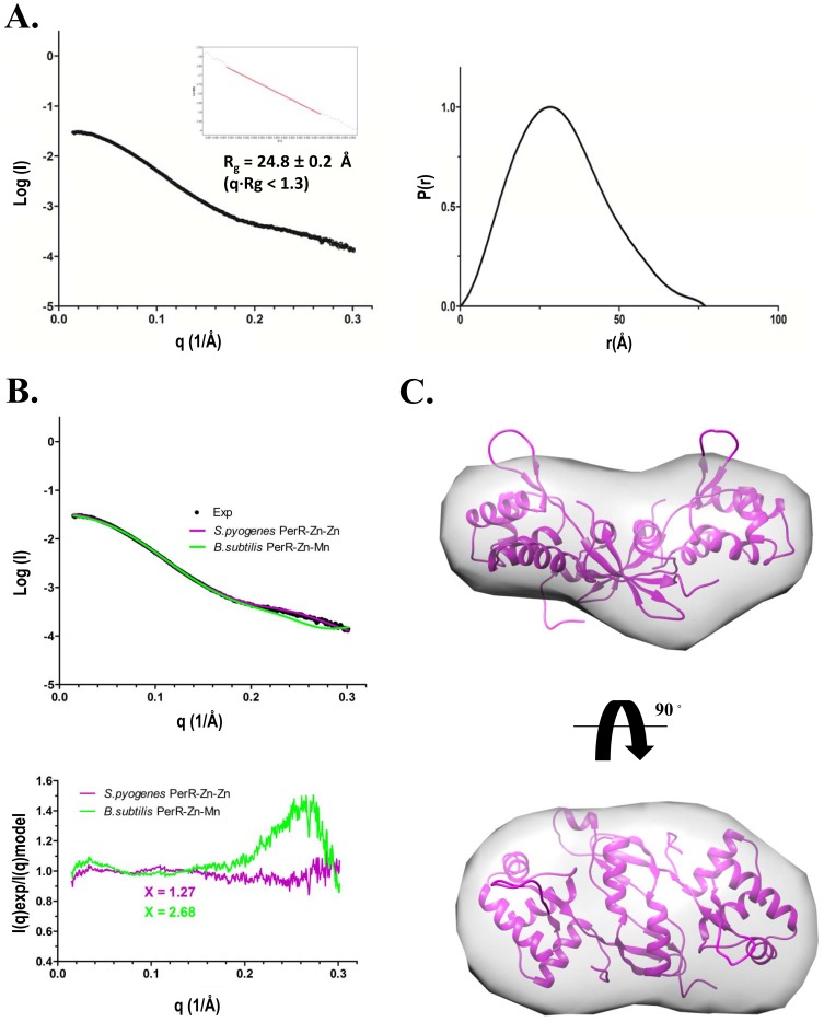
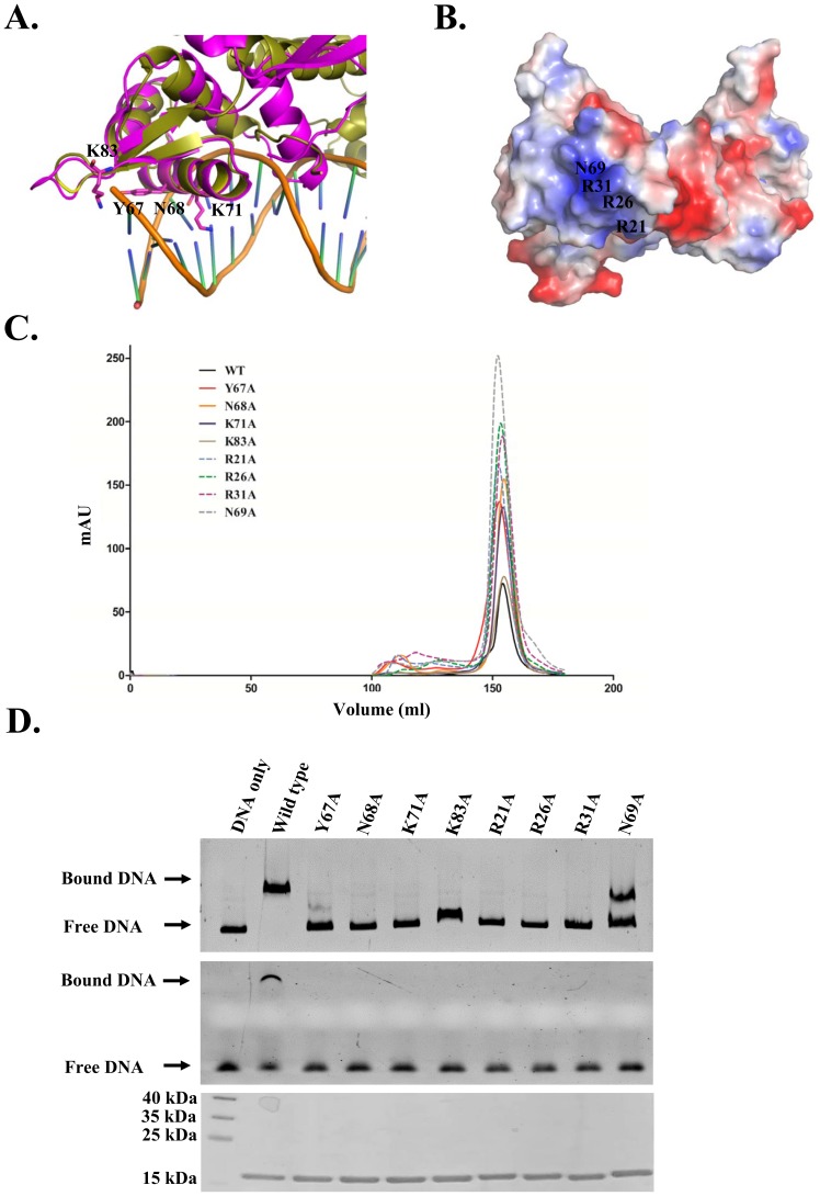
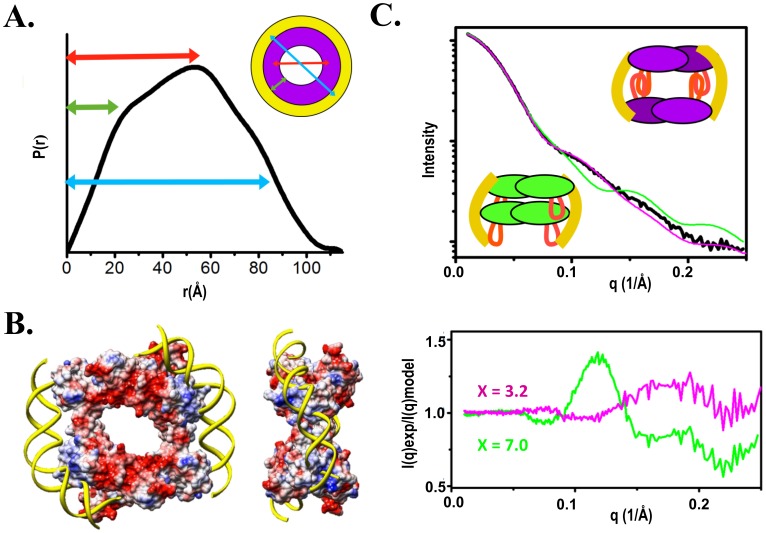
Group A streptococcus (GAS, Streptococcus pyogenes) is a strict human pathogen that causes severe, invasive diseases. GAS does not produce catalase, but has an ability to resist killing by reactive oxygen species (ROS) through novel mechanisms. The peroxide response regulator (PerR), a member of ferric uptake regulator (Fur) family, plays a key role for GAS to cope with oxidative stress by regulating the expression of multiple genes. Our previous studies have found that expression of an iron-binding protein, Dpr, is under the direct control of PerR. To elucidate the molecular interactions of PerR with its cognate promoter, we have carried out structural studies on PerR and PerR-DNA complex. By combining crystallography and small-angle X-ray scattering (SAXS), we confirmed that the determined PerR crystal structure reflects its conformation in solution. Through mutagenesis and biochemical analysis, we have identified DNA-binding residues suggesting that PerR binds to the dpr promoter at the per box through a winged-helix motif. Furthermore, we have performed SAXS analysis and resolved the molecular architecture of PerR-DNA complex, in which two 30 bp DNA fragments wrap around two PerR homodimers by interacting with the adjacent positively-charged winged-helix motifs. Overall, we provide structural insights into molecular recognition of DNA by PerR and define the hollow structural arrangement of PerR-30bpDNA complex, which displays a unique topology distinct from currently proposed DNA-binding models for Fur family regulators.


Conflict of interest statement

Competing Interests: The authors have declared that no competing interests exist.

Figures

Figure 1
Figure 1. EMSA analysis of PerR DNA-binding ability.
(A) The dpr promoter from sequence −403 to +16 was incubated with wide-type PerR at the concentrations indicated for 15 min at room temperature. The mixture were separated by 6% native PAGE, and the mobility shift of the promoter was resolved by ethidium bromide staining. (B) Seven DNA fragments were derived from dpr promoter. Segments A to G represents sequences as follows: A (−84 to +16), B (−134 to −34), C (−184 to −84), D (−234 to −134), E (−284 to −184), F (−334 to −234) and G (−403 to −284). Putative per box was located at sequence between −157 to −143. (C) PerR binding to dpr promoter fragments was analyzed by EMSA as described in Figure 1A. (D) PerR binds to dpr promoter through sequence −185 to −135. A per box similar sequence with opposite direction was found at −185 to −174.
Figure 2
Figure 2. Crystal structure of GAS PerR.
(A) Ribbon diagram of the dimeric PerR structure. Each of the monomer is colored in red and magenta. Two zinc metals bound to each PerR subunit are shown as yellow spheres. (B) Superposition of two GAS PerR structures. The PerR-Zn-Zn structure is colored in magenta (PDB code 4LMY); PerR-Zn-Ni is in green (PDB code 4I7H). (C) Magnified view of zinc binding sites on PerR subunit. Left panel, zinc-finger motif coordinated by four cysteine residues (Cys104, Cys107, Cys144 and Cys147). Right panel, zinc-coordinated regulatory site compose of His4, His6, Asn15, His19, His97 and His99 in pseudo octahedral geometry.
Figure 3
Figure 3. EMSA analysis of PerR mutants.
EMSA shows that the residues at regulatory site mutated to alanine abolished the DNA binding of PerR. Mutation of cysteine to serine in the C-terminal zinc-finger motif also abolished the DNA-binding of PerR. H44A and N101A that are not coordinated by zinc are used as controls.
Figure 4
Figure 4. Solution structure of PerR.
(A) Experimental scattering profile of GAS PerR was merged from scattering curves of indicated protein concentrations. The Rg is derived from linear region of Guinier plot (left panel). Pair-distance distribution function P(r) was computed from program GNOM. The maximal dimension estimated from P(r) is ∼77 Å (right panel). (B) Comparison of experimental scattering curve (black) with theoretical curves calculated from full-atomic models derived from crystal structure of, S. pyogenes PerR-Zn-Zn (purple) and, B. subtilis PerR-Zn-Mn (green). Residual calculated as I(q)experimental/I(q)model is shown below the scattering curves. (C) Low-resolution SAXS envelope of PerR and the crystallographic PerR structure was fitted into the envelope by the program Chimera .
Figure 5
Figure 5. Analysis of PerR DNA-binding residues.
(A) Superposition of the winged-helix motif of S. aureus BlaI-DNA complex (PDB code 1XSD) with GAS PerR. Yellow, S. aureus BlaI-DNA complex; Magenta, GAS PerR. Predicted residues (Y67, N68, K71 and K83) related to DNA-binding are labeled and shown as sticks. (B) Surface electrostatic potential representation of PerR. Residues (R21, R26, R31 and N69) with strong positive charge are labeled. (C) Size-exclusion chromatographic profile of PerR (black) and PerR mutants (red, Y67A; orange N68A; blue, K71A; brown, K83A; blue dashed line, R21A; green dashed line, R26A; purple dashed line, R31A; grey dashed line, N69A). (D) EMSA analysis of binding activity of the mutant PerR proteins to dpr promoter (−403 to +16, top panel) and dpr segment (−185 to −135, middle panel). A coomassie blue stained SDS-PAGE with equal amounts of wide-type or mutant PerR is shown below EMSA gels.
Figure 6
Figure 6. Molecular architecture of PerR-DNA complex.
(A) The P(r) function calculated from experimental SAXS profile of a PerR:DNA 1∶1 complex indicates multilayers-hollow structural assembly. (B) Two PerR homodimers satisfy this hollow assembly. Surface electrostatic potential representation of assembled PerR tetramer indicates strong positive charges (blue) where the 30 bp DNA was manually docked. (C) The theoretical SAXS curve calculated from the built PerR-30bpDNA complex showed good fit (magenta) to the experimental SAXS data (black) (χ = 3.2). An alternative complex model, with a compact assembly in which the head-to-tail PerR tetramer was wrapped by DNA, showed poor fit to the experimental data (χ = 7.0) (green).

Similar articles

Cited by

References

    1. Carapetis JR, Steer AC, Mulholland EK, Weber M (2005) The global burden of group A streptococcal diseases. Lancet Infect Dis 5: 685–694. - PubMed
    1. Cunningham MW (2000) Pathogenesis of group A streptococcal infections. Clin Microbiol Rev 13: 470–511. - PMC - PubMed
    1. Troillet N LA, de Werra P, Praz G (1994) Invasive Streptococcus pyogenes infection (P-hemolytic streptococcus of group A). Schweiz Med Wochenschr 124: 1064–1069. - PubMed
    1. King KY, Horenstein JA, Caparon MG (2000) Aerotolerance and peroxide resistance in peroxidase and PerR mutants of Streptococcus pyogenes . J Bacteriol 182: 5290–5299. - PMC - PubMed
    1. Brenot A, King KY, Caparon MG (2005) The PerR regulon in peroxide resistance and virulence of Streptococcus pyogenes . Mol Microbiol 55: 221–234. - PubMed

Publication types

MeSH terms

LinkOut - more resources