. 2014 Feb 21;9(2):e89051.
doi: 10.1371/journal.pone.0089051. eCollection 2014.

Major membrane protein TDE2508 regulates adhesive potency in Treponema denticola

Affiliations

Major membrane protein TDE2508 regulates adhesive potency in Treponema denticola

Yuki Abiko et al. PLoS One. .

Abstract

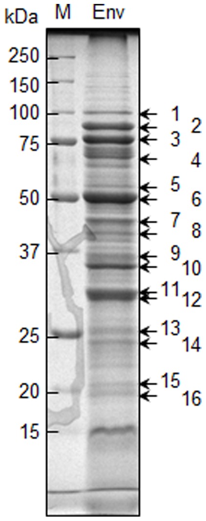
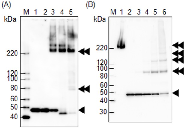
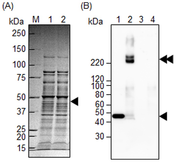
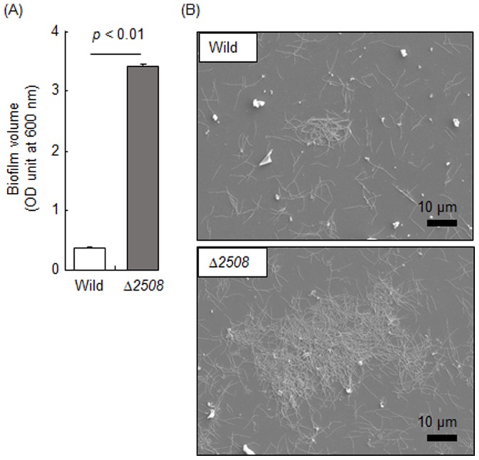
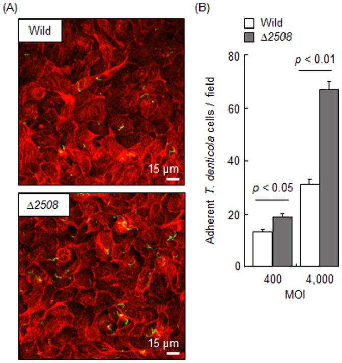
The cultivation and genetic manipulation of Treponema denticola, a Gram-negative oral spirochaeta associated with periodontal diseases, is still challenging. In this study, we formulated a simple medium based on a commercially available one, and established a transformation method with high efficiency. We then analyzed proteins in a membrane fraction in T. denticola and identified 16 major membrane-associated proteins, and characterized one of them, TDE2508, whose biological function was not yet known. Although this protein, which exhibited a complex conformation, was presumably localized in the outer membrane, we did not find conclusive evidence that it was exposed on the cell surface. Intriguingly, a TDE2508-deficient mutant exhibited significantly increased biofilm formation and adherent activity on human gingival epithelial cells. However, the protein deficiency did not alter autoaggregation, coaggregation with Porphyromonas gingivalis, hemagglutination, cell surface hydrophobicity, motility, or expression of Msp which was reported to be an adherent molecule in this bacteria. In conclusion, the major membrane protein TDE2508 regulates biofilm formation and the adhesive potency of T. denticola, although the underlying mechanism remains unclear.


Conflict of interest statement

Competing Interests: The authors have declared that no competing interests exist.

Figures

Figure 1
Figure 1. Gene map of tde2508 and its neighbors on the chromosomal DNA of T. denticola ATCC 35405.
The small arrows show primers which are presented in Table 1. The tde2508 and tde2509 genes were co-transcribed (data not shown).
Figure 2
Figure 2. Growth curves of T. denticola.
T. denticola ATCC 35405 was inoculated in mGAM-TS (5% serum, circle), TYGVS (10% serum, triangle), and mNOS (10% serum, square), and anaerobically incubated at 37°C. The optical density (OD) at 600 nm was monitored. The figure shows a representative image from one of three independent experiments.
Figure 3
Figure 3. Major membrane proteins on an SDS-PAGE gel.
Ten micrograms of the envelope fraction (Env) of T. denticola ATCC 35405 was separated by SDS-PAGE and stained with CBB. The numbers (1–16) indicate proteins identified by mass spectrometry. Annotation of each protein is shown in Table 2. M denotes a standard marker.
Figure 4
Figure 4. Subcellular localization of TDE2508.
The whole cell lysate (WCL), soluble fraction (Sol) and envelope fraction (Env) of T. denticola ATCC 35405 were separated by SDS-PAGE with CBB-staining (A) and analyzed by Western blotting with anti-TDE2508 antiserum (B). The envelope fraction was further fractionated by differential solubilization in 0, 0.25, 0.5, 1, 2, 4, and 8% Triton X-100, and the soluble fractions (lanes 1–7, respectively) and insoluble fractions (lanes 8–14, respectively) were subjected to Western blot analysis with anti-T. denticola whole cell antiserum (C) and anti-TDE2508 antiserum (D). The grey, white and black arrowheads denote Msp, TmpC and TDE2508, respectively. M denotes a standard marker.
Figure 5
Figure 5. TDE2508 complex formation.
(A) The whole cell lysate of T. denticola ATCC 35405 was subjected to Western blot analysis with anti-TDE2508 antiserum. Samples in lanes 1–5 were denatured by heating at 100, 80, 60, 37, and 24°C, respectively for 10 min. (B) T. denticola intact cells in lanes 1–6 were treated with 0, 0, 10, 50, 100, and 200 mM of a chemical cross-linker, respectively. The treated cells were sonicated and denatured by heating at 60°C for lane 1 or 100°C for lanes 2–6 for 10 min, and subjected to Western blot analysis with anti-TDE2508 antiserum. The single and double black arrowheads denote a monomer and a complex form of TDE2508, respectively. M denotes a standard marker.
Figure 6
Figure 6. Confirmation of TDE2508 abolition in the tde2508-deletion mutant.
The whole cell lysates of T. denticola ATCC 35405 (lanes 1 and 1–2 in panels A and B, respectively) and tde2508-deletion mutant (lanes 2 and 3–4 in panels A and B, respectively) were subjected to SDS-PAGE with CBB-staining (A) and Western blot analysis with anti-TDE2508 antiserum (B). Samples were denatured by heating at 100°C for 10 min, except for lanes 2 and 4 in panel B; these two were treated without heating. The single and double black arrowheads denote a monomer and a complex forms of TDE2508, respectively. M denotes a standard marker.
Figure 7
Figure 7. Biofilm formation. T. denticola ATCC 35405 (Wild) and tde2508-deletion mutant (Δ2508) were incubated on a polystyrene plate for 2 days.
After washing, the adherent cells were collected and quantified using the optical density (OD) at 600 nm (A). Data represent means and standard deviations of two experiments performed in triplicate. Additionally, the adherent cells were observed by scanning electron microscopy (B).
Figure 8
Figure 8. Adherence to human gingival epithelial cells.
T. denticola ATCC 35405 (Wild) and tde2508-deletion mutant (Δ2508) were incubated with Ca9-22 epithelial cells for 1 h at 400 and 4,000 MOI. After immunostaining, the cells were observed by the confocal laser scanning microscopy (A). T. denticola cells and actin of the epithelial cells are shown as green and red, respectively. Each field corresponds to 0.045 mm2. Panel (A) shows the adherence at 4,000 MOI. We performed experiments independently twice in duplicate and counted the number of adherent T. denticola cells in 10 fields. Panel (B) shows the means and standard deviations of the number.

Similar articles

Cited by

References

    1. Dashper SG, Seers CA, Tan KH, Reynolds EC (2011) Virulence factors of the oral spirochete Treponema denticola . J Dent Res 90: 691–703. - PMC - PubMed
    1. Holt SC, Ebersole JL (2005) Porphyromonas gingivalis, Treponema denticola, and Tannerella forsythia: the “red complex”. a prototype polybacterial pathogenic consortium in periodontitis. Periodontol 2000 38: 72–122. - PubMed
    1. Okuda K, Ishihara K, Nakagawa T, Hirayama A, Inayama Y, et al. (2001) Detection of Treponema denticola in atherosclerotic lesions. J Clin Microbiol 39: 1114–1117. - PMC - PubMed
    1. Nobbs AH, Jenkinson HF, Jakubovics NS (2011) Stick to your gums: mechanisms of oral microbial adherence. J Dent Res 90: 1271–1278. - PMC - PubMed
    1. Visser MB, Ellen RP (2011) New insights into the emerging role of oral spirochaetes in periodontal disease. Clin Microbiol Infect 17: 502–512. - PubMed

Publication types

MeSH terms

Substances

LinkOut - more resources