. 2014 Feb 25;9(2):e89135.
doi: 10.1371/journal.pone.0089135. eCollection 2014.

Twisted gastrulation, a BMP antagonist, exacerbates podocyte injury

Affiliations

Twisted gastrulation, a BMP antagonist, exacerbates podocyte injury

Sachiko Yamada et al. PLoS One. .

Abstract

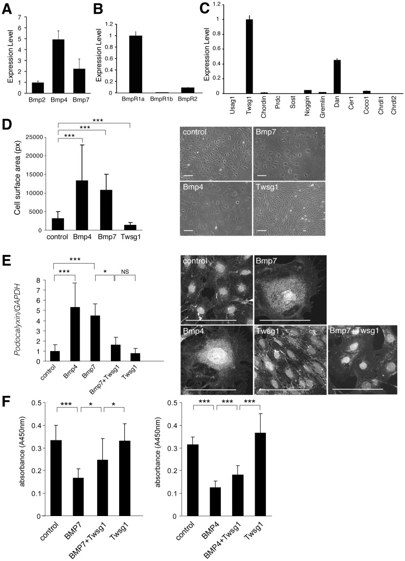
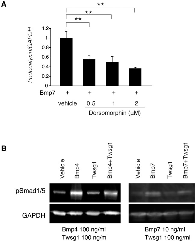
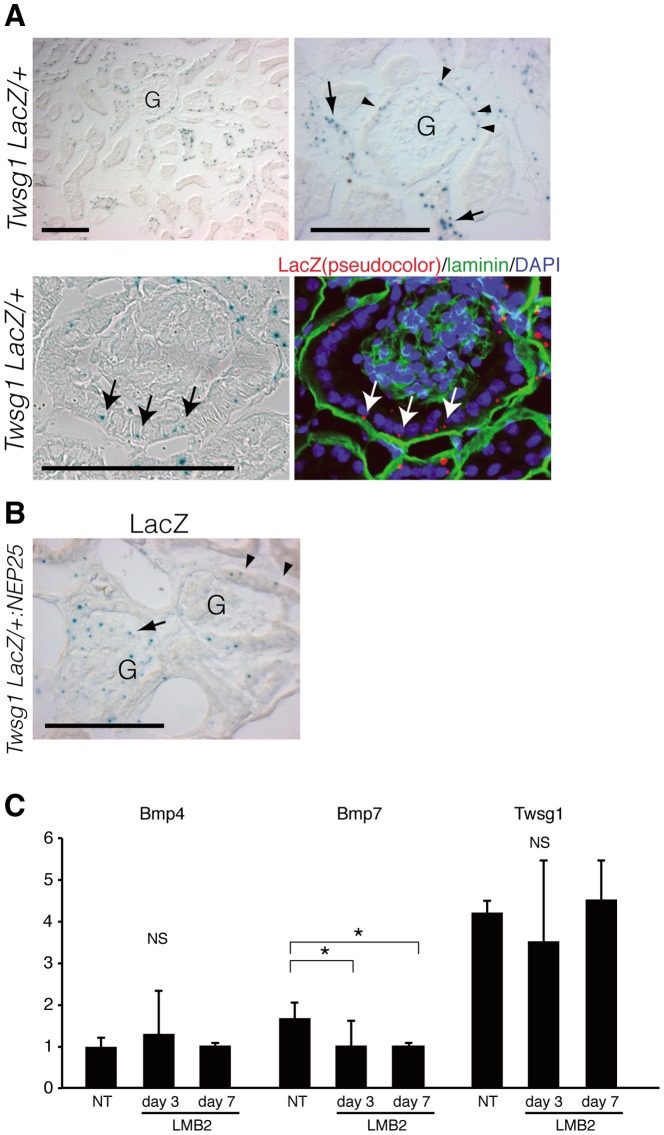
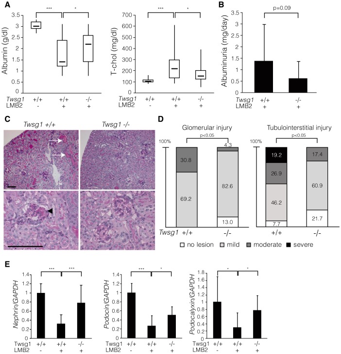
Podocyte injury is the first step in the progression of glomerulosclerosis. Previous studies have demonstrated the beneficial effect of bone morphogenetic protein 7 (Bmp7) in podocyte injury and the existence of native Bmp signaling in podocytes. Local activity of Bmp7 is controlled by cell-type specific Bmp antagonists, which inhibit the binding of Bmp7 to its receptors. Here we show that the product of Twisted gastrulation (Twsg1), a Bmp antagonist, is the central negative regulator of Bmp function in podocytes and that Twsg1 null mice are resistant to podocyte injury. Twsg1 was the most abundant Bmp antagonist in murine cultured podocytes. The administration of Bmp induced podocyte differentiation through Smad signaling, whereas the simultaneous administration of Twsg1 antagonized the effect. The administration of Bmp also inhibited podocyte proliferation, whereas simultaneous administration of Twsg1 antagonized the effect. Twsg1 was expressed in the glomerular parietal cells (PECs) and distal nephron of the healthy kidney, and additionally in damaged glomerular cells in a murine model of podocyte injury. Twsg1 null mice exhibited milder hypoalbuminemia and hyperlipidemia, and milder histological changes while maintaining the expression of podocyte markers during podocyte injury model. Taken together, our results show that Twsg1 plays a critical role in the modulation of protective action of Bmp7 on podocytes, and that inhibition of Twsg1 is a promising means of development of novel treatment for podocyte injury.


Conflict of interest statement

Competing Interests: Aris N. Economides is employed by Regeneron Pharmaceuticals, Inc. There are no patents, products in development or marketed products to declare. This does not alter the authors' adherence to all the PLOS ONE policies on sharing data and materials, as detailed online in the guide for authors.

Figures

Figure 1
Figure 1. Expression and function of Bmps and Bmp antagonists in cultured podocytes.
(A–C) Murine podocytes cultured under non-permissive conditions were subjected to quantitative real-time PCR with various primers for Bmps, Bmp receptors and Bmp antagonists using vector control (see method)(n = 3). (A) Expression of Bmps in cultured podocytes. The expression of each Bmp normalized by vector control was shown relative to that of Bmp2. (B) Expression of Bmp receptors in cultured podocytes. Expression of Bmp receptor type Ib and Bmp receptor type II normalized by vector control was shown relative to that of Bmp receptor type Ia. (C) Expression of Bmp antagonists in cultured podocytes. The expression of Bmp antagonists normalized by vector control was shown relative to that of Twsg1. (D–E) The effects of Bmps and Twsg1 administration on cultured podocytes. Podocytes cultured under non-permissive conditions were stimulated with Bmp and Twsg1, and incubated for an additional 14 days. Recombinant Bmp7 (300 ng/ml), Bmp4 (300 ng/ml), Twsg1 (300 ng/ml) were added to the cultured podocytes at day 1 and day 7. (D) Morphological changes in podocytes after the administration of Bmp4, Bmp7 and Twsg1. Surface area (pixel) of each podocyte was analyzed (n = 30 in each group). (E) The expression of podocalyxin in podocytes treated with Bmp4, Bmp7, and Twsg1. The results of quantitative real-time PCR analysis (n = 7) and representative immunostaining were shown. (F) Proliferation assay of podocyte in the presence of Bmp and Twsg1. We utilized recombinant Bmp7 (300 ng/ml), Bmp4 (300 ng/ml), and Twsg1 (300 ng/ml). (n = 11 in Bmp7 experiment, and n = 8 in Bmp4 experiment) Data are presented as means ± SD. *p<0.05, ***p<0.001; NS, no significant difference. Scale bars: 100 µm.
Figure 2
Figure 2. Smad signaling in podocyte differentiation and Twsg1 antagonism.
(A) The results of quantitative real-time PCR analysis of podocalyxin in the presence or absence of dorsomprohin. Bmp7 (10 ng/ml) and each concentration of dorsomphin were added to the podocyte. Data are presented as means ± SD. n = 3. **, p<0.01. (B) Western blotting of podocytes treated with Bmp proteins and Twsg1. Podocytes treated with these proteins were subjected for western blotting. Representative data is shown. GAPDH was utilized as a loading control.
Figure 3
Figure 3. Localization of Twsg1 in healthy and diseased kidney.
(A, upper row) X-gal staining of the kidney of Twsg1+/LacZ mice. Twsg1 was expressed in glomerular parietal cells (PECs)(arrowheads), and distal nephrons (arrows). Twsg1 expression was not observed in podocytes of healthy kidneys. G: glomeruli. (A, lower row) X-gal staining and laminin immunostaining to show the expression of Twsg1 in PECs (arrows). Scale bars: 100 µm. (B) X-gal staining of the kidney of Twsg1+/LacZ:NEP25 mice treated with immunotoxin. LacZ staining emerged within glomerular cells of some glomeruli (arrows), whereas LacZ staining in other glomeruli was confined in PECs (arrowheads). Scale bars: 100 µm. (C) Glomerular RNA of NEP25 mice was collected before and after the administration of LMB2, and was analyzed for the relative expression of Bmp and Twsg1 by real-time PCR. (n = 3) NT, non treat. Data are presented as means ± SD. * p<0.05; NS, no significant difference.
Figure 4
Figure 4. Twsg1 null mice were resistant to podocyte injury.
(A) Twsg1+/+:NEP25 and Twsg1LacZ/LacZ:NEP25 mice were injected with immunotoxin and analyzed seven days later. While Twsg1+/+:NEP25 mice injected with immunotoxin (Tswg1+/+LMB2+, n = 30) showed decreased levels of serum albumin and increased levels of total cholesterol compared to the controls (Tswg1+/+LMB2-, n = 13), these changes in serum parameters were significantly attenuated in Twsg1LacZ/LacZ:NEP25 mice (Tswg1−/−LMB2+, n = 24). Data are given in boxplots, in which the bottom and top of each box represent the 25th and 75th percentile, respectively, and the band near the middle of each box indicates the median. The ends of the whiskers represent the minimum and maximum of all data. (B) Urinary albumin levels were analyzed four days after the injection. Twsg1LacZ/LacZ:NEP25 mice (Tswg1−/−LMB2+, n = 17) showed lower levels of urinary albumin compared to Twsg1+/+:NEP25 mice (Tswg1+/+LMB2+, n = 15), although this difference was not statistically significant. Data are presented as means ± SD. (C) Representative histological findings of both genotypes seven days after the injection. Histological changes such as tubular degeneration (arrows) and glomerulosclerosis (arrowhead) were less severe in Twsg1LacZ/LacZ:NEP25 mice (Tswg1−/−) than in Twsg1+/+:NEP25 mice (Tswg1+/+). Scale bars: 100 µm. (D) Glomerular and tubulointerstitial injury was analyzed semi-quantitatively in both genotypes. The severity of both glomerular and tubulointerstitial damage was significantly lower in Twsg1LacZ/LacZ:NEP25 mice (Tswg1−/−, n = 23) than in Twsg1+/+:NEP25 mice (Tswg1+/+, n = 26). (E) Quantitative real-time PCR analysis of podocyte-specific genes in the whole kidneys seven days after the injection. Expression levels of nephrin, podocin and podocalyxin were severely reduced due to the injection of the immunotoxin in Twsg1+/+:NEP25 mice (Tswg1+/+LMB2+, n = 15) compared to the controls (Tswg1+/+LMB2-, n = 6), whereas this reduction was significantly attenuated in Twsg1LacZ/LacZ:NEP25 mice (Tswg1−/−LMB2+, n = 9). Data are presented as means ± SD. *p<0.05, ***p<0.001.

Similar articles

Cited by

References

    1. LeHir M, Kriz W (2007) New insights into structural patterns encountered in glomerulosclerosis. Curr Opin Nephrol Hypertens 16: 184–191. - PubMed
    1. D'Agati VD (2008) Podocyte injury in focal segmental glomerulosclerosis: Lessons from animal models (a play in five acts). Kidney Int 73: 399–406. - PubMed
    1. Wiggins RC (2007) The spectrum of podocytopathies: a unifying view of glomerular diseases. Kidney Int 71: 1205–1214. - PubMed
    1. Barisoni L, Schnaper HW, Kopp JB (2007) A proposed taxonomy for the podocytopathies: a reassessment of the primary nephrotic diseases. Clin J Am Soc Nephrol 2: 529–542. - PubMed
    1. Mundel P, Shankland SJ (2002) Podocyte biology and response to injury. J Am Soc Nephrol 13: 3005–3015. - PubMed

Publication types

LinkOut - more resources