

Repair of microdamage in osteonal cortical bone adjacent to bone screw
- PMID: 24586702
- PMCID: PMC3930719
- DOI: 10.1371/journal.pone.0089343
Repair of microdamage in osteonal cortical bone adjacent to bone screw
Abstract
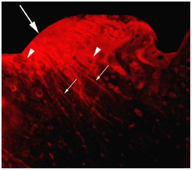
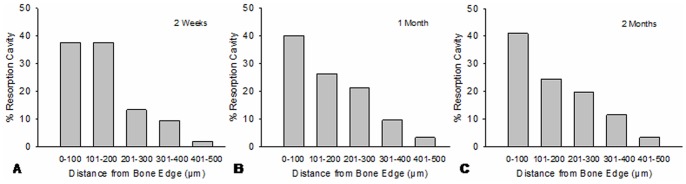
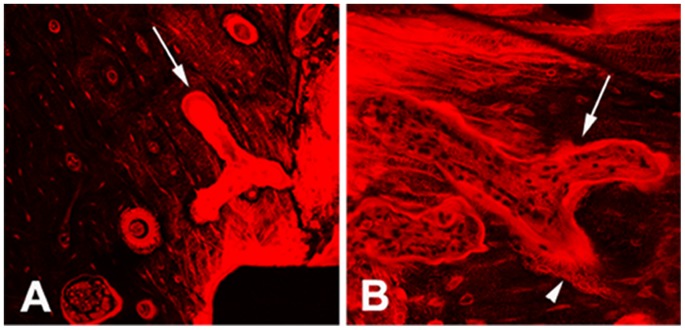
Up to date, little is known about the repair mode of microdamage in osteonal cortical bone resulting from bone screw implantation. In this study, self-tapping titanium cortical bone screws were inserted into the tibial diaphyses of 24 adult male rabbits. The animals were sacrificed at 1 day, 2 weeks, 1 month and 2 months after surgery. Histomorphometric measurement and confocal microscopy were performed on basic fuchsin stained bone sections to examine the morphological characteristics of microdamage, bone resorption activity and spatial relationship between microdamage and bone resorption. Diffuse and linear cracks were coexisted in peri-screw bone. Intracortical bone resorption was significantly increased 2 weeks after screw installation and reach to the maximum at 1 month. There was no significant difference in bone resorption between 1-month and 2-months groups. Microdamage was significantly decreased within 1 month after surgery. Bone resorption was predisposed to occur in the region of <100 µm from the bone-screw interface, where had extensive diffuse damage mixed with linear cracks. Different patterns of resorption cavities appeared in peri-screw bone. These data suggest that 1) the complex microdamage composed of diffuse damage and linear cracks is a strong stimulator for initiating targeted bone remodeling; 2) bone resorption activities taking place on the surfaces of differently oriented Haversian and Volkmann canals work in a team for the repair of extensive microdamage; 3) targeted bone remodeling is a short-term reaction to microdamage and thereby it may not be able to remove all microdamage resulting from bone screw insertion.
Conflict of interest statement
Figures


References
-
- Yadav S, Upadhyay M, Liu S, Roberts E, Neace WP, et al. (2012) Microdamage of the cortical bone during mini-implant insertion with self-drilling and self-tapping techniques: A randomized controlled trial. Am J Orthod Dentofacial Orthop 141: 538–546. - PubMed
-
- Xu C, Wei Z, Liu N, Sun F, Chen H, et al.. (2013) The Effect of Implant Shape and Screw Pitch on Microdamage in Mandibular Bone. Clin Implant Dent Relat Res. - PubMed
-
- Burr D (2003) Microdamage and bone strength. Osteoporos Int 14 Suppl 567–72. - PubMed
-
- Schaffler MB, Radin EL, Burr DB (1990) Long-term fatigue behavior of compact bone at low strain magnitude and rate. Bone 11: 321–326. - PubMed
Publication types
MeSH terms
LinkOut - more resources
Full Text Sources
Other Literature Sources
Medical
