. 2014 Feb 27;9(2):e89350.
doi: 10.1371/journal.pone.0089350. eCollection 2014.

PD-1 blockade and OX40 triggering synergistically protects against tumor growth in a murine model of ovarian cancer

Affiliations

PD-1 blockade and OX40 triggering synergistically protects against tumor growth in a murine model of ovarian cancer

Zhiqiang Guo et al. PLoS One. .

Erratum in

Abstract

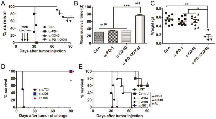
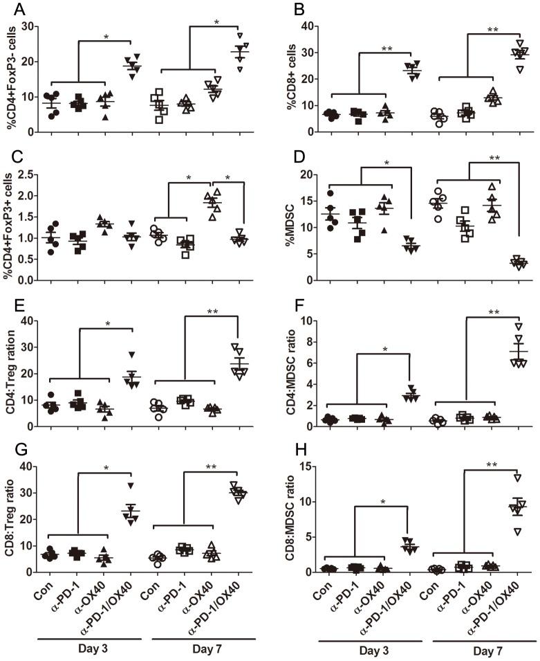
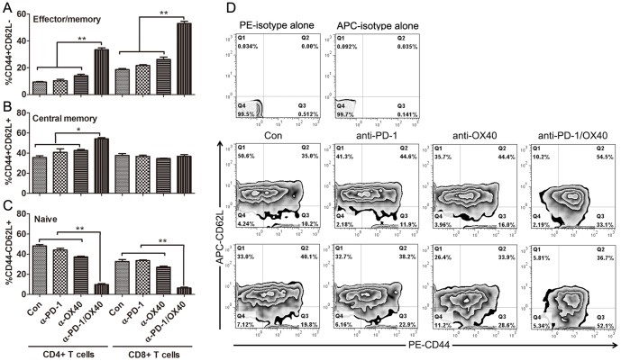
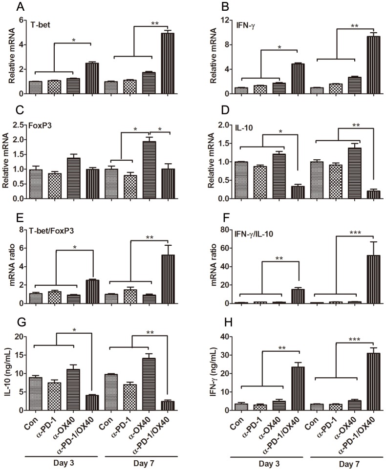
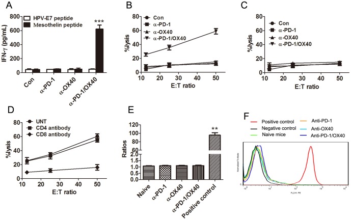
The co-inhibitory receptor Programmed Death-1 (PD-1) curtails immune responses and prevent autoimmunity, however, tumors exploit this pathway to escape from immune destruction. The co-stimulatory receptor OX40 is upregulated on T cells following activation and increases their clonal expansion, survival and cytokine production when engaged. Although antagonistic anti-PD-1 or agonistic anti-OX40 antibodies can promote the rejection of several murine tumors, some poorly immunogenic tumors were refractory to this treatment. In the present study, we evaluated the antitumor effects and mechanisms of combinatorial PD-1 blockade and OX40 triggering in a murine ID8 ovarian cancer model. Although individual anti-PD-1 or OX40 mAb treatment was ineffective in tumor protection against 10-day established ID8 tumor, combined anti-PD-1/OX40 mAb treatment markedly inhibited tumor outgrowth with 60% of mice tumor free 90 days after tumor inoculation. Tumor protection was associated with a systemic immune response with memory and antigen specificity and required CD4(+) cells and CD8(+) T cells. The anti-PD-1/OX40 mAb treatment increased CD4(+) and CD8(+) cells and decreased immunosuppressive CD4(+)FoxP3(+) regulatory T (Treg) cells and CD11b(+)Gr-1(+) myeloid suppressor cells (MDSC), giving rise to significantly higher ratios of both effector CD4(+) and CD8(+) cells to Treg and MDSC in peritoneal cavity; Quantitative RT-PCR data further demonstrated the induction of a local immunostimulatory milieu by anti-PD-1/OX40 mAb treatment. The splenic CD8(+) T cells from combined mAb treated mice produced high levels of IFN-γ upon tumor antigen stimulation and exhibited antigen-specific cytolytic activity. To our knowledge, this is the first study testing the antitumor effects of combined anti-PD-1/OX40 mAb in a murine ovarian cancer model, and our results provide a rationale for clinical trials evaluating ovarian cancer immunotherapy using this combination of mAb.


Conflict of interest statement

Competing Interests: The authors have declared that no competing interests exist.

Figures

Figure 1
Figure 1. Treatment of combined anti-PD-1/OX40 mAb induced tumor-specific long-lasting immunity against ID8 ovarian cancer.
A, Mice (10 mice per group) transplanted i.p. with 1×10⁶ ID8 cells 10 day before were treated thrice with 200 µg of control, anti-PD-1, anti-OX40 and anti-PD-1/OX40 mAb at 4 days interval and overall survival of mice was recorded. B, Mean survival time of mice with tumor growth was calculated. C, The peritoneal tumor masses was weighed when mice were euthanized with each dot representing each mouse. D, Long-term surviving (90 days after first tumor challenge) mice (4 mice per group) from 2 mAb treatment group were rechallenged with ID8 cells given i.p. or s.c. or with syngeneic unrelated TC1 cells transplanted s.c., and then overall survival of mice was recorded. E, Mice (5 mice per group) treated with combined anti-PD-1/OX40 mAb were also injected with an anti-CD4, anti-CD8, anti-NK1.1, or control mAb with 500 µg of each mAb per mouse 1 day before and two days after tumor challenge followed by injection of 200 µg every 5 days thereafter for the duration of the experiments. Tumor-bearing untreated mice were as negative controls (UNT). Overall survival of mice was recorded. Data are representative of three independent experiments except for D and E, which was from one experiment. ***P<0.001, combined mAb vs control or single mAb treated mice in A-C; ***P<0.001, s.c. or i.p.ID8 vs s.c.TC1 in C; * P<0.05, control or NK1.1 vs UNT, CD4 or CD8 in D.
Figure 2
Figure 2. Composition analysis of peritoneal immune cells (PIC) from treated mice.
Mice (5 mice per group) injected i.p. with 1×10⁶ ID8 cells 10 day earlier were injected thrice at 4 days interval with 200 µg of control, single or combined anti-PD-1/OX40 mAb. Three or seven days later, peritoneal lavages from treated mice were analyzed by flow cytometry for the composition of various immune subsets. The percentages of CD4+FoxP3 T cells, CD8+ T cells, CD4+FoxP3+ Treg and CD11b+GR-1+ MDSC in CD45+ peritoneal immune cells are shown in A, B, C and D respectively with each dot representing data from each mouse. The ratios of both CD4+ and CD8+ T cells to Treg and MDSC are shown in E, F, G and H respectively with each dot representing data from each mouse. Data are representative of 2 independent experiments, *P<0.05, **P<0.01.
Figure 3
Figure 3. Phenotypic analysis of peritoneal CD4+ and CD8+ T cells from treated mice.
Mice (5 mice per group) injected i.p. with 1×10⁶ ID8 cells 10 day earlier were injected thrice at 4 days interval with 200 µg of control, single or combined anti-PD-1/OX40 mAb. Seven days later, the expression of CD44 and CD62L on peritoneal CD4+ and CD8+ T cells from treated mice was analyzed by flow cytometry. The frequencies of CD44+CD62L effector/memory, CD44+CD62L+ central memory and CD44CD62L+ naïve cells in peritoneal CD4+ and CD8+ T cells are shown in A, B and C respectively. The representative dotplots are shown in D with upper, middle and bottom panels displaying isotype, CD44 and CD62L staining in gated CD4+ and CD8+ T cells respectively. Data are representative of 2 independent experiments, *P<0.05, **P<0.01.
Figure 4
Figure 4. Immune-related gene expression in PIC from treated mice.
Mice (5 mice per group) injected i.p. with 1×10⁶ ID8 cells 10 day earlier were injected thrice at 4 days interval with 200 µg of control, single or combined anti-PD-1/OX40 mAb. Three or seven days later, the expression of Th1-associated T-bet and IFN-γ genes and immunoregulatory FoxP3 and IL-10 genes in PIC from treated mice was analyzed by quantitative RT-PCR. The expression of T-bet, IFN-γ, FoxP3 and IL-10 genes are shown in A, B, C and D respectively. The ratios of both T-bet/FoxP3 and IFN-γ/IL-10 are shown in E and F respectively. Freshly isolated PIC from treated mice were stimulated with 50 ng/ml PMA and 1 µg/ml ionomycin for 6 hours (as described in Materials and Methods) and supernatants were assessed for levels of IL-10 (G) and IFN-γ (H). Data were expressed as M±SEM of 5 mice and representative of 2 independent experiments, *P<0.05, **P<0.01, ***P<0.001.
Figure 5
Figure 5. Mice treated with anti-PD-1/OX40 mAbs developed a tumor antigen-specific CTL response.
A, Mice injected i.p. with 1×10⁶ ID8 cells 10 day earlier were injected thrice at 4 days interval with 200 µg of control, single or combined anti-PD-1/OX40 mAb. Seven days after the last mAb injection, splenocytes from treated mice were cultured in the presence or absence of H-2Db-restricted mesothelin-derived peptides or control HPV-E7-derived peptide for 3 days and IFN-γ production in the supernatants were assayed by ELISA. B, Pooled splenocytes (5×10⁶) from all mice treated with control, single or combined anti-PD-1/OX40 mAb were incubated with 5×10⁵ UV-irradiated ID8 cells for 4 days prior to subject to analysis of antigen-specific CTL activity by CytoTox 96 Non-radioactive cytotoxicity assay using EL4 cells pulsed with H-2Db-restricted mesothelin peptide as target cells. C, As a specific control, pooled splenocytes were tested cytotoxicity against EL4 target cells pulsed with control HPV-E7 peptide. D, The killing activity of splenocytes from combined mAb treated mice on mesothelin-pulsed EL4 cells was also evaluated in the presence of blocking anti-CD4, anti-CD8 or control antibody. Data were expressed as M±SEM of triplicate wells in A-D. E, Sera were obtained from naïve mice (5 mice), single mAb treated mice (5 mice each group) or 2 mAb treated long-term surviving mice (4 mice). The presence of mesothelin-specific antibodies was determined by flow cytometry via staining mesothelin-expressing ID8 cells with collected sera in a 1∶200 dilution, followed by staining with secondary PE-conjugated anti-mouse IgG antibody and MFI was calculated. Staining with commercially available anti-mesothelin antibody plus secondary antibody or secondary antibody alone was used as positive or negative control. The ratios of MFI from positive control or sera to MFI from negative control were shown. F, A representative histogram in E was shown.

References

    1. Siegel R, DeSantis C, Virgo K, Stein K, Mariotto A, et al. (2012) Cancer treatment and survivorship statistics, 2012. CA Cancer J Clin 62: 220–241. - PubMed
    1. Bast RC Jr, Hennessy B, Mills GB (2009) The biology of ovarian cancer: new opportunities for translation. Nat Rev Cancer 9: 415–428. - PMC - PubMed
    1. Jemal A, Tiwari RC, Murray T, Ghafoor A, Samuels A, et al. (2004) Cancer statistics, 2004. CA Cancer J Clin 54: 8–29. - PubMed
    1. Ozols RF, Bundy BN, Greer BE, Fowler JM, Clarke-Pearson D, et al. (2003) Phase III trial of carboplatin and paclitaxel compared with cisplatin and paclitaxel in patients with optimally resected stage III ovarian cancer: a Gynecologic Oncology Group study. J Clin Oncol 21: 3194–3200. - PubMed
    1. Kandalaft LE, Powell DJ Jr, Singh N, Coukos G (2011) Immunotherapy for ovarian cancer: what's next? J Clin Oncol 29: 925–933. - PMC - PubMed

Publication types

MeSH terms