

Comparative study of sex differences in distal femur morphology in osteoarthritic knees in a Chinese population
- PMID: 24586746
- PMCID: PMC3929686
- DOI: 10.1371/journal.pone.0089394
Comparative study of sex differences in distal femur morphology in osteoarthritic knees in a Chinese population
Abstract
Objectives: The purpose of this study was to investigate sex differences in resected distal femoral morphology in Chinese osteoarthritic knees.
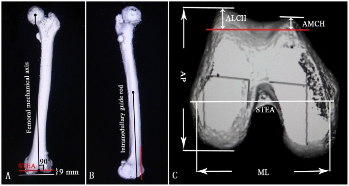
Methods: The study included 130 osteoarthritic knees in 65 men and 65 women in China. None had anterior femoral osteophyte or serious patellar femoral joint degeneration. The following were measured using computed tomography and analyzed to identify morphological differences according to sex in the resected distal femurs: anterior lateral condylar height (ALCH), anterior medial condylar height (AMCH), and mediolateral (ML) and anteroposterior (AP) dimensions. The ML/AP aspect ratio was calculated.
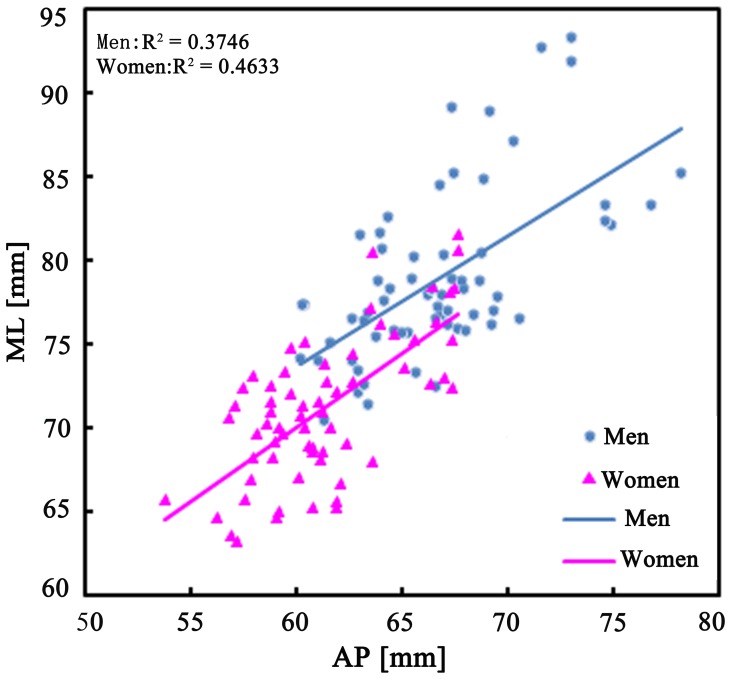
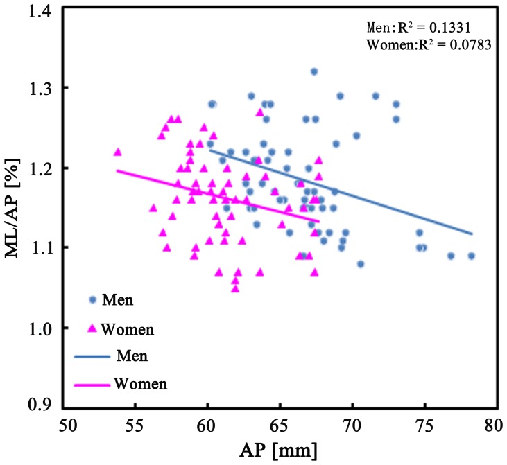
Results: The average ALCH and AMCH were 8.2±1.8 mm, 3.1±1.5 mm for men and 7.4±1.7 mm, 3.6±1.5 mm for women. There were significant differences between men and women in ALCH values (P = 0.014) but not in AMCH values (P = 0.09). Women had smaller ML/AP aspect ratios than men for a given AP dimension. This indicated that the femoral ML dimension of a prosthesis with a given AP dimension may have overhang in women.
Conclusions: This study suggested that sex differences should be taken into account in the design of femoral prosthesis for Chinese men and women.
Conflict of interest statement
Figures
Similar articles
-
Femoral anterior condyle height decreases as the distal anteroposterior size increases in total knee arthroplasty: A comparative study.PLoS One. 2024 Feb 26;19(2):e0297634. doi: 10.1371/journal.pone.0297634. eCollection 2024. PLoS One. 2024. PMID: 38408088 Free PMC article.
-
Highly Variable Femoral Morphology in Osteoarthritic Chinese: Are Prostheses Today Sufficiently Suitable?J Knee Surg. 2017 Nov;30(9):936-942. doi: 10.1055/s-0037-1599250. Epub 2017 Mar 14. J Knee Surg. 2017. PMID: 28293922
-
Morphological Measurement and Clinical Significance of Abnormal Development of Distal Femur with Hemophilia Knee Arthritis: A Consideration on the Renewal of Total Knee Prosthesis.Orthop Surg. 2024 Nov;16(11):2661-2670. doi: 10.1111/os.14170. Epub 2024 Aug 6. Orthop Surg. 2024. PMID: 39105304 Free PMC article.
-
Intraoperative morphometric study of gender differences in Asian femurs.J Arthroplasty. 2011 Oct;26(7):984-8. doi: 10.1016/j.arth.2010.11.012. Epub 2011 Feb 12. J Arthroplasty. 2011. PMID: 21316917
-
Gender-based differences in the dimensions of the femoral trochlea and condyles in the Chinese population: correlation to the risk of femoral component overhang.Knee. 2014 Jan;21(1):252-6. doi: 10.1016/j.knee.2012.11.005. Epub 2012 Dec 12. Knee. 2014. PMID: 23245733
Cited by
-
Morphometric evaluation of the knee in Chinese population reveals sexual dimorphism and age-related differences.Int Orthop. 2018 Oct;42(10):2349-2356. doi: 10.1007/s00264-018-3826-x. Epub 2018 Feb 20. Int Orthop. 2018. PMID: 29464370
-
Analysis of Gender Differences in the Rotational Alignment of the Distal Femur in Kinematically Aligned and Mechanically Aligned Total Knee Arthroplasty.J Clin Med. 2021 Aug 20;10(16):3691. doi: 10.3390/jcm10163691. J Clin Med. 2021. PMID: 34441989 Free PMC article.
-
What Differences in Morphologic Features of the Knee Exist Among Patients of Various Races? A Systematic Review.Clin Orthop Relat Res. 2017 Jan;475(1):170-182. doi: 10.1007/s11999-016-5097-4. Epub 2016 Oct 4. Clin Orthop Relat Res. 2017. PMID: 27704318 Free PMC article.
-
Bone shape mediates the relationship between sex and incident knee osteoarthritis.BMC Musculoskelet Disord. 2018 Sep 12;19(1):331. doi: 10.1186/s12891-018-2251-z. BMC Musculoskelet Disord. 2018. PMID: 30208910 Free PMC article.
-
A Morphometric Study of the Distal Femoral Resected Surface In Osteoarthritis Knees of the Patients in Southwest China and a Comparison with Femoral Components in Six Total Knee Arthroplasty Systems.Orthop Surg. 2023 Apr;15(4):953-960. doi: 10.1111/os.13647. Epub 2023 Jan 31. Orthop Surg. 2023. PMID: 36718658 Free PMC article.
References
-
- Cheng FB, Ji XF, Lai Y, Feng JC, Zheng WX, et al. (2009) Three dimensional morphometry of the knee to design the total knee arthroplasty for Chinese population. Knee 16: 341–347. - PubMed
-
- Hitt K, Shurman JR, Greene K, McCarthy J, Moskal J, et al. (2003) Anthropometric measurements of the human knee: correlation to the sizing of current knee arthroplasty systems. J Bone Joint Surg Am 85A: 115–122. - PubMed
-
- Conley S, Rosenberg A, Crowninshield R (2007) The female knee: anatomic variations. J Am Acad Orthop Surg 15: S31–36. - PubMed
-
- Chin PL, Tey TT, Ibrahim MY, Chia SL, Yeo SJ, et al. (2011) Intraoperative morphometric study of gender differences in Asian femurs. J Arthroplasty 26: 984–988. - PubMed
Publication types
MeSH terms
LinkOut - more resources
Full Text Sources
Other Literature Sources
