. 2014 Feb 27;9(2):e89397.
doi: 10.1371/journal.pone.0089397. eCollection 2014.

Mice carrying a hypomorphic Evi1 allele are embryonic viable but exhibit severe congenital heart defects

Affiliations

Mice carrying a hypomorphic Evi1 allele are embryonic viable but exhibit severe congenital heart defects

Emilie A Bard-Chapeau et al. PLoS One. .

Abstract

The ecotropic viral integration site 1 (Evi1) oncogenic transcription factor is one of a number of alternative transcripts encoded by the Mds1 and Evi1 complex locus (Mecom). Overexpression of Evi1 has been observed in a number of myeloid disorders and is associated with poor patient survival. It is also amplified and/or overexpressed in many epithelial cancers including nasopharyngeal carcinoma, ovarian carcinoma, ependymomas, and lung and colorectal cancers. Two murine knockout models have also demonstrated Evi1's critical role in the maintenance of hematopoietic stem cell renewal with its absence resulting in the death of mutant embryos due to hematopoietic failure. Here we characterize a novel mouse model (designated Evi1(fl3)) in which Evi1 exon 3, which carries the ATG start, is flanked by loxP sites. Unexpectedly, we found that germline deletion of exon3 produces a hypomorphic allele due to the use of an alternative ATG start site located in exon 4, resulting in a minor Evi1 N-terminal truncation and a block in expression of the Mds1-Evi1 fusion transcript. Evi1(δex3/δex3) mutant embryos showed only a mild non-lethal hematopoietic phenotype and bone marrow failure was only observed in adult Vav-iCre/+, Evi1(fl3/fl3) mice in which exon 3 was specifically deleted in the hematopoietic system. Evi1(δex3/δex3) knockout pups are born in normal numbers but die during the perinatal period from congenital heart defects. Database searches identified 143 genes with similar mutant heart phenotypes as those observed in Evi1(δex3/δex3) mutant pups. Interestingly, 42 of these congenital heart defect genes contain known Evi1-binding sites, and expression of 18 of these genes are also effected by Evi1 siRNA knockdown. These results show a potential functional involvement of Evi1 target genes in heart development and indicate that Evi1 is part of a transcriptional program that regulates cardiac development in addition to the development of blood.


Conflict of interest statement

Competing Interests: The authors have declared that no competing interests exist.

Figures

Figure 1
Figure 1. Deletion of Evi1 exon3 generates a hypomorphic allele.
(A) Sequenced products obtained after 5′RACE from wild type or Evi1δex3/δex3 mutant embryos. (B) Table showing the fraction of embryos of each genotype detected at different stages of embryonic development. The Mendelian ratios were not affected by the Evi1 exon3 deletion. (C) Pictures of 28 hr-old littermates highlight the poor health of dying Evi1δex3/δex3 pups. (D) Kaplan-Meyer curves for wild type, Evi1δex3/+ and Evi1δex3/δex3 progeny indicate lethality of all Evi1δex3/δex3 pups by three days after birth (n = 5 to 16 per genotype). Log rank test, Chi square p value <0.0001. (E) RT-qPCR from cDNA of E14.5 embryos. The primers used amplified the regions between Evi1 exons 2 and 3, 3 and 4 or 13 and 14. Mean of three different samples per condition. The standard deviation is shown. (F) Expression of Evi1 and γ-tubulin protein products in E14.5 wild type or E17.5 Evi1δex3/+ and Evi1δex3/δex3 mutant embryos (100 µg protein/lane). (G) Nucleotide sequence of Evi1 cDNA in the exon 3 and 4 genomic region. Two ATG sites are present in exon 3 and one in exon 4. All ATGs are conserved in higher vertebrates.
Figure 2
Figure 2. Disruption of hematopoiesis in Evi1δex3/δex3 newborn mice.
(A,B) Flow cytometric profiles of wild type, Evi1δex3/+ and Evi1δex3/δex3 littermate fetal livers at E14.5. (A) HSC and progenitor cell subpopulations were detected by a combination of markers (KSL: c-Kit+, S: Sca-1+, L: lineage, or KL-CD34+). We found a significant reduction of cells in the Evi1-deleted samples; p values are from an unpaired t-test between +/+ and Evi1δex3/δex3 fetal livers. (B) Bar graph shows the number of granulocytes (Gr1), B-lymphocytes (B220) and erythroid cells (Tert119) in fetal livers of various different genotypes. (C) Colony forming counts from cells of 3 fetal livers of each genotype at E14.5 We observed a significant reduction in colony formation between +/+ and Evi1δex3/δex3 fetal livers, p = 0.0057 (unpaired t-test). No BFU-E or CFU-Mix colonies were identified. (D) Hemogram results for 4 hr- to 24 hr-old wild type (N = 17), Evi1δex3/+, (N = 30) and Evi1δex3/δex3 (N = 16) littermate pups. Mean ± SEM is indicated. *p<0.05, **p<0.01, ***p<0.001, unpaired t-test. Leukocyte counts in peripheral blood and white blood cell differentials reveal a mild leucopenia in Evi1δex3/δex3 newborn mice. Platelet (PLT) counts and mean platelet volume (MPV) results show a mild hypoproliferative thrombocytopenia in Evi1δex3/δex3 pups. Normal erythrocyte counts, hemoglobin quantification and hematocrit assessment in the peripheral blood of Evi1δex3/δex3 animals. Mean corpuscular volume (MCV), mean corpuscular hemoglobin (MCH), mean corpuscular hemoglobin concentration (MCHC) and red cell distribution widths (RDW) are shown. (E) Hematoxylin and eosin staining of 5 µm sections of 24 hr- to 48 hr-old Evi1δex3/δex3 pups. Mild hemorrhages were seen in 31% of the mice (4 out of 13 pups).
Figure 3
Figure 3. Profound depletion of hematopoietic cells in adult mice carrying an Evi1 exon3 deletion.
(A) Kaplan-Meyer survival curves indicate significant lethality in Vav-iCre; Evi1fl3/fl3 mice, with a median survival of 7.7 weeks (Log rank test, Chi square p value <0.0001). (B) Hemograms for 6 to 9 week-old Vav-iCre; Evi1fl3/fl3 mice. These adult mice displayed leucopenia, severe anemia and thrombocytopenia. Mean ± SEM is indicated. *p<0.05, **p<0.01, ***p<0.001, unpaired t-test. (C) Flow cytometric profiles of bone marrow cells from Vav-iCre/+;Evi1fl3/fl3 and littermate control mice (Evi1fl3/+ or Evi1fl3/fl3). HSC and progenitor cell subpopulations were detected by a combination of markers (KSL: c-Kit+, S: Sca-1+, L: lineage). We found a significant reduction of cells in Evi1-deleted samples, p = 0.00011 and p = 0.0024, for KSL and KL, respectively (unpaired t-test). (D) Colony forming counts for cells from bone marrow of Vav-iCre;Evi1fl3/fl3 and littermate control mice (Evi1fl3/+ or Evi1fl3/fl3). N = 3 for each group, p = 0.0019 (unpaired t-test). No BFU-E and CFU-Mix colonies were identified.
Figure 4
Figure 4. Spontaneous lethal bone marrow depletion in mice harboring an Evi1 exon3 deletion in the hematopoietic system.
(A) Histology was performed on sick Vav-iCre; Evi1fl3/fl3 and littermate control mice. Bone marrow depletion was observed in the mutant mice. Adipose tissue replaced the hematopoietic cells in the bone marrow. (B) Increased erythropoiesis in the spleen of Vav-iCre; Evi1fl3/fl3 mice. No visible border was found between the red pulp and white pulp. Erythroid cells are shown by the arrows. Excess erythropoiesis in spleen likely happens to compensate for bone marrow loss. (C) H&E stained sections of the brain of a dying Vav-iCre; Evi1fl3/fl3 mouse. Hemorrhages (red areas) were visible at several locations (also see Fig. S3E in File S1). (D) Histological sections of tissues from dying Vav-iCre; Evi1fl3/fl3 animals showing bacteremia. Red arrows indicate the presence of bacteria in alveolar capillaries. Giemsa stains reveal the presence of cocci or small rods within glomerular capillaries. No sign of immune system defense (inflammatory cells) was observed despite the infection.
Figure 5
Figure 5. Cardiac malformations and failure in Evi1δex3/δex3 mice.
(A) Transverse sections and (B) 3D reconstruction (left-ventral oblique view) of hearts from Evi1δex3/δex3 or wild type littermate (+/+) E15.5 embryos analyzed by magnetic resonance imaging (MRI). The aorta (Ao), right ventricle (RV), left ventricle (LV), ventricular septum (VS), trachea (Tr), aortic arch (AoA) and ductus arteriosus (DA) are indicated. Ventricular septal defect (VSD), interrupted aortic arch (IAA) and common arterial trunk (CAT) were observed in Evi1δex3/δex3 hearts. (C) List of the congenital heart defects identified in fifteen E15.5 embryos of various different genotypes by MRI and 3D reconstruction. (D) Hematoxylin and eosin staining of 5 µm sections of a sick Evi1δex3/δex3 pup. Subcutaneous and other tissue edema (white spaces) was present, consistent with heart failure.
Figure 6
Figure 6. Expression of Mecom mRNA in cardiac structures of wild type embryos.
(A–D) Whole mount mRNA in situ hybridization to show Mecom expression. A–C) Expression during subsequent stages of heart tube formation E8.5 (black brackets). D) At E9.5 Evi1 is expressed in the endothelial cells and in the endocardium of the heart and in the mesenchyme of the aortic arches. Expression also includes a population of migrating neural crest cells (white arrowhead). E–J) E10.5 Sagittal sections (from right to left) showing Evi1 in the aortic arches (a), mesenchyme of the secondary heart field (black arrowheads), outflow and atrio-ventricular canal endocardium including the cushions.
Figure 7
Figure 7. Evi1 regulates the expression of other CHD genes during embryonic heart development.
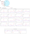
(A) The number of CHD genes represented in Evi1 ChIP-Seq data (Evi1 bound genes) or in the list of genes regulated by Mecom. An enriched number of CHD genes were found bound or regulated by Mecom (50 out of 143 genes), p = 0.0453 and p = 0.0276, respectively. These genes represent potential Mecom target genes in heart development. (B) Mecom regulates the expression of 23 CHD genes, which contain Evi1-binding sites specifically in heart. Heart and head (neural crest) tissues were harvested from WT and Evi1δex3/δex3 embryos of somite number 9 to 18. RT-qPCR assays were performed. Genes considered to be mis-regulated in Evi1δex3/δex3 hearts were increased or decreased in expression by at least three fold in average for all samples of the same time-point. These graphs are representative of two to five independent experiments.

Similar articles

Cited by

References

    1. Koonin EV, Wolf YI (2010) Constraints and plasticity in genome and molecular-phenome evolution. Nat Rev Genet 11: 487–498. - PMC - PubMed
    1. Nerlov C (2010) Transcriptional and translational control of C/EBPs: the case for “deep” genetics to understand physiological function. Bioessays 32: 680–686. - PubMed
    1. Lugthart S, van Drunen E, van Norden Y, van Hoven A, Erpelinck CA, et al. (2008) High EVI1 levels predict adverse outcome in acute myeloid leukemia: prevalence of EVI1 overexpression and chromosome 3q26 abnormalities underestimated. Blood 111: 4329–4337. - PubMed
    1. Ogawa S, Kurokawa M, Tanaka T, Mitani K, Inazawa J, et al. (1996) Structurally altered Evi-1 protein generated in the 3q21q26 syndrome. Oncogene 13: 183–191. - PubMed
    1. Goyama S, Kurokawa M (2009) Pathogenetic significance of ecotropic viral integration site-1 in hematological malignancies. Cancer Sci 100: 990–995. - PMC - PubMed

Publication types

MeSH terms

Substances