

The association between smoking and ectopic pregnancy: why nicotine is BAD for your fallopian tube
- PMID: 24586750
- PMCID: PMC3930728
- DOI: 10.1371/journal.pone.0089400
The association between smoking and ectopic pregnancy: why nicotine is BAD for your fallopian tube
Abstract
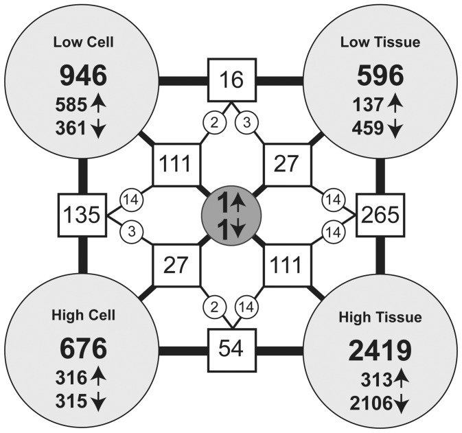
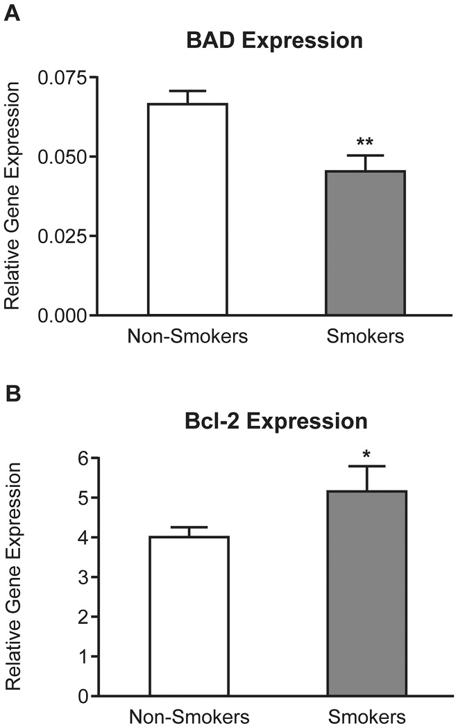
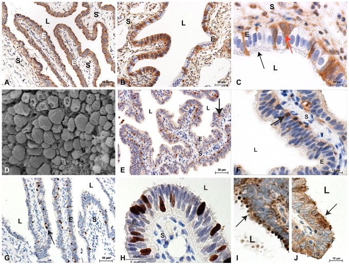
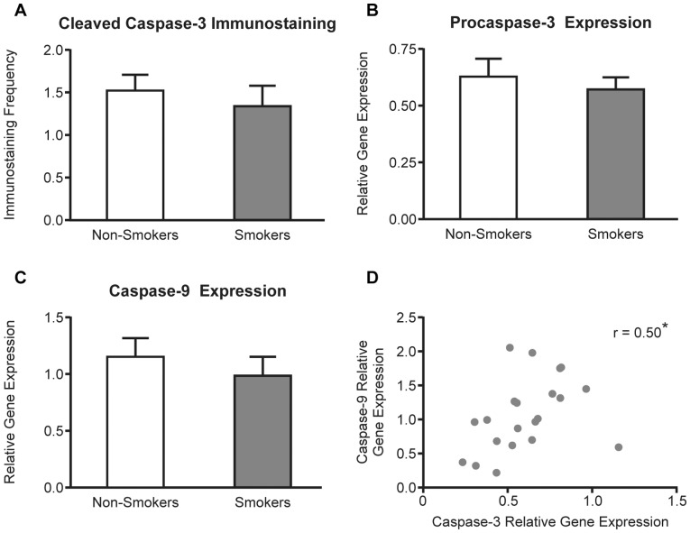
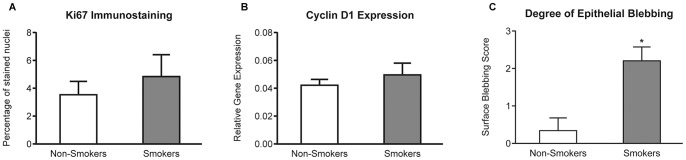
Epidemiological studies have shown that cigarette smoking is a major risk factor for tubal ectopic pregnancy but the reason for this remains unclear. Here, we set out to determine the effect of smoking on Fallopian tube gene expression. An oviductal epithelial cell line (OE-E6/E7) and explants of human Fallopian tubes from non-pregnant women (n = 6) were exposed to physiologically relevant concentrations of cotinine, the principle metabolite of nicotine, and changes in gene expression analyzed using the Illumina Human HT-12 array. Cotinine sensitive genes identified through this process were then localized and quantified in Fallopian tube biopsies from non-pregnant smokers (n = 10) and non-smokers (n = 11) using immunohistochemistry and TaqMan RT-PCR. The principle cotinine induced change in gene expression detected by the array analysis in both explants and the cell line was significant down regulation (P<0.05) of the pro-apoptotic gene BAD. We therefore assessed the effect of smoking on cell turnover in retrospectively collected human samples. Consistent with the array data, smoking was associated with decreased levels of BAD transcript (P<0.01) and increased levels of BCL2 transcript (P<0.05) in Fallopian tube biopsies. BAD and BCL2 specific immunolabelling was localized to Fallopian tube epithelium. Although no other significant differences in levels of apoptosis or cell cycle associated proteins were observed, smoking was associated with significant changes in the morphology of the Fallopian tube epithelium (P<0.05). These results suggest that smoking may alter tubal epithelial cell turnover and is associated with structural, as well as functional, changes that may contribute to the development of ectopic pregnancy.
Conflict of interest statement
Figures
Similar articles
-
Cotinine exposure increases Fallopian tube PROKR1 expression via nicotinic AChRalpha-7: a potential mechanism explaining the link between smoking and tubal ectopic pregnancy.Am J Pathol. 2010 Nov;177(5):2509-15. doi: 10.2353/ajpath.2010.100243. Epub 2010 Sep 23. Am J Pathol. 2010. PMID: 20864676 Free PMC article.
-
Cigarette smoking alters sialylation in the Fallopian tube of women, with implications for the pathogenesis of ectopic pregnancy.Mol Reprod Dev. 2016 Dec;83(12):1083-1091. doi: 10.1002/mrd.22747. Epub 2016 Oct 20. Mol Reprod Dev. 2016. PMID: 27704662 Clinical Trial.
-
Chlamydia trachomatis infection increases fallopian tube PROKR2 via TLR2 and NFκB activation resulting in a microenvironment predisposed to ectopic pregnancy.Am J Pathol. 2011 Jan;178(1):253-60. doi: 10.1016/j.ajpath.2010.11.019. Epub 2010 Dec 23. Am J Pathol. 2011. PMID: 21224062 Free PMC article.
-
Mechanisms of disease: the endocrinology of ectopic pregnancy.Expert Rev Mol Med. 2012 Mar 2;14:e7. doi: 10.1017/erm.2011.2. Expert Rev Mol Med. 2012. PMID: 22380790 Review.
-
Mechanism of Human Tubal Ectopic Pregnancy Caused by Cigarette Smoking.Reprod Sci. 2023 Apr;30(4):1074-1081. doi: 10.1007/s43032-022-00947-6. Epub 2022 Aug 12. Reprod Sci. 2023. PMID: 35962304 Review.
Cited by
-
Ectopic pregnancy and epithelial to mesenchymal transition: is there a link?Reproduction. 2021 Mar;161(3):V11-V14. doi: 10.1530/REP-20-0542. Reproduction. 2021. PMID: 33275118 Free PMC article.
-
Genetic and Epigenetic Alterations Associated With Human Prenatal Tobacco and Environmental Tobacco Smoke Exposure: Protocol for a Systematic Evidence Map.Public Health Rev. 2025 Jul 14;46:1606742. doi: 10.3389/phrs.2025.1606742. eCollection 2025. Public Health Rev. 2025. PMID: 40726774 Free PMC article. Review.
-
BTEB2 prevents neuronal apoptosis via promoting bad phosphorylation in rat intracerebral hemorrhage model.J Mol Neurosci. 2015 Jan;55(1):206-216. doi: 10.1007/s12031-014-0305-8. Epub 2014 Apr 27. J Mol Neurosci. 2015. PMID: 24770868 Free PMC article.
-
Demographic, lifestyle, and reproductive risk factors for ectopic pregnancy.Fertil Steril. 2018 Dec;110(7):1328-1337. doi: 10.1016/j.fertnstert.2018.08.022. Fertil Steril. 2018. PMID: 30503132 Free PMC article.
-
Substance Use in the Perinatal Period.Curr Psychiatry Rep. 2015 Nov;17(11):91. doi: 10.1007/s11920-015-0626-5. Curr Psychiatry Rep. 2015. PMID: 26386836 Free PMC article. Review.
References
-
- Jurkovic D, Wilkinson H (2011) Diagnosis and management of ectopic pregnancy. BMJ 342: d3397. - PubMed
-
- Jansen RP (1984) Endocrine response in the fallopian tube. Endocr Rev 5: 525–551. - PubMed
-
- Lindblom B, Hamberger L, Ljung B (1980) Contractile patterns of isolated oviductal smooth muscle under different hormonal conditions. Fertil Steril 33: 283–287. - PubMed
Publication types
MeSH terms
Substances
Grants and funding
LinkOut - more resources
Full Text Sources
Other Literature Sources
Medical
Research Materials
