. 2014 Feb 21;9(2):e89451.
doi: 10.1371/journal.pone.0089451. eCollection 2014.

The ubiquitin ligase ASB4 promotes trophoblast differentiation through the degradation of ID2

Affiliations

The ubiquitin ligase ASB4 promotes trophoblast differentiation through the degradation of ID2

W H Davin Townley-Tilson et al. PLoS One. .

Abstract

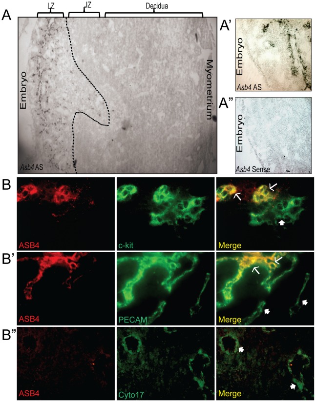
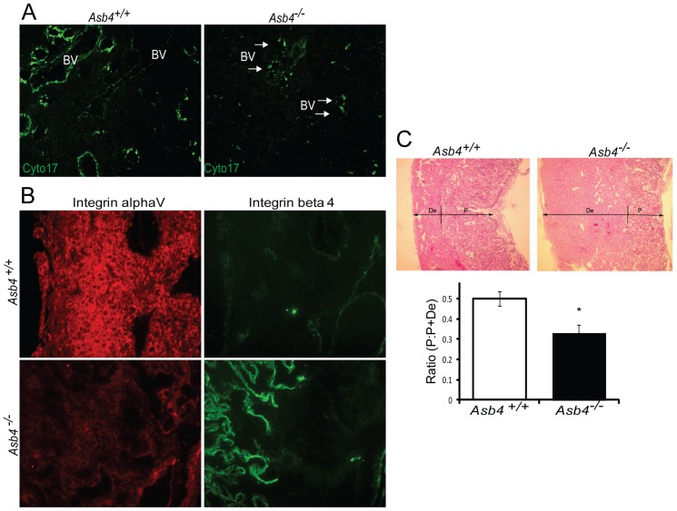
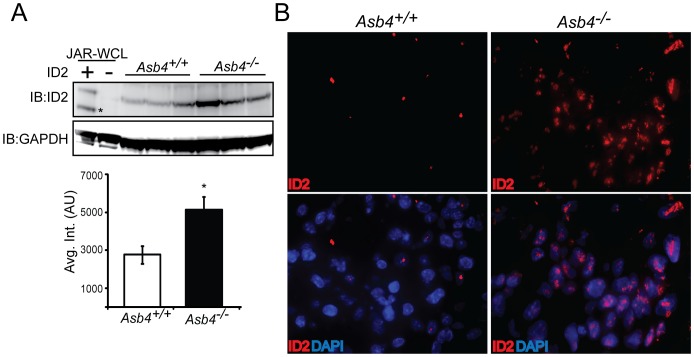
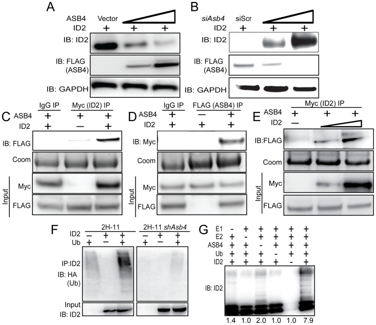
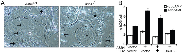
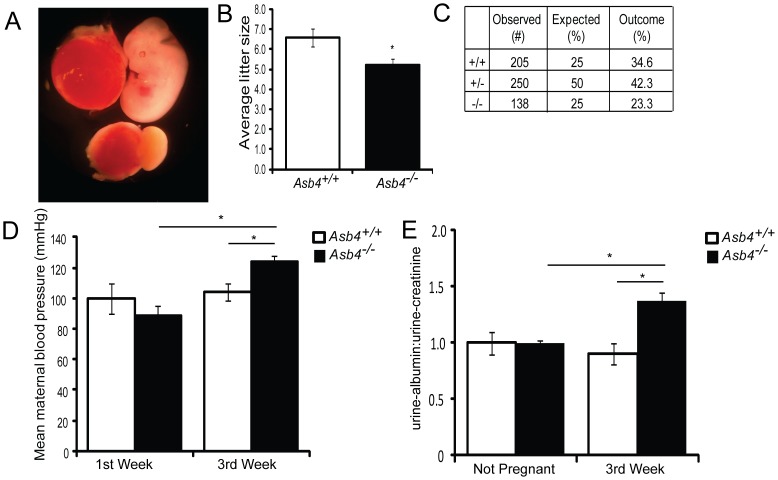
Vascularization of the placenta is a critical developmental process that ensures fetal viability. Although the vascular health of the placenta affects both maternal and fetal well being, relatively little is known about the early stages of placental vascular development. The ubiquitin ligase Ankyrin repeat, SOCS box-containing 4 (ASB4) promotes embryonic stem cell differentiation to vascular lineages and is highly expressed early in placental development. The transcriptional regulator Inhibitor of DNA binding 2 (ID2) negatively regulates vascular differentiation during development and is a target of many ubiquitin ligases. Due to their overlapping spatiotemporal expression pattern in the placenta and contrasting effects on vascular differentiation, we investigated whether ASB4 regulates ID2 through its ligase activity in the placenta and whether this activity mediates vascular differentiation. In mouse placentas, ASB4 expression is restricted to a subset of cells that express both stem cell and endothelial markers. Placentas that lack Asb4 display immature vascular patterning and retain expression of placental progenitor markers, including ID2 expression. Using JAR placental cells, we determined that ASB4 ubiquitinates and represses ID2 expression in a proteasome-dependent fashion. Expression of ASB4 in JAR cells and primary isolated trophoblast stem cells promotes the expression of differentiation markers. In functional endothelial co-culture assays, JAR cells ectopically expressing ASB4 increased endothelial cell turnover and stabilized endothelial tube formation, both of which are hallmarks of vascular differentiation within the placenta. Co-transfection of a degradation-resistant Id2 mutant with Asb4 inhibits both differentiation and functional responses. Lastly, deletion of Asb4 in mice induces a pathology that phenocopies human pre-eclampsia, including hypertension and proteinuria in late-stage pregnant females. These results indicate that ASB4 mediates vascular differentiation in the placenta via its degradation of ID2.


Conflict of interest statement

Competing Interests: The authors have declared that no competing interests exist.

Figures

Figure 1
Figure 1. ASB4 is expressed in the developing placental vasculature.
A) Asb4 mRNA is expressed only in the labyrinth zone of developing placentas. In situ hybridization was performed on E11.5 placental sections and imaged with bright field microscopy. Wide-field (4x, A) and higher magnification (20x, A’) anti-sense (AS)-probed sections illustrate Asb4 localized to the labyrinth zone. A sense probe was used as a negative control (A”). B) ASB4 is expressed in a subset of c-kit-positive and PECAM-positive cells but not mature cytokeratin 17-expressing cells. E11.5 placental sections were probed with markers of stem cells (c-kit, B), endothelial cells (PECAM, B’), and differentiated TB cells (cytokeratin 17, B”) and fluorescently imaged at 20x magnification. These images were then merged to show co-localization. ASB4 only co-localizes with cells expressing c-kit and PECAM (arrows) but not cytokeratin 17 (filled arrows). There are also subsets of c-kit or PECAM positive cells that ASB4 did not co-localize at this stage (filled arrows).
Figure 2
Figure 2. Asb4−/− placentas express markers of undifferentiated vasculature and TB cells.
A) Placentas lacking Asb4 have reduced cytokeratin 17 expression in near-term placentas. E17.5 placental sections from wild-type and Asb4−/− mice were labeled with cytokeratin 17 (cyto17), a marker of terminally differentiated endothelial-like TB cells. Blood vessels (BVs) in Asb4−/− placentas display reduced cytokeratin 17 labeling compared with BVs in wild-type placentas. B) Placentas from E15.5 wild-type and Asb4−/− placentas were labeled for integrin alpha V, a marker of mature, terminally differentiated TB cells and integrin beta 4, a marker of immature, undifferentiated TB cells. Wild-type placentas express alpha V but not beta 4 integins. Cells in Asb4−/− placentas retain integrin beta 4 expression and fail to express integrin alpha V. C) Placental disc invasion is reduced in Asb4−/− mothers at E17.5, indicating restricted trophoblast expansion. The ratio of the placental disc (P) to the total placenta (the sum of the decidua (De) and the placental disc) is decreased in Asb4−/− placentas compared to wild-type placentas, indicating a defect in TB cell invasion and migration.
Figure 3
Figure 3. ID2 expression increases in placentas that lack Asb4.
A) Lysates from three E13.5 wild-type and Asb4−/− placentas were immunoblotted against ID2 (top panel, asterisk denotes nonspecific band) and quantified (bottom panel). p<0.01. JAR-WCL =  whole cell lysates transfected with Id2 or vector and run as a positive immunoblotting control. B) E12.5 sections from wild-type and Asb4−/− placentas were labeled for ID2 (left panel), confirming that wild-type TB cells at this stage have low ID2 expression while ID2 expression is dramatically greater in Asb4−/− placentas. 100x magnification.
Figure 4
Figure 4. ASB4 negatively regulates ID2 expression through polyubiquitination and associates with ID2 in JAR cells.
A) ASB4 represses ID2 expression in a dose-dependent manner. Wild-type Id2 and vector, 0.5, or 2 µg of Asb4 were co-transfected in JAR cells. ID2 expression decreases as the ASB4 expression increases. B) ID2 expression increases as ASB4 expression decreases. 2H-11 cells that constitutively express high levels of ectopic ASB4 were transfected with Id2 and either a scrambled nucleotide siRNA duplex (siScr) or increasing doses (0.15 nM, 0.5 nM) of siAsb4 duplex. As ASB4 expression decreases, ID2 expression concurrently increases. C and D) ID2 and ASB4 associate in JAR cells. 3xFLAG-tagged Asb4 and 6xmyc-tagged Id2 were co-transfected in JAR cells. Lysates were pre-cleared with species-specific IgG and Protein A/G agarose beads were run with these reactions as a control against non-specific binding. Pre-cleared lysates were either immunoprecipitated with anti-myc- or anti-FLAG -conjugated agarose beads and blotted for FLAG or myc, respectively (C, D). Gels were stained with coomassie post-transfer as a loading control for immunoprecipitations. Input represents 2.5% of total lysate. ASB4 is detected in ID2 immunoprecipitation; conversely, ID2 is detected with ASB4 immunoprecipitation. E) 2H-11 cells that stably express FLAG-tagged Asb4 were transfected with increasing amounts of myc-tagged Id2. Cells were lysed and pre-cleared as in C and D, then immunoprecipitated with anti-myc conjugated agarose beads and then blotted for FLAG. FLAG expression increases in parallel with myc expression, indicating specific interaction between ID2 and ASB4. F) ID2 ubiquitination increases in cells with ASB4 expression. Wild-type Id2 and HA-tagged ubiquitin were transfected into either 2H-11 cells that express endogenous Asb4 or 2H-11 cells that have Asb4 constitutively knocked down. ID2 was immunoprecipitated using anti-ID2 and then blotted against HA. Reactions were blotted on the same membrane. Input represents 2.5% of total lysate. Ubiquitination of ID2 increases in endothelial cells that express ASB4 compared with cells that do not. G) ASB4 directly ubiquitinates ID2 in vitro. Recombinant ID2 was incubated with recombinant ASB4, and components of the reaction as indicated. Reactions were resolved on SDS-PAGE gels and immunoblotted against ID2. ID2 is ubiquitinated approximately four-fold more with ASB4 than without (lane 3). Quantification of ubiquitination is fold change relative to lane 5 (without ID2).
Figure 5
Figure 5. ASB4 promotes JAR cell-mediated endothelial apoptosis and stabilization of endothelial cell networks.
A) JAR cells expressing ASB4 promote 2H-11 cell apoptosis. JAR cells were transfected with vector, Asb4, Asb4 and wild-type Id2, or Asb4 and DR-Id2 prior to being seeded on top of 2H-11 monolayers. TUNEL-positive cells were counted and are presented as the percent of total endothelial cells within the field in panel B. Asb4-transfected cells increase apoptosis of the underlying endothelial cells, even when transfected with wild-type Id2. DR-Id2 co-transfected with Asb4 inhibits JAR-mediated 2H-11 apoptosis. * p<0.01 as compared to vector/vector. † p<0.01 compared to Asb4-only transfection. C) JAR cells transfected with Asb4 promote endothelial tube stability. 2H-11 cells were placed on Matrigel and allowed to form tube-like networks. JAR cells transfected as in A were then plated on the networks, and total network area was measured at the times indicated. JAR cells expressing DR-ID2 destabilize 2H-11 cell networks at 16 hours, while cells expressing ASB4 or ASB4 and wild-type ID2 maintained the size of these 2H-11 cell networks compared to vector transfected cells at 48 hours after plating (D).* p<0.05, *** p<0.01 as compared to vector/vector.
Figure 6
Figure 6. ASB4 promotes TB cell differentiation in vitro.
A) TB stem cells (TBSCs) were isolated from wild-type and Asb4−/− extraembryonic ectoderm at E7.5. Cells isolated from each conceptus were cultured in isolation, and these data represent 4 unique populations of cells for each genotype. Serum withdrawal induces the formation of large, multinucleated trophoblast giant cells (TGCs, arrows) that differentiate from TBSCs (asterisks). As shown, wild-type TBSCs largely differentiate into TGCs (left panel) while Asb4−/− cells remain in undifferentiated embryoid bodies (right panel). MEF-feeder cells are indicated by filled arrows. Dashed outlines indicate the border of non-MEF cell clusters. B) JAR cells were transfected to express vector, Asb4, or Asb4 co-transfected with vector, wild-type Id2, or degradation-resistant Id2 (DR-Id2). ASB4 induced hCG secretion, and co-expression of wild-type ID2 with ASB4 did not change hCG secretion compared to ASB4 expression alone. DR-ID2 prevented dcbAMP-induced hGC section, with concentrations of hCG no different than vector/vector transfected cells. * p<0.01 compared with vector/vector.
Figure 7
Figure 7. Pregnant Asb4−/− mice display symptoms of pre-eclampsia.
A) A subset of Asb4−/− embryos dies in utero. Asb4−/− littermates are shown at E12.5, illustrating the lack of placental vasculature and dramatically reduced fetal growth in a subset of Asb4−/− embryos. The resultant average litter size, taken from more than 25 litters from each group, is quantified in B. C) Heterozygous breeding results in a lower than expected number of Asb4−/− pups (p<0.01, Fisher’s exact test). Pregnant Asb4−/− mice have significantly elevated mean blood pressure (D) and urine-albumin:urine-creatinine (E) in the third trimester of pregnancy compared with both Asb4−/− mice in the first week of pregnancy and wild-type mice in the third week of pregnancy. * p<0.01.

Similar articles

Cited by

References

    1. Ferguson JE 3rd, Kelley RW, Patterson C (2005) Mechanisms of endothelial differentiation in embryonic vasculogenesis. Arterioscler Thromb Vasc Biol 25: 2246–2254. - PubMed
    1. Drake CJ, Fleming PA (2000) Vasculogenesis in the day 6.5 to 9.5 mouse embryo. Blood 95: 1671–1679. - PubMed
    1. Downs KM (1998) The murine allantois. Curr Top Dev Biol 39: 1–33. - PubMed
    1. Conway SJ, Kruzynska-Frejtag A, Kneer PL, Machnicki M, Koushik SV (2003) What cardiovascular defect does my prenatal mouse mutant have, and why? Genesis 35: 1–21. - PubMed
    1. Kam EP, Gardner L, Loke YW, King A (1999) The role of trophoblast in the physiological change in decidual spiral arteries. Hum Reprod 14: 2131–2138. - PubMed

Publication types

Substances

LinkOut - more resources