

Decreased plasma brain-derived neurotrophic factor and vascular endothelial growth factor concentrations during military training
- PMID: 24586790
- PMCID: PMC3933459
- DOI: 10.1371/journal.pone.0089455
Decreased plasma brain-derived neurotrophic factor and vascular endothelial growth factor concentrations during military training
Abstract
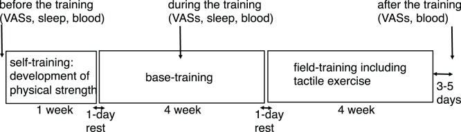
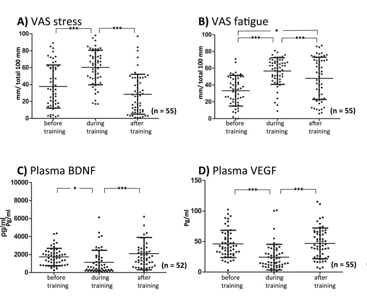
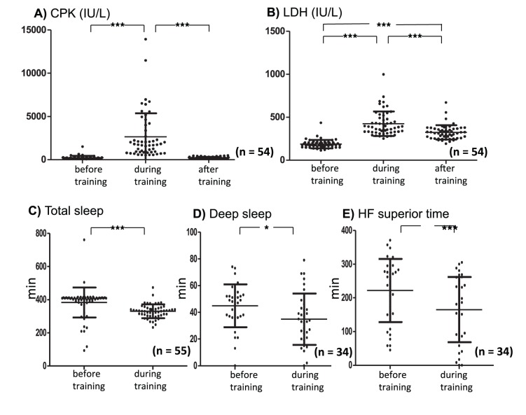
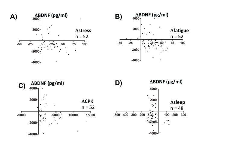
Decreased concentrations of plasma brain-derived neurotrophic factor (BDNF) and serum BDNF have been proposed to be a state marker of depression and a biological indicator of loaded psychosocial stress. Stress evaluations of participants in military mission are critically important and appropriate objective biological parameters that evaluate stress are needed. In military circumstances, there are several problems to adopt plasma BDNF concentration as a stress biomarker. First, in addition to psychosocial stress, military missions inevitably involve physical exercise that increases plasma BDNF concentrations. Second, most participants in the mission do not have adequate quality or quantity of sleep, and sleep deprivation has also been reported to increase plasma BDNF concentration. We evaluated plasma BDNF concentrations in 52 participants on a 9-week military mission. The present study revealed that plasma BDNF concentration significantly decreased despite elevated serum enzymes that escaped from muscle and decreased quantity and quality of sleep, as detected by a wearable watch-type sensor. In addition, we observed a significant decrease in plasma vascular endothelial growth factor (VEGF) during the mission. VEGF is also neurotrophic and its expression in the brain has been reported to be up-regulated by antidepressive treatments and down-regulated by stress. This is the first report of decreased plasma VEGF concentrations by stress. We conclude that decreased plasma concentrations of neurotrophins can be candidates for mental stress indicators in actual stressful environments that include physical exercise and limited sleep.
Conflict of interest statement
Figures

Similar articles
-
Rapid antidepressant changes with sleep deprivation in major depressive disorder are associated with changes in vascular endothelial growth factor (VEGF): a pilot study.Brain Res Bull. 2011 Aug 10;86(1-2):129-33. doi: 10.1016/j.brainresbull.2011.06.003. Epub 2011 Jun 16. Brain Res Bull. 2011. PMID: 21704134 Free PMC article. Clinical Trial.
-
BDNF in sleep, insomnia, and sleep deprivation.Ann Med. 2016;48(1-2):42-51. doi: 10.3109/07853890.2015.1131327. Epub 2016 Jan 13. Ann Med. 2016. PMID: 26758201 Review.
-
Neurotrophic factors in depression in response to treatment.J Affect Disord. 2015 Sep 1;183:287-94. doi: 10.1016/j.jad.2015.05.027. Epub 2015 May 21. J Affect Disord. 2015. PMID: 26047306
-
Improved Sleep Quality is Associated with Reductions in Depression and PTSD Arousal Symptoms and Increases in IGF-1 Concentrations.J Clin Sleep Med. 2015 Jun 15;11(6):615-23. doi: 10.5664/jcsm.4770. J Clin Sleep Med. 2015. PMID: 25766717 Free PMC article.
-
The Brain-Derived Neurotrophic Factor: Missing Link Between Sleep Deprivation, Insomnia, and Depression.Neurochem Res. 2020 Feb;45(2):221-231. doi: 10.1007/s11064-019-02914-1. Epub 2019 Nov 28. Neurochem Res. 2020. PMID: 31782101 Review.
Cited by
-
Depressive Mood Assessment Method Based on Emotion Level Derived from Voice: Comparison of Voice Features of Individuals with Major Depressive Disorders and Healthy Controls.Int J Environ Res Public Health. 2021 May 19;18(10):5435. doi: 10.3390/ijerph18105435. Int J Environ Res Public Health. 2021. PMID: 34069609 Free PMC article.
-
Sleep Deprivation-Induced Blood-Brain Barrier Breakdown and Brain Dysfunction are Exacerbated by Size-Related Exposure to Ag and Cu Nanoparticles. Neuroprotective Effects of a 5-HT3 Receptor Antagonist Ondansetron.Mol Neurobiol. 2015 Oct;52(2):867-81. doi: 10.1007/s12035-015-9236-9. Epub 2015 Jul 2. Mol Neurobiol. 2015. Retraction in: Mol Neurobiol. 2025 Jan;62(1):1318. doi: 10.1007/s12035-024-04678-4. PMID: 26133300 Retracted.
-
Low-Cost, Disposable Biosensor for Detection of the Brain-Derived Neurotrophic Factor Biomarker in Noninvasively Collected Saliva toward Diagnosis of Mental Disorders.ACS Polym Au. 2025 Aug 1;5(4):420-431. doi: 10.1021/acspolymersau.5c00038. eCollection 2025 Aug 13. ACS Polym Au. 2025. PMID: 40821874 Free PMC article.
-
Performance Evaluation of a Voice-Based Depression Assessment System Considering the Number and Type of Input Utterances.Sensors (Basel). 2021 Dec 23;22(1):67. doi: 10.3390/s22010067. Sensors (Basel). 2021. PMID: 35009610 Free PMC article.
-
Effectiveness of a Voice-Based Mental Health Evaluation System for Mobile Devices: Prospective Study.JMIR Form Res. 2020 Jul 20;4(7):e16455. doi: 10.2196/16455. JMIR Form Res. 2020. PMID: 32554367 Free PMC article.
References
-
- Lommatzsch M, Zingler D, Schuhbaeck K, Schloetcke K, Zingler C, et al. (2005) The impact of age, weight and gender on BDNF levels in human platelets and plasma. Neurobiol Aging 26: 115–123. - PubMed
-
- Marano CM, Phatak P, Vemulapalli UR, Sasan A, Nalbandyan MR, et al. (2007) Increased plasma concentration of brain-derived neurotrophic factor with electroconvulsive therapy: a pilot study in patients with major depression. J Clin Psychiatry 68: 512–517. - PubMed
-
- Pan W, Banks WA, Fasold MB, Bluth J, Kastin AJ (1998) Transport of brain-derived neurotrophic factor across the blood-brain barrier. Neuropharmacology 37: 1553–1561. - PubMed
Publication types
MeSH terms
Substances
LinkOut - more resources
Full Text Sources
Other Literature Sources
Medical
