. 2014 Feb 24;9(2):e89485.
doi: 10.1371/journal.pone.0089485. eCollection 2014.

Oncogenic H-Ras up-regulates acid β-hexosaminidase by a mechanism dependent on the autophagy regulator TFEB

Affiliations

Oncogenic H-Ras up-regulates acid β-hexosaminidase by a mechanism dependent on the autophagy regulator TFEB

Lorena Urbanelli et al. PLoS One. .

Abstract

The expression of constitutively active H-RasV12 oncogene has been described to induce proliferative arrest and premature senescence in many cell models. There are a number of studies indicating an association between senescence and lysosomal enzyme alterations, e.g. lysosomal β-galactosidase is the most widely used biomarker to detect senescence in cultured cells and we previously reported that H-RasV12 up-regulates lysosomal glycohydrolases enzymatic activity in human fibroblasts. Here we investigated the molecular mechanisms underlying lysosomal glycohydrolase β-hexosaminidase up-regulation in human fibroblasts expressing the constitutively active H-RasV12. We demonstrated that H-Ras activation increases β-hexosaminidase expression and secretion by a Raf/extracellular signal-regulated protein kinase dependent pathway, through a mechanism that relies on the activity of the transcription factor EB (TFEB). Because of the pivotal role of TFEB in the regulation of lysosomal system biogenesis and function, our results suggest that this could be a general mechanism to enhance lysosomal enzymes activity during oncogene-induced senescence.


Conflict of interest statement

Competing Interests: The authors have declared that no competing interests exist.

Figures

Figure 1
Figure 1. Analysis of HuDe fibroblasts transfected with H-RasV12, H-RasV12S35, H-RasV12G37, H-RasV12C40 mutants.
A, Immunoblotting analysis. Cell extracts were incubated with an anti-H-Ras antibody. As internal control an anti-β actin antibody was used. The increased expression of H-Ras with respect to empty vector transfected cells is shown. B, Growth curve. Cells were counted after the end of the selection with blasticidin-S (4 µg/ml) and cell number is reported. C, Cells morphology was examined by light microscopy, 200× total magnification. Arrows indicate flattened cells and cytosolic vacuolization associated with senescence.
Figure 2
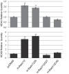
Figure 2. HEXA and HEXB gene expression analysis by qRT-PCR.
About 10-Ras mutants, with respect to those infected with the vector alone as control, is represented. The value is expressed as Relative Quantity (RQ). Analysis was repeated three times. The mean±s.d. of a representative experiment is reported. ** P<0.01 vs empty vector.
Figure 3
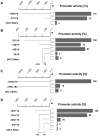
Figure 3. HEXA and HEXB gene promoter active segments.
Deletions of lysosomal HEXA and HEXB promoters were tested in luciferase reporter assay. A and B, Promoter active segments of the (−1594/+9 bp) and (−321/+9 bp) deletion constructs of HEXA promoter. A, Promoter activity is given in percent of the segment −1594/+9 (set 100). B, Promoter activity is given in percent of HEXA −321/+9 segment (set 100). C and D. Promoter active segments of the (−1570/+15 bp) and (−183/+15 bp) deletion constructs of HEXB promoter. C, Promoter activity is given in percent of the segment −1570/+15 (set 100). D, Promoter activity is given in percent of the segment −183/+15 (set 100). Given are mean values of at least three separate assays, each conducted in duplicates. S.d. of duplicates and separate assays was below 15%.
Figure 4
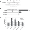
Figure 4. Identification of factors relevant for HEXA gene promoter activity.
A, Sequence of the segment with the highest promoter activity −100/−78. The E-box/CLEAR motif is indicated. B, Reporter activity of the wild type sequence (set 100) compared with the sequence mutated in the E-box. Measures are mean of three separate assays, each performed in duplicate. S.d. of duplicates and separate assays was below 15%. C, Wild type and mutated segment −100/−78 co-transfected in HuDe fibroblasts with an excess of H-Ras mutants and the empty vector as control. Vertical bars indicate reporter activity fold induction of wild type and mutated constructs versus empty vector (set 1). Measures are mean values±s.d. of three separate assays, each one in duplicate. ** P<0.01 vs wild type sequence.
Figure 5
Figure 5. Binding of TFEB to the HEXA gene promoter in vitro and in vivo.
A, Protein binding analysis by EMSA. Controls were run either without NE or with an additional 100-fold molar excess of unbiotinylated promoter segment as competitor DNA. B, Characterization of protein binding by super-shift analysis. HuDe NE was incubated with the segment −71/−104. The addition of NE, unspecific (anti-USF; 1 µg/assay) or specific antibody (anti-TFEB; 1 µg/assay) is indicated. C, ChIP assay using anti-TFEB or IgG control antibodies was performed on chromatin isolated from HuDe starved cells. An equivalent amount of chromatin was used as ‘input’ DNA. PCR products of the HEXA promoter region (left panel) and HEXA exon 11 control region (right panel) run on a 2% agarose gel are shown.
Figure 6
Figure 6. Analysis of HuDe fibroblasts over-expressing TFEB.
A, Immunoblotting of cells transfected with TFEB. Extracts from cells transfected with TFEB or empty vector as control were incubated with an anti-TFEB antibody. As internal control, an anti-βactin antibody was used. B, Reporter activity of HEXA promoter in the presence of TFEB. The wild type and E-box mutated segments −78/−100 were co-transfected with an excess of TFEB expressing plasmid. Vertical bars indicate reporter activity fold induction in the presence of TFEB, with respect to empty vector (set 1). Measures are the mean ± s.d. of three separate experiments, each one in duplicate. C, Hex A and Total Hex enzymatic activity in cell extracts and culture medium of HuDe fibroblasts expressing TFEB. Results are indicated as mU/mg of proteins (specific activity) for cell extracts and in mU/106 cells for cell culture medium. ** P<0.01.
Figure 7
Figure 7. Down-regulation of HEXA gene expression by TFEB knock down.
HuDe fibroblasts were transfected with shRNA for TFEB (shTFEB) or scrambled shRNA (shContr) as control. A, Analysis of TFEB transcript level by qRT-PCR. Reactions were performed using SYBR green, GADPH gene was used as endogenous control. The value is expressed as Relative Quantity (RQ). Each measure was repeated at least three times, each one in triplicate. The mean±s.d.of a representative experiment is reported. B, Analysis of TFEB expression by immunoblotting. Nuclear extracts were tested with an anti-TFEB antibody. As internal control, an anti-H3 histone antibody was used. C, Analysis of HEXA transcript level by qRT-PCR. Reactions were performed and elaborated as described in panel A. D, Reporter activity of HEXA promoter in TFEB knocked down cells. The segment −78/−100 (set 100) was co-transfected with an excess of shTFEB or shContr vector. Measures are the mean ± s.d. of three separate experiments, each one in duplicate. E, Hex A and Total Hex enzymatic activity in cell extracts and culture medium of HuDe transfected with shTFEB or shContr. Results are indicated as mU/mg of proteins (specific activity) for cell extracts and in mU/106 cells for cell culture medium. * P<0.05; ** P<0.01.
Figure 8
Figure 8. Immunoblotting analysis of TFEB expression in HuDe fibroblasts transfected with H-Ras mutants.
A, Cytoplasm extracts were tested with anti-H-Ras antibody. As internal control an anti-β actin antibody was used. Nuclear extracts were tested with an anti-TFEB antibody. As internal control, an anti-H3 histone antibody was used. B, Model of lysosomal HEXA gene regulation by TFEB through a Raf/ERK dependent pathway.

Similar articles

Cited by

References

    1. Kornfeld S, Mellman I (1989) The biogenesis of lysosomes. Annu Rev Cell Biol 5: 483–525. - PubMed
    1. Reddy A, Caler E, Andrews NW (2001) Plasma membrane repair is mediated by Ca(2+)-regulated exocytosis of lysosomes. Cell 106: 157–69. - PubMed
    1. Premzl A, Puizdar V, Zavasnik-Bergant V, Kopitar-Jerala N, Lah TT, et al. (2001) Invasion of ras-transformed breast epithelial cells depends on the proteolytic activity of cysteine and aspartic proteinases. Biol Chem 382: 853–7. - PubMed
    1. Urbanelli L, Emiliani C, Massini C, Persichetti E, Orlacchio A, et al. (2008) Cathepsin D expression is decreased in Alzheimer’s disease fibroblasts. Neurobiol Aging 29: 12–22. - PubMed
    1. Fehrenbacher N, Bastholm L, Kirkegaard-Sorensen T, Rafn B, Bottzauw T, et al. (2008) Sensitization to the lysosomal cell death pathway by oncogene-induced down-regulation of lysosome-associated membrane proteins 1 and 2. Cancer Res 68: 6623–33. - PubMed

Publication types

MeSH terms