. 2014 Feb 20;9(2):e89487.
doi: 10.1371/journal.pone.0089487. eCollection 2014.

Assembly of the novel five-component apicomplexan multi-aminoacyl-tRNA synthetase complex is driven by the hybrid scaffold protein Tg-p43

Affiliations

Assembly of the novel five-component apicomplexan multi-aminoacyl-tRNA synthetase complex is driven by the hybrid scaffold protein Tg-p43

Jason M van Rooyen et al. PLoS One. .

Abstract

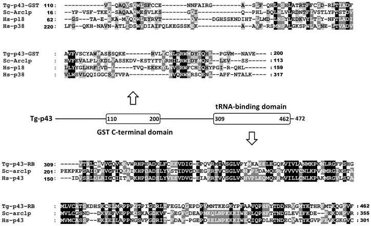
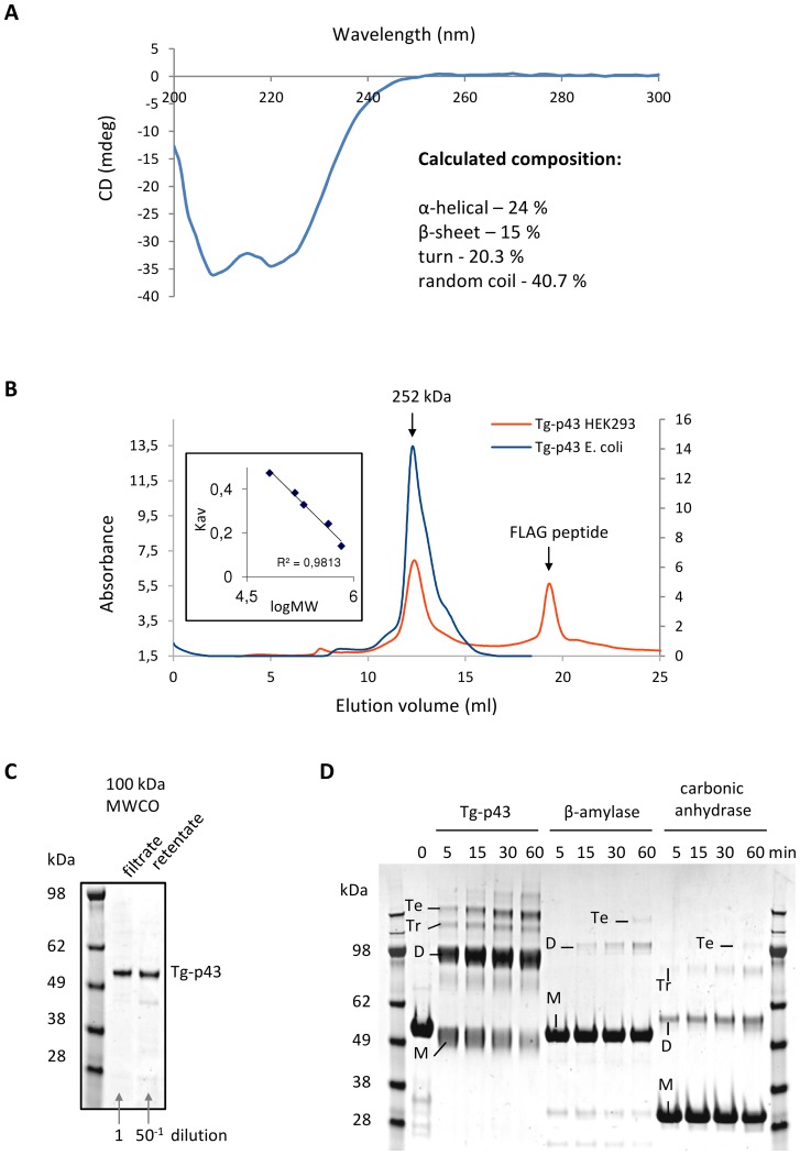
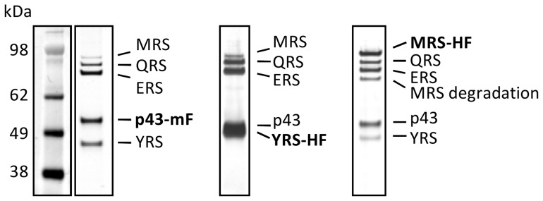
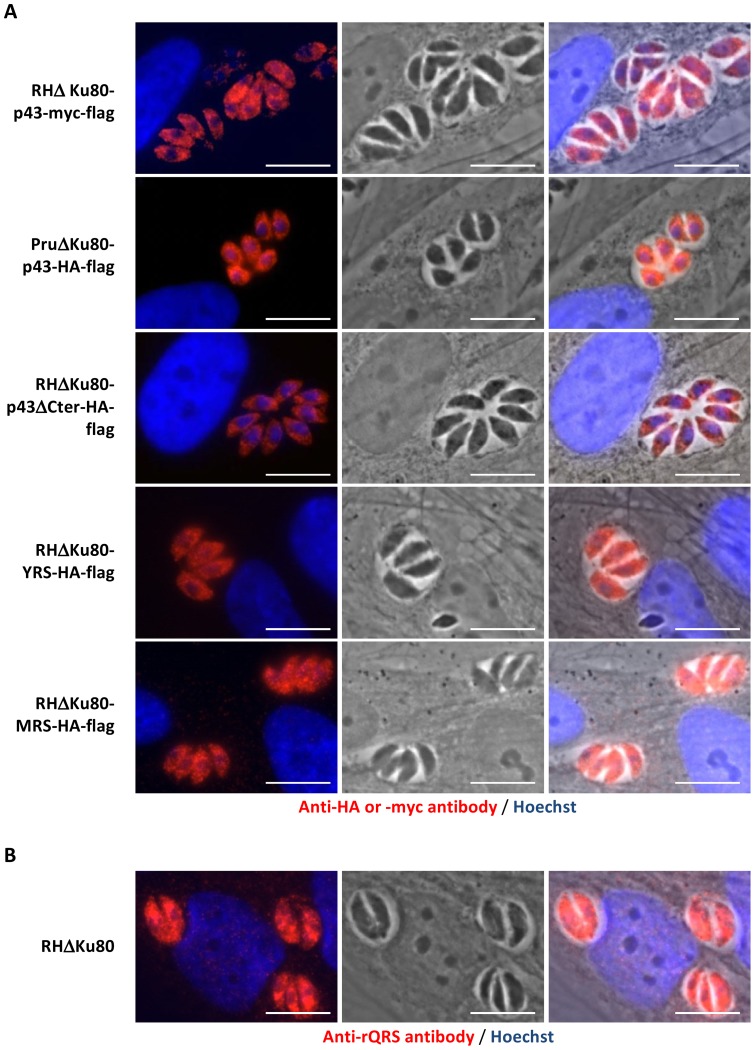
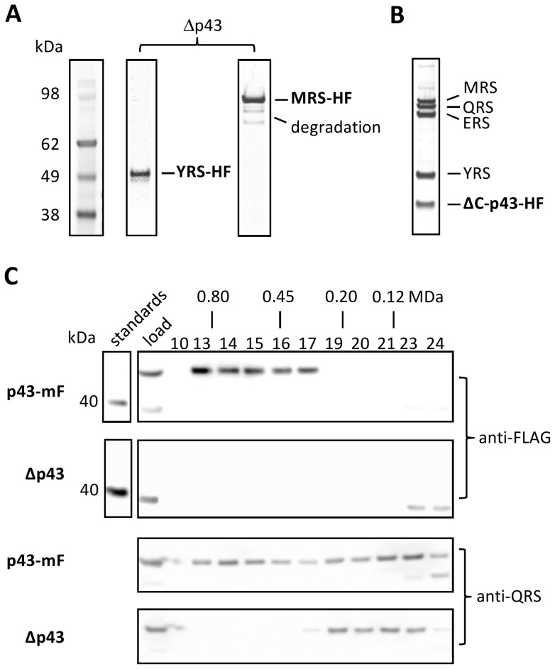
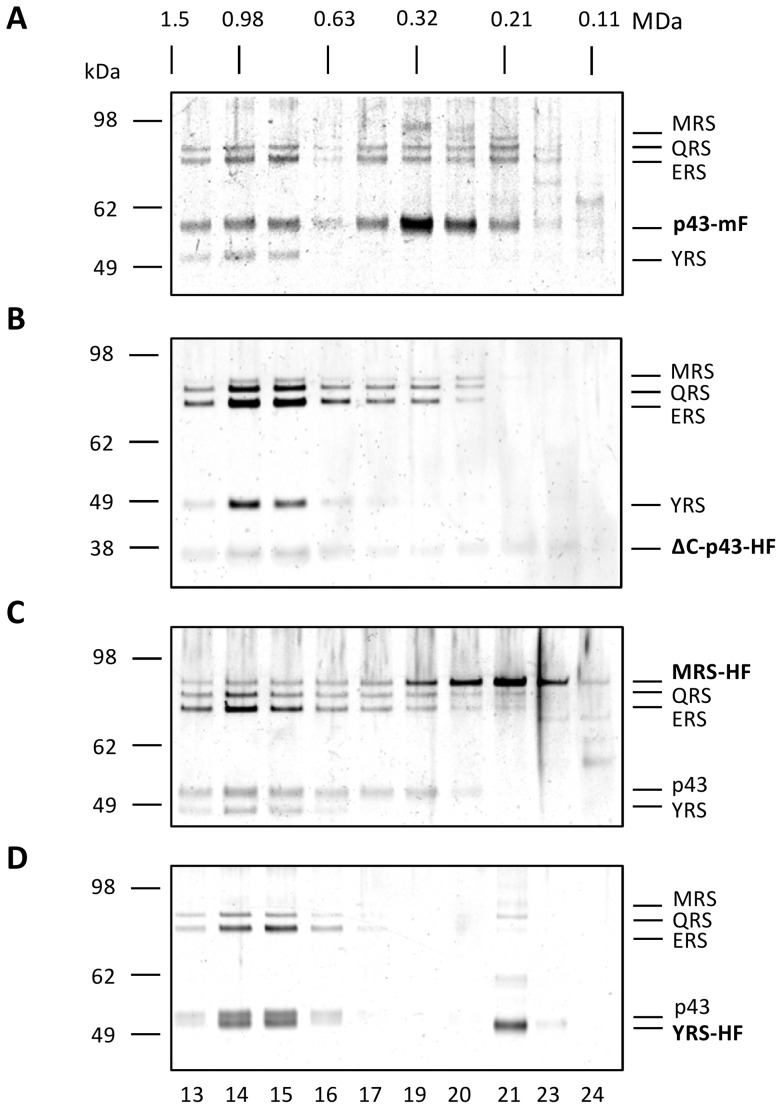
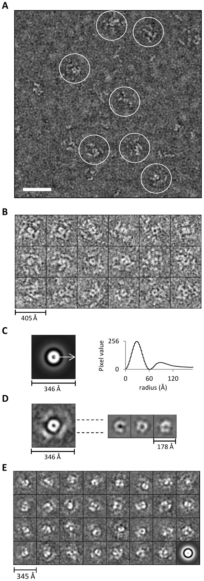
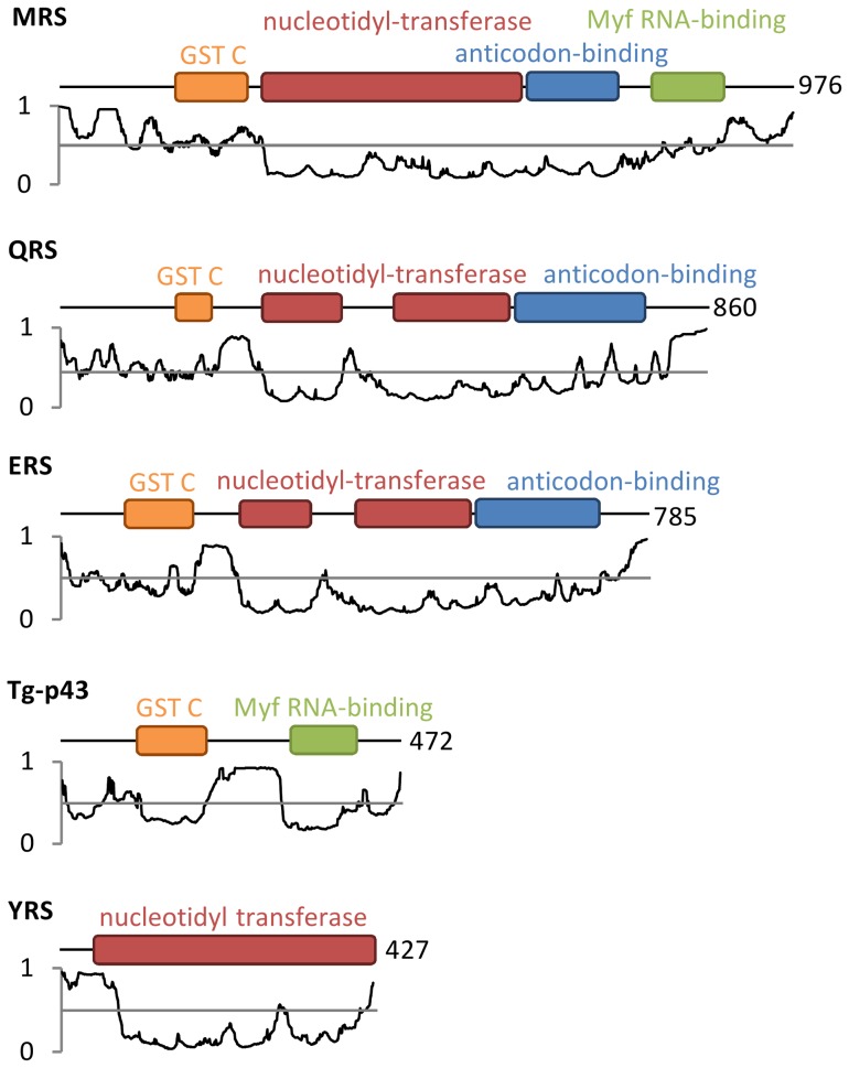
In Toxoplasma gondii, as in other eukaryotes, a subset of the amino-acyl-tRNA synthetases are arranged into an abundant cytoplasmic multi-aminoacyl-tRNA synthetase (MARS) complex. Through a series of genetic pull-down assays, we have identified the enzymes of this complex as: methionyl-, glutaminyl-, glutamyl-, and tyrosyl-tRNA synthetases, and we show that the N-terminal GST-like domain of a partially disordered hybrid scaffold protein, Tg-p43, is sufficient for assembly of the intact complex. Our gel filtration studies revealed significant heterogeneity in the size and composition of isolated MARS complexes. By targeting the tyrosyl-tRNA synthetases subunit, which was found exclusively in the complete 1 MDa complex, we were able to directly visualize MARS particles in the electron microscope. Image analyses of the negative stain data revealed the observed heterogeneity and instability of these complexes to be driven by the intrinsic flexibility of the domain arrangements within the MARS complex. These studies provide unique insights into the assembly of these ubiquitous but poorly understood eukaryotic complexes.


Conflict of interest statement

Competing Interests: The coauthor Mohamed Hakimi, is a PLOS ONE Editorial Board member. This does not alter the authors' adherence to all the PLOS ONE policies on sharing data and materials.

Figures

Figure 1
Figure 1. Relationship of Tg-p43 to similar amino-acyl tRNA synthetase-interacting multifunctional proteins from other species.
Multiple sequence alignments of the GST-C (GST) and RNA-binding (RB) domains of Tg-p43 and select homologs (Tg = Toxoplasma gondii, Sc = Saccharomyces cerevisiae, Hs = Homo sapiens) are shown above and below a schematic diagram of the Tg-p43 domain arrangement. The residue numbers of domain boundaries are listed in the schematic and at the beginning and end of the individual sequences.
Figure 2
Figure 2. Characterization of the folding and oligomeric state of recombinant Tg-p43.
(A) Circular dichroism spectrum of recombinant Tg-p43 expressed and purified from E. coli. The percentages of the calculated secondary structures are given in the inset. (B) SEC profile of pure recombinant E. coli (blue) and HEK293 (orange)-derived Tg-p43 protein. The MW of the eluted proteins, calculated by calibration with HMW standards (inset left), is shown above the trace. (C) Coomassie blue-stained SDS-PAGE gel image of E.coli-derived Tg-p43 retained and passed by a 100 kDa MWCO ultrafiltration device. (D) Coomassie blue-stained SDS-PAGE gel comparing the susceptibility to cross-linking (glutaraldehyde) of E.coli-derived Tg-p43 vs. beta-amylase (tetrameric oligomer) and carbonic anhydrase (monomeric) proteins. Based on size considerations, bands have been labeled as either monomers (M), dimers (D), trimers (Tr), or tetramers (Te).
Figure 3
Figure 3. Composition of the Toxoplasma MARS complex.
Silver-stained PAGE of MARS complex proteins (identified by MS-MS) isolated by FLAG immuno-precipitation of endogenously tagged subunits (indicated in boldface: mF = Myc-FLAG and HF = HA-FLAG epitope tags) from extracellular parasites.
Figure 4
Figure 4. Localization of the Tg-MARS complex.
(A) Fluorescent, light, and overlay of the two channels of endogenously tagged subunits of Tg-MARS visualised by in situ immunofluorescence in intracellular parasites with anti-Myc or anti-HA (red) antibodies and Hoechst DNA-specific dye (blue). (B) Fluorescent, light, and overlay of two channels of the QRS subunit of Tg-MARS visualised by in situ immunofluorescence in intracellular parasites with an anti-rQRS (red) antibody and Hoechst DNA-specific dye (blue). Scale bar  = 10 µm.
Figure 5
Figure 5. Dependence of MARS complex assembly on the scaffold protein Tg-p43.
(A) Silver-stained PAGE gel of immunoprecipitations of HA-FLAG-tagged (HF) YRS and MRS subunits in Tg-p43 knockout strains. (B) Isolation of the MARS complex by immunoprecipitation of a C-terminal-truncated (ΔC) form of the Tg-p43 protein (a.a. 1–296). (C) Western blots of eluted fractions from SEC analyses of crude cell lysates of Tg-p43-containing and knock-out strains (all RHΔku80). Corresponding fractions (numbered above the first gel image) are vertically aligned and their solution molecular weights, as determined by calibration with high molecular weight standards, are also given. Molecular weight standards are shown for the anti-FLAG Western only as a reference for evaluation of the knock-out blot.
Figure 6
Figure 6. Compositional and size heterogeneity of MARS complex populations.
Silver-stained PAGE gel of eluted fractions of immunoprecipitated MARS complexes separated by SEC. Each panel represents a separate SEC analysis of MARS complexes purified from strains harbouring C-terminal tags on different subunits of the complex: (A) Myc-FLAG-tagged Tg-p43 (mF = Myc-FLAG), (B) HA-FLAG-tagged C-terminal-truncated (ΔC) form of the Tg-p43 (residues 1–296) (HF = HA-FLAG), (C) HA-FLAG-tagged methionyl-tRNA synthetase, and (D) HA-FLAG-tagged tyrosyl-tRNA synthetase. Corresponding fractions (numbered below each gel image, and calibrated by the elution peak of the FLAG peptide) are vertically aligned and their solution molecular weights, as determined by calibration with high molecular weight standards, are given. Grey-levels of the images were adjusted to enhance the contrast but no bands were masked by this process.
Figure 7
Figure 7. Appearance of the MARS complex as revealed by electron microscopy.
(A) Electron micrograph of negatively-stained YRS-HA-FLAG MARS complexes. The scale bar represents 50 nm. (B) Subset of representative propeller views (18/137) of negatively-stained immunoprecipitated YRS-HA-FLAG MARS complexes windowed from images such as shown in (A). (C) Left – Rotational average of all views (1030 particle including the 137 propeller views) following pre-centring. Right – Corresponding radial profile from which the central core (120 Å) and peripheral domain's (240 Å) maximum average diameters can be determined. (D) An average image of aligned representative particles (137 propeller views from B) based on the entire image is shown on the left. On the right, three different class averages (homogenous sub-classes) generated by re-aligning only the central regions of the 137 views on the left are presented. (E) Reference-free class averages (homogenous sub-classes) derived from classification of all windowed particles (1030 in total) not just the propeller views shown in (B). The last image is the rotational average of all particles that went into classification.
Figure 8
Figure 8. Disorder potential and domain structures of Toxoplasma MARS complex subunit proteins.
Genesilico MetaDisorder2 disorder predictions for each peptide are displayed below domain arrangement schematics derived from NCBI conserved domain searches . Values above 0.5 are predicted to be disordered (coloured red) and values below 0.5 correspond to folded domains (coloured green). The number of residues in each protein is given after each schematic.

References

    1. Ibba M, Soll D (2000) Aminoacyl-tRNA synthesis. Annu Rev Biochem 69: 617–650 10.1146/annurev.biochem.69.1.617 - DOI - PubMed
    1. Szymański M, Deniziak M, Barciszewski J (2000) The new aspects of aminoacyl-tRNA synthetases. Acta Biochim Pol 47: 821–834. - PubMed
    1. Hausmann CD, Ibba M (2008) Aminoacyl-tRNA synthetase complexes: molecular multitasking revealed. FEMS Microbiol Rev 32: 705–721 10.1111/j.1574-6976.2008.00119.x - DOI - PMC - PubMed
    1. Guo M, Schimmel P, Yang X-L (2010) Functional expansion of human tRNA synthetases achieved by structural inventions. FEBS Lett 584: 434–442 10.1016/j.febslet.2009.11.064 - DOI - PMC - PubMed
    1. Lee SW, Cho BH, Park SG, Kim S (2004) Aminoacyl-tRNA synthetase complexes: beyond translation. J Cell Sci 117: 3725–3734 10.1242/jcs.01342 - DOI - PubMed

Publication types

MeSH terms