. 2014 Feb 19;9(2):e89492.
doi: 10.1371/journal.pone.0089492. eCollection 2014.

Macrophage/epithelium cross-talk regulates cell cycle progression and migration in pancreatic progenitors

Affiliations

Macrophage/epithelium cross-talk regulates cell cycle progression and migration in pancreatic progenitors

Kristin Mussar et al. PLoS One. .

Abstract

Macrophages populate the mesenchymal compartment of all organs during embryogenesis and have been shown to support tissue organogenesis and regeneration by regulating remodeling of the extracellular microenvironment. Whether this mesenchymal component can also dictate select developmental decisions in epithelia is unknown. Here, using the embryonic pancreatic epithelium as model system, we show that macrophages drive the epithelium to execute two developmentally important choices, i.e. the exit from cell cycle and the acquisition of a migratory phenotype. We demonstrate that these developmental decisions are effectively imparted by macrophages activated toward an M2 fetal-like functional state, and involve modulation of the adhesion receptor NCAM and an uncommon "paired-less" isoform of the transcription factor PAX6 in the epithelium. Over-expression of this PAX6 variant in pancreatic epithelia controls both cell motility and cell cycle progression in a gene-dosage dependent fashion. Importantly, induction of these phenotypes in embryonic pancreatic transplants by M2 macrophages in vivo is associated with an increased frequency of endocrine-committed cells emerging from ductal progenitor pools. These results identify M2 macrophages as key effectors capable of coordinating epithelial cell cycle withdrawal and cell migration, two events critical to pancreatic progenitors' delamination and progression toward their differentiated fates.


Conflict of interest statement

Competing Interests: The authors declared that no competing interests exist.

Figures

Figure 1
Figure 1. Increased frequency of PDX-1+ pancreatic progenitors emerging from the ducts in transplant microenvironments enriched in M2 macrophages.
Tissue sections of E14–15.5 pancreatic epithelial grafts transplanted in BM-reconstituted Id1+/−Id3−/− mice (A–C) or WT controls (D–F), stained by two-color immuno-fluorescence for PDX-1 (green) and E-cadherin (red). Numerous PDX-1+ cells are comprised within or emerge from ductal structures in the grafts of BM-reconstituted Id1+/−Id3−/− mice (arrowheads). (G–H) Morphometric analysis of E-Cadherin+ grafts areas (G) and PDX-1+ cells counted within the grafts (H). Bars represent mean ± SEM of n = 4 experiments including a total of 8 grafts in Id1+/−Id3−/− and 6 grafts in WT mice. Scale bar = 60 µm; * p≤0.001.
Figure 2
Figure 2. Frequency and proliferative potential of endocrine-committed progenitors in pancreatic transplants.
(A–B) Tissue sections of E14–15.5 pancreatic epithelial grafts transplanted in BM-reconstituted Id1+/−Id3−/− mice (A) or WT controls (B), stained by two-color immuno-fluorescence for Insulin (green) and the proliferation marker PCNA (red). Dotted lines delineate boundaries of ductal structures. (C–E) Morphometric analysis of Insulin+ areas (C), proliferating Insulin+PCNA+ cells (D) and Glucagon+ areas (E), assessed in the grafts. Bars represent mean ± SEM of n = 3 experiments, including a total of 7 grafts in Id1+/−Id3−/− and 5 grafts in WT mice. Scale bar = 120 µm; *p = 0.01 and ** p<0.002.
Figure 3
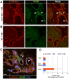
Figure 3. Modulation of NCAM expression in the ductal epithelium of embryonic pancreatic transplants.
(A–B) Tissue sections of E14–15.5 pancreatic grafts transplanted in BM-reconstituted Id1+/−Id3−/− mice (A) or WT (B) hosts, immuno-stained for NCAM (green) and E-cadherin (red). Numerous patches of NCAM+ epithelial cells are present in ductal structures of grafts from Id1+/−Id3−/− mice (arrowheads) but not in those from WT hosts. C) NCAM+E-cadherin+ ductal domains comprise PDX-1+ progenitors (blue, arrowheads). D) Morphometric analysis of NCAM+ areas measured in the grafts. Bars represent mean ± SEM of n = 3 experiments, including a total of 7 grafts in Id1+/−Id3−/− and 5 grafts in WT mice. Scale bars = 90 µm in A–B and 45 µm in C.
Figure 4
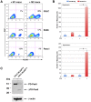
Figure 4. Differential induction of NCAM and PAX6 by M2 versus M1 macrophages in organ cultures, and in select pancreatic epithelial subpopulations.
A) E14–15.5 pancreatic epithelial cells, cultured for 3 days in medium alone (Ep) or in the presence of M1- (Ep+M1 macs) or M2- (Ep+M2 macs) macrophages, were immuno-stained for NCAM and the pan-leukocyte marker CD45. Flow cytometric contour plots of cells gated as CD45 negative (i.e. epithelial cells only) are shown, and percentages of NCAM+ cells are indicated in the right quadrants. Representative of n = 4 experiments. B) qPCR analysis of the indicated gene transcripts detected in macrophage/embryonic epithelium organ cultures. Values from each experimental condition were normalized to E-cadherin mRNA. Bars are mean ± SEM of n = 4 experiments; ** p<0.01. C) Flow cytometric hystogram of epithelial cells from E14–E15.5 pancreata stained for E-cadherin (red) or IgG control (blue). The sorting windows used for the isolation of E-cadherin High, Intermediate and Low populations are shown. (D–E) qPCR analysis of E-cadherin, SOX9 and PAX6-specific transcripts expressed in the indicated sorted populations. Representative of 2 experiments, each from a pool of 28–30 embryos with values presented as mean ± SEM of triplicate samples.
Figure 5
Figure 5. Expression of PAX6 transcripts and protein isoforms in the grafts and cultures of embryonic pancreatic epithelium.
(A) qPCR analysis of paired (PD) and paired-less (ΔPD) PAX6 transcripts detected in epithelial grafts from WT or Id1+/−Id3−/− hosts 2 weeks post-transplant, as compared to those detected in epithelial clusters isolated from E14.5 pancreata. Bars represent mean ± SEM of triplicate values normalized to E-cadherin mRNA. Representative of n = 2 experiments. (B) Western blotting analysis of cytoplasmic and nuclear cell lysates from organ cultures of E14.5–15.5 pancreatic epithelium with M1 (EP+M1) or M2 (EP+M2) macrophages. Membranes were probed with an anti-PAX6 antibody or with an anti-β-actin antibody, as loading control. Representative of n = 3 experiments. (C–D) Tissue sections of E14–15.5 pancreatic grafts transplanted in BM-reconstituted Id1+/−Id3−/− mice, immuno-stained for PAX6 (green) and either E-cadherin or NCAM (red). Scale bars = 25 µm in C and 15 µm in D. (C) PAX6 immunoreactivity in ductal structures is observed in individual E-cadherin+ cells, within NCAM+ ductal domains and with a prevalent cytoplasmic pattern (arrowheads). (D) In contrast, PAX6 immunoreactivity in islet-like cell clusters shows a predominant nuclear localization.
Figure 6
Figure 6. Induction of NCAM and ΔPD-PAX6 by M2 macrophages are overlapping phenotypes associated with enhanced migration.
(A) Flow cytometric analysis of G3LC, SU86 and PANC-1 cell lines co-cultured with either M1 (M1 macs) or M2 (M2 macs) macrophages, immuno-stained for NCAM and the pan-leukocyte marker CD45. The contour plots shows cells gated as CD45neg (i.e. epithelial cells only), and the percentage of NCAM+ cells are indicated in the right lower quadrant. (B) Migration of G3LC cells sorted from co-cultures with M2 macrophages as CD45negNCAMNeg and CD45negNCAMPos subsets, in response to 20% FCS. Bars represent the mean ± SD of duplicate samples in each experiment. *p<0.001. (C) Western blotting analysis of PAX6 isoforms expressed in cytoplasmic cell lysates from sorted NCAMNeg and NCAMPos G3LC cells derived from the M2 macrophages co-cultures shown in panel A.
Figure 7
Figure 7. Over-expression of ΔPD-PAX6 enhances migration of pancreatic epithelial cells but does not induce NCAM.
(A) Flow cytometric analysis of G3LC cells transduced with either empty (G3LC-Mock) or ΔPD-PAX6 expressing lenti-viruses at 30 MOI (G3LC-ΔPD-PAX6High) immunostained for NCAM. Over-expression of ΔPD-PAX6 is not sufficient to induce NCAM expression. (B–C) Migratory responses of Low (ΔPD-PAX6Low) and High (ΔPD-PAX6High) ΔPD-PAX6 expressors, generated by lentivirus transduction at 5 and 30 MOI, respectively, in response to chemotactic stimuli provided by FCS and HGF/SF as compared to medium alone. Bars represent mean ± SEM of n = 3 experiments. * p≤0.001.
Figure 8
Figure 8. Modulation of epithelial cell cycle progression by ΔPD-PAX6.
(A) Cell cycle analysis of mock-transduced, Low (ΔPD-PAX6Low) and High (ΔPD-PAX6High) ΔPD-PAX6 G3LC, and SU.86 cells, as measured by flow cytometric analysis of DNA content and BrdU incorporation. In each plot, percentages of cells in G0/G1, S and G2/M are indicated in regions R1, R2 and R3 respectively. Representative of n = 2 experiments. (B) Growth curves of G3LC and SU86 lines as a function of ΔPD-PAX6 expression levels. Representative of n = 3 experiments. (C) Western blotting analysis of PAX6, p27kip, p21cip, cyclin E, Retinoblastoma (Rb) protein and β-actin expressed in cell lysates obtained from Mock, Low (ΔPD-PAX6Low) and High ΔPD-PAX6 (ΔPD-PAX6High) G3LC. Representative of n = 2 experiments.
Figure 9
Figure 9. Hypothetical model of macrophages-induced developmental effects on embryonic pancreatic epithelium.
(A) Interactions of M2 macrophages with proliferating ductal epithelial cells induce expression of NCAM in delaminating progenitors. Concomitantly, high levels of ΔPD-PAX6 are induced. This latter change results in slowing-down the cell cycle, allowing for the initiation of differentiative events in progenitors committed to endocrine lineages. At the same time, NCAM induction, through disruption of E-cadherin and/or potentiation of FGF signaling, facilitates sorting of committed progenitors out of the ductal lining. (B) Motility of the committed progenitors increases as high levels of ΔPD-PAX6 progressively wear off, allowing their delamination and migration away from the ducts. Eventually, expression of ΔPD-PAX6 returns to very low levels in endocrine-committed cells, permitting migration arrest and resumption of proliferation in response to pro-proliferative cues provided by stromal cells, leading to the formation of islet cell clusters. We suggest that regulation of such ΔPD-PAX6-driven functions, reliant on cell-cell interactions with M2 macrophages and exquisitely sensitive to gene dosage, provides a novel mechanism explaining how cell cycle withdrawal and initiation of cell migration may be functional linked and coordinated in epithelial cells committed to leave ductal progenitor pools.

Similar articles

Cited by

References

    1. Stappenbeck TS, Miyoshi H (2009) The role of stromal stem cells in tissue regeneration and wound repair. Science 324: 1666–1669. - PubMed
    1. Morris L, Graham CF, Gordon S (1991) Macrophages in haemopoietic and other tissues of the developing mouse detected by the monoclonal antibody F4/80. Development 112: 517–526. - PubMed
    1. Pollard JW (2009) Trophic macrophages in development and disease. Nat Rev Immunol 9: 259–270. - PMC - PubMed
    1. Dai XM, Ryan GR, Hapel AJ, Dominguez MG, Russell RG, et al. (2002) Targeted disruption of the mouse colony-stimulating factor 1 receptor gene results in osteopetrosis, mononuclear phagocyte deficiency, increased primitive progenitor cell frequencies, and reproductive defects. Blood 99: 111–120. - PubMed
    1. Michaelson MD, Bieri PL, Mehler MF, Xu H, Arezzo JC, et al. (1996) CSF-1 deficiency in mice results in abnormal brain development. Development 122: 2661–2672. - PubMed

Publication types

MeSH terms