. 2014 Feb 21;9(2):e89534.
doi: 10.1371/journal.pone.0089534. eCollection 2014.

Exosomes: decreased sensitivity of lung cancer A549 cells to cisplatin

Affiliations

Exosomes: decreased sensitivity of lung cancer A549 cells to cisplatin

Xia Xiao et al. PLoS One. .

Abstract

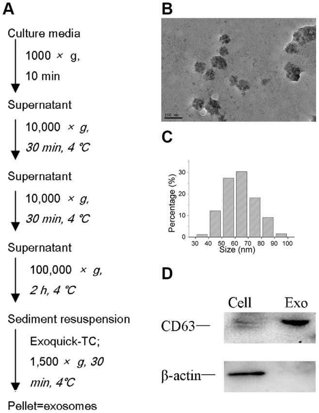
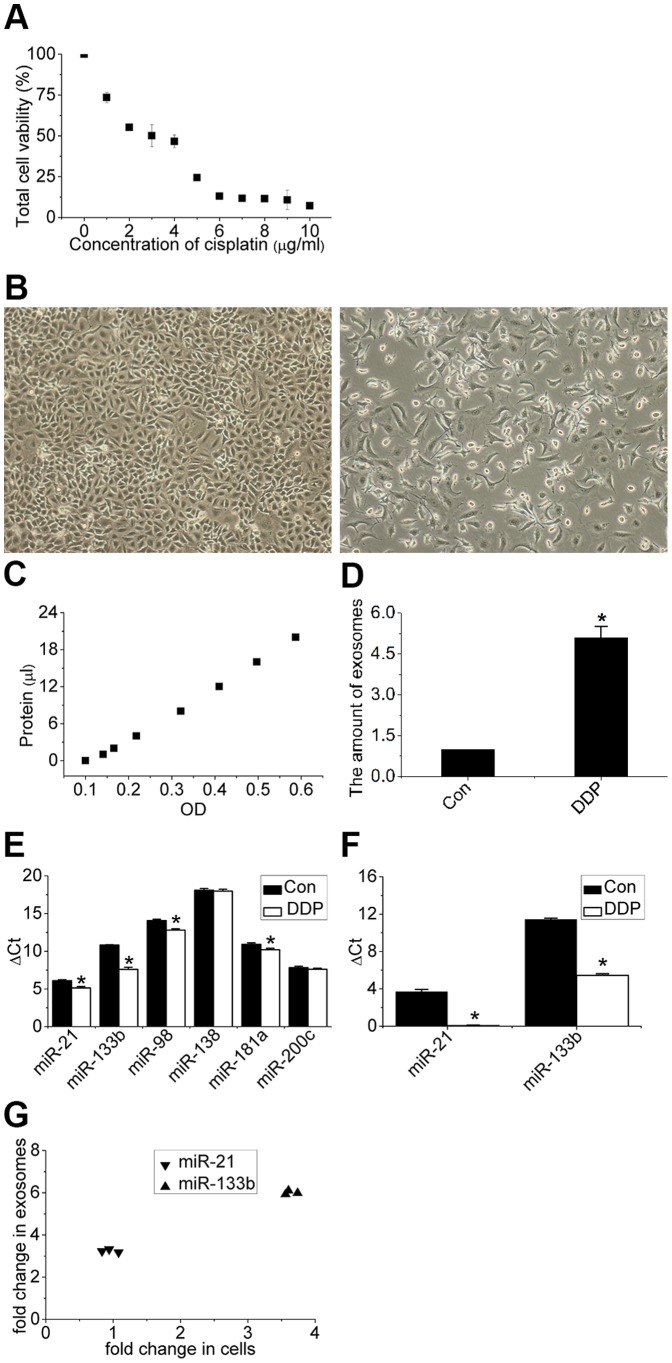
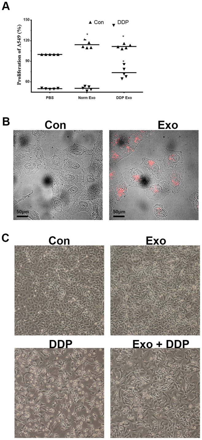
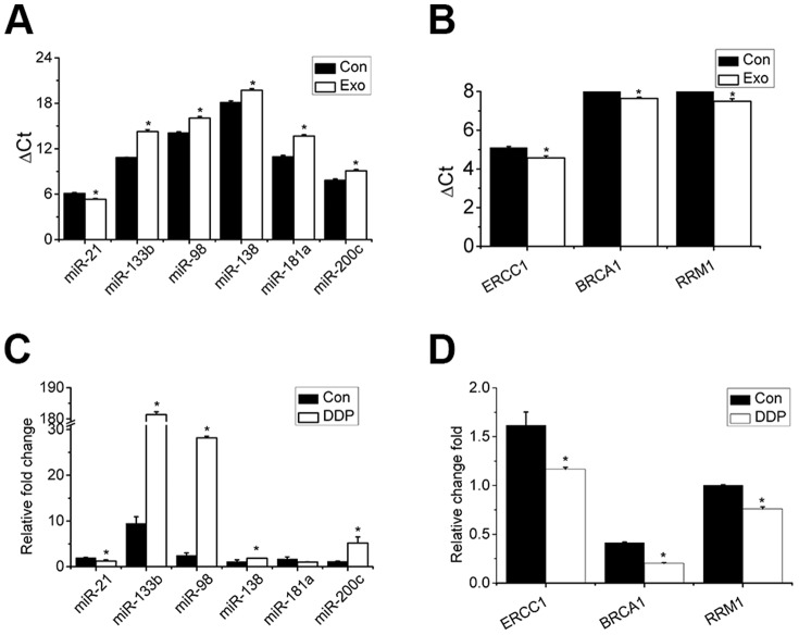
Exosomes are small extracellular membrane vesicles of endocytic origin released by many cells that could be found in most body fluids. The main functions of exosomes are cellular communication and cellular waste clean-up. This study was conducted to determine the involvement of exosomes in the regulation of sensitivity of the lung cancer cell line A549 to cisplatin (DDP). When DDP was added to A549 cells, exosomes secretion was strengthened. Addition of the secreted exosomes to other A549 cells increased the resistance of these A549 cells to DDP. Upon exposure of A549 to DDP, the expression levels of several miRNAs and mRNAs reportedly associated with DDP sensitivity changed significantly in exosomes; these changes may mediate the resistance of A549 cells to DDP. Exosomes released by A549 cells during DDP exposure decreased the sensitivity of other A549 cells to DDP, which may be mediated by miRNAs and mRNAs exchange by exosomes via cell-to-cell communication. Although the detailed mechanism of resistance remains unclear, we believed that inhibition of exosomes formation and release might present a novel strategy for lung cancer treatment in the future.


Conflict of interest statement

Competing Interests: The authors have declared that no competing interests exist.

Figures

Figure 1
Figure 1. Isolation and characterization of exosomes.
(A) Exosome isolation method used in this study. (B, C) Transmission electron microscopic image of the exosomes. Exosomes are small vesicles measuring from 30nm to 100 nm in diameter. (D) Western blot of CD63 and β-actin in exosomes and cells.
Figure 2
Figure 2. Response of A549 cells to cisplatin.
(A) Dose response relationship between viability of A549 cells (%) and concentration of cisplatin (1–10 µg/mL) for 48 h. (B) Microscopic image of A549 cells treated with 0 and 3 µg/mL cisplatin. Images were obtained at a magnification of 100×. (C, D) Quantity of exosomes determined via the BCA standard curve and enhanced secretion of exosomes normalized to cell numbers after exposure of A549 cells to cisplatin. (E) Differential expression levels of the six miRNAs in cells after exposure of A549 cells to cisplatin. (F) Differential expression levels of the two miRNAs in exosomes after exposure of A549 cells to cisplatin. (G) Fold-changes in expression of miR-21 and miR-133b in exosomes are higher than those in cells. Bars indicate mean ± SD of three replicates. *indicates p<0.05 versus control groups.
Figure 3
Figure 3. Influence of exosomes on sensitivity of cells to cisplatin.
(A) Pretreatment of A549 cells with exosomes released during cisplatin (3 µg/mL) exposure decreases the sensitivity of cells to cisplatin by about 20% compared with those without pretreatment for 48 h. (B) Uptake of exosomes (red) by A549 cells after incubation for 3 h was visualized by microscopy. Scale bar: 50 µm. (C) Microscopic image of A549 cells cultured under four conditions [control, exosomes, DDP (3 µg/mL), exosomes+DDP] for 48 h. Images were obtained at a magnification of 100×. *in (A) indicates p<0.05 versus control groups.
Figure 4
Figure 4. Influence of exosomes on expression of miRNAs and mRNAs detected in this study.
Expression levels of (A) miRNAs and (B) mRNAs under normal conditions with and without exosome pretreatment. Fold-changes in expression of (C) miRNAs and (D) mRNAs under cisplatin with and without exosome pretreatment. Bars indicate mean ± SD of three replicates. *indicates p<0.05 versus control groups.

References

    1. Hall MD, Okabe M, Shen DW, Liang XJ, Gottesman MM (2008) The role of cellular accumulation in determining sensitivity to platinum-based chemotherapy. Annu Rev Pharmacol Toxicol 48: 495–535. - PubMed
    1. Eastman A (1987) The formation, isolation and characterization of DNA adducts produced by anticancer platinum complexes. Pharmacol Ther 34: 155–166. - PubMed
    1. Halazonetis TD, Gorgoulis VG, Bartek J (2008) An oncogene-induced DNA damage model for cancer development. Science 319: 1352–1355. - PubMed
    1. Zhang G, Sun L, Lu X, Chen Z, Duerksen-Hughes PJ, et al. (2012) Cisplatin treatment leads to changes in nuclear protein and microRNA expression. Mutat Res 746: 66–77. - PubMed
    1. Gately DP, Howell SB (1993) Cellular accumulation of the anticancer agent cisplatin: a review. Br J Cancer 67: 1171–1176. - PMC - PubMed

Publication types