. 2014 Feb 20;9(2):e89553.
doi: 10.1371/journal.pone.0089553. eCollection 2014.

Mutation or loss of p53 differentially modifies TGFβ action in ovarian cancer

Affiliations

Mutation or loss of p53 differentially modifies TGFβ action in ovarian cancer

Eoghainín Ó hAinmhire et al. PLoS One. .

Abstract

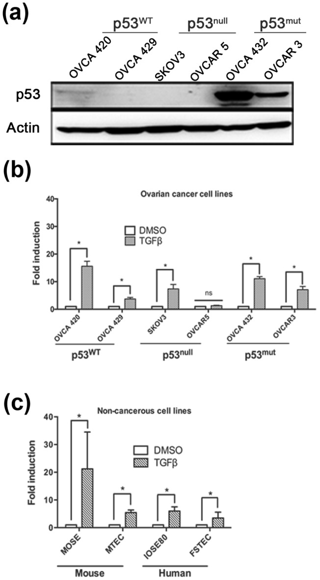
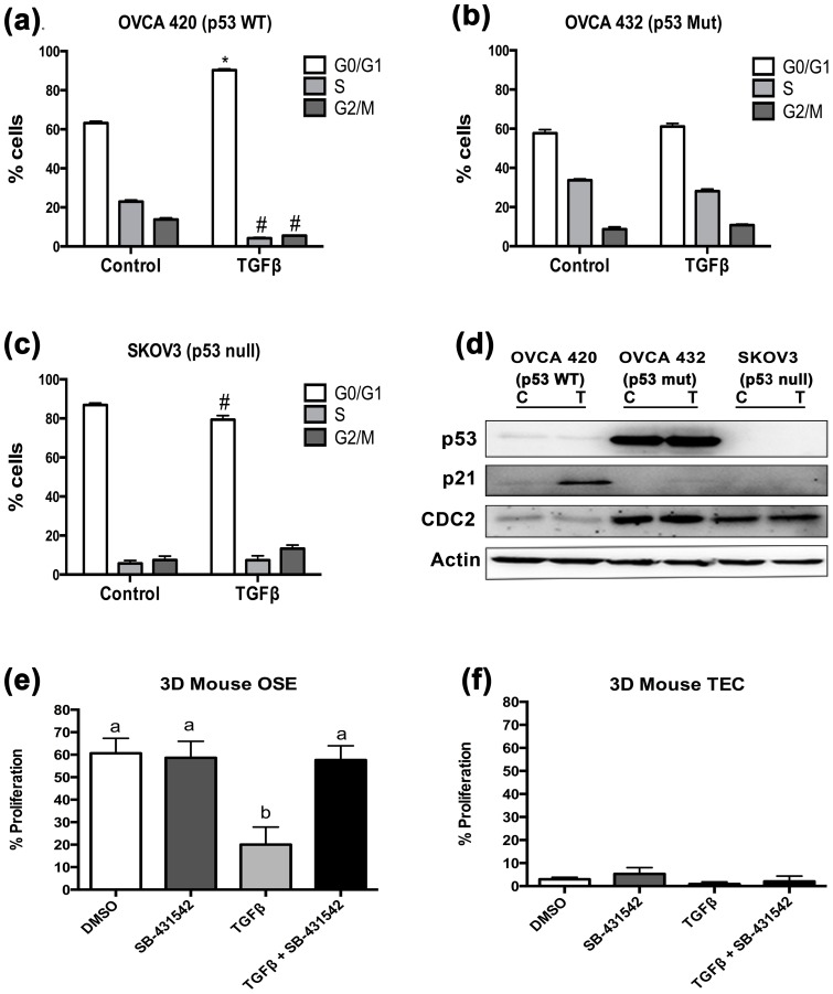
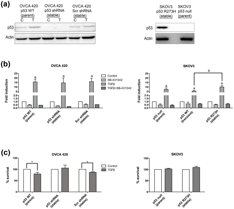
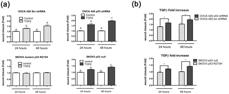
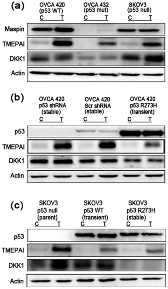
Ovarian cancer is the most lethal gynecological disease affecting women in the US. The Cancer Genome Atlas Network identified p53 mutations in 96% of high-grade serous ovarian carcinomas, demonstrating its critical role. Additionally, the Transforming Growth Factor Beta (TGFβ) pathway is dysfunctional in various malignancies, including ovarian cancer. This study investigated how expression of wild-type, mutant, or the absence of p53 alters ovarian cancer cell response to TGFβ signaling, as well as the response of the ovarian surface epithelium and the fallopian tube epithelium to TGFβ. Only ovarian cancer cells expressing wild-type p53 were growth inhibited by TGFβ, while ovarian cancer cells that were mutant or null p53 were not. TGFβ induced migration in p53 null SKOV3 cells, which was not observed in SKOV3 cells with stable expression of mutant p53 R273H. Knockdown of wild-type p53 in the OVCA 420 ovarian cancer cells enhanced cell migration in response to TGFβ. Increased protein expression of DKK1 and TMEPAI, two pro-invasive genes with enhanced expression in late stage metastatic ovarian cancer, was observed in p53 knockdown and null cells, while cells stably expressing mutant p53 demonstrated lower DKK1 and TMEPAI induction. Expression of mutant p53 or loss of p53 permit continued proliferation of ovarian cancer cell lines in the presence of TGFβ; however, cells expressing mutant p53 exhibit reduced migration and decreased protein levels of DKK1 and TMEPAI.


Conflict of interest statement

Competing Interests: The authors have declared that no competing interests exist.

Figures

Figure 1
Figure 1. Ovarian cancer cell lines respond to TGFβ regardless of p53 status.
(a) Western blot analysis of ovarian cancer cell lines demonstrating their p53 status. Actin used as a loading control. (b) Six ovarian cancer cell lines (OVCA 420, 429, SKOV3, OVCAR 5, OVCA 432, and OVCAR 3), along with four primary, non-cancerous cell lines (MOSE, MTEC, IOSE80, and FTSEC) were treated with or without TGFβ (10 ng/mL) using the SBE-luc plasmid. ANOVA was performed separately for fold induction (TGFβ) and fold repression (inhibitor and TGFβ + inhibitor) to analyze significance compared to untreated. Data represented as mean ± SEM, *p≤0.05.
Figure 2
Figure 2. p53 wild-type ovarian cancer cell lines undergo cell cycle arrest in response to TGFβ.
(a–c) OVCA 420, OVCA 432, and SKOV3 cell lines were treated with 20 ng/mL TGFβ for 24 hours and subjected to flow cytometry analysis. Distributions of cells in the three phases of the cell cycle are represented by mean percentages +/− SEM. Statistical significance represents a difference between number of cells in each cycle between treated and untreated and represented with * for an increase of treated cells compared to untreated; #represents decrease of treated cells compared to untreated; p≤0.05 (d) Western blot analysis of the three ovarian cancer cell lines probed for cell cycle proteins p21 and CDC2. Actin was used as a loading control. (e–f) Proliferation assay performed using BrdU incorporation in 3D organ culture of mouse ovaries and tubes. One-way ANOVA was performed. Data represented as mean ± SEM *p≤0.05.
Figure 3
Figure 3. Wild-type p53 cells, but not p53 null or mutant p53 cells, are growth inhibited by TGFβ.
(a) Western blot analysis of stable cell lines to knockdown of p53 by shRNA plasmid or expression of mutant p53 R273H. C = control, T = TGFβ treated. (b) SB-431542 (5 μM) was used to inhibit TGFβ signaling. For each panel, data represents mean ± SEM p≤0.05 increase over untreated for groups labeled with a, or between treated groups labeled with b. (c) Cell survival. Percentage of TGFβ-treated cell survival compared to untreated. Data represent mean ± SEM, *p≤0.05.
Figure 4
Figure 4. TGFβ induces migration in p53 null cells in comparison to p53 wild-type or mutant cells.
(a) Wound healing assays were performed on SKOV3 and OVCA 420 stable cell lines. Cell monolayers were scratched and treated with or without TGFβ at 20 ng/mL for 48 hours. Wound closure was measured as a fold increase or decrease compared to no treatment control. Paired t-test was used with a p≤0.05. (b) Comparison of the fold increase of TGFβ samples from 5(a). Unpaired t-test was used to analyze significance. Significance is represented by * and signifies a statistical difference between cell lines. Data represented as mean ± SEM, *p≤0.05.
Figure 5
Figure 5. TGFβ-induced expression of pro-metastatic proteins is upregulated in p53 null cells.
(a) OVCA 420 (p53 wild-type), OVCA 432 (p53 mutant), and SKOV3 (null p53) cells were treated with 10 ng/mL TGFβ for 24 hours and analyzed by western blotting. Membranes were probed with Maspin, TMEPAI and DKK1 primary antibodies. Actin was used as an internal loading control. (b) OVCA 420 cell lines were analyzed by western blot and probed for pro-metastatic factors TMEPAI, and DKK1. Actin was used as an internal loading control. (c) SKOV3 cell lines were analyzed by western blot and probed for pro-metastatic factors TMEPAI and DKK1. SKOV3 p53 WT was transiently transfected with 100 ng/mL of p53 wild-type plasmid. Actin was used as an internal loading control.

References

    1. Siegel R, Naishadham D, Jemal A (2013) Cancer statistics, 2013. CA Cancer J Clin 63: 11–30 10.3322/caac.21166 - DOI - PubMed
    1. Cho KR, Shih I-M (2009) Ovarian cancer. Annu Rev Pathol Mech Dis 4: 287–313 10.1146/annurev.pathol.4.110807.092246 - DOI - PMC - PubMed
    1. Whibley C, Pharoah PDP, Hollstein M (2009) P53 Polymorphisms: Cancer Implications. Nat Rev Cancer 9: 95–107 10.1038/nrc2584 - DOI - PubMed
    1. Cordenonsi M, Dupont S, Maretto S, Insinga A, Imbriano C, et al. (2003) Links between tumor suppressors: p53 is required for TGF-beta gene responses by cooperating with Smads. Cell 113: 301–314. - PubMed
    1. Bast RC, Hennessy B, Mills GB (2009) The biology of ovarian cancer: new opportunities for translation. Nat Rev Cancer 9: 415–428 10.1038/nrc2644 - DOI - PMC - PubMed

Publication types

MeSH terms

Substances