. 2014 Feb 21;9(2):e89599.
doi: 10.1371/journal.pone.0089599. eCollection 2014.

Serum and glucocorticoid-inducible kinase1 increases plasma membrane wt-CFTR in human airway epithelial cells by inhibiting its endocytic retrieval

Affiliations

Serum and glucocorticoid-inducible kinase1 increases plasma membrane wt-CFTR in human airway epithelial cells by inhibiting its endocytic retrieval

Jennifer M Bomberger et al. PLoS One. .

Abstract

Background: Chloride (Cl) secretion by the Cystic Fibrosis Transmembrane Conductance Regulator (CFTR) located in the apical membrane of respiratory epithelial cells plays a critical role in maintenance of the airway surface liquid and mucociliary clearance of pathogens. Previously, we and others have shown that the serum and glucocorticoid-inducible kinase-1 (SGK1) increases wild type CFTR (wt-CFTR) mediated Cl transport in Xenopus oocytes by increasing the amount of wt-CFTR protein in the plasma membrane. However, the effect of SGK1 on the membrane abundance of wt-CFTR in airway epithelial cells has not been examined, and the mechanism whereby SGK1 increases membrane wt-CFTR has also not been examined. Thus, the goal of this study was to elucidate the mechanism whereby SGK1 regulates the membrane abundance of wt-CFTR in human airway epithelial cells.

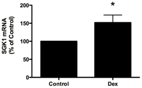
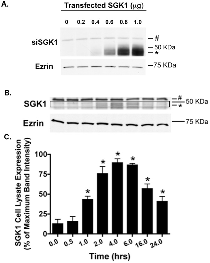
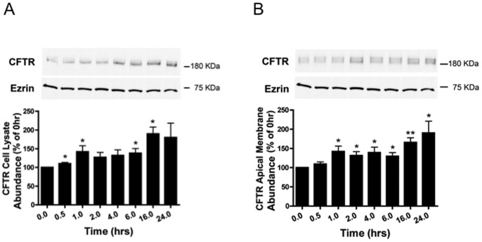
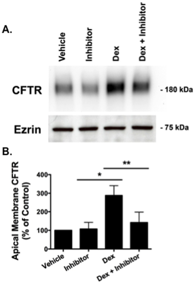
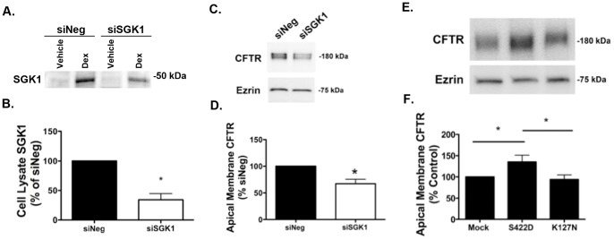
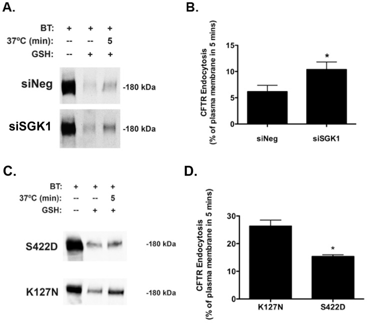
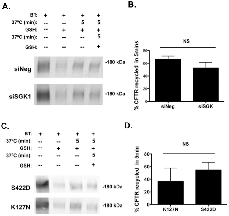
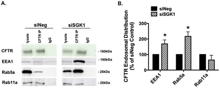
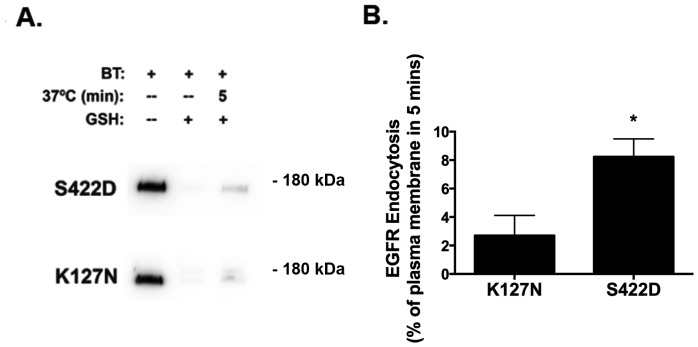
Methods and results: We report that elevated levels of SGK1, induced by dexamethasone, increase plasma membrane abundance of wt-CFTR. Reduction of SGK1 expression by siRNA (siSGK1) and inhibition of SGK1 activity by the SGK inhibitor GSK 650394 abrogated the ability of dexamethasone to increase plasma membrane wt-CFTR. Overexpression of a constitutively active SGK1 (SGK1-S422D) increased plasma membrane abundance of wt-CFTR. To understand the mechanism whereby SGK1 increased plasma membrane wt-CFTR, we examined the effects of siSGK1 and SGK1-S442D on the endocytic retrieval of wt-CFTR. While siSGK1 increased wt-CFTR endocytosis, SGK1-S442D inhibited CFTR endocytosis. Neither siSGK1 nor SGK1-S442D altered the recycling of endocytosed wt-CFTR back to the plasma membrane. By contrast, SGK1 increased the endocytosis of the epidermal growth factor receptor (EGFR).

Conclusion: This study demonstrates for the first time that SGK1 selectively increases wt-CFTR in the plasma membrane of human airway epithelia cells by inhibiting its endocytic retrieval from the membrane.


Conflict of interest statement

Competing Interests: The authors have declared that no competing interests exist.

Figures

Figure 1
Figure 1. Dexamethasone increased SGK1 mRNA expression in CFBE cells.
Dexamethasone (50 nM for 30 minutes) increased SGK1 mRNA versus control, as determined by Q-RT-PCR. Experiments performed three times. *p<0.05.
Figure 2
Figure 2. Dexamethasone increased SGK1 protein abundance in a time-dependent manner.
(A) Western blot demonstrating that the Sigma SGK1 antibody recognizes SGK1. CFBE cells were transfected with cDNA (0, no SGK1 insert), or SGK1 cDNA (0.2, 0.4, 0.6, 0.8 and 1.0 µg) and 48 hours later cells were lysed and SGK1 and ezrin were analyzed by Western blot. The SGK1 antibody recognized a protein (∼48 kDa, the predicted molecular mass of SGK1) that increases in intensity as the amount of SGK1 cDNA transfected into CFBE cells was increased. By contrast, the intensity of a non-specific protein (>50 kDa, marked by a # sign in Figure 2A), did not change as a function of the amount of SGK1 cDNA transfected in CFBE cells. (B) Representative western blot of an experiment in which CFBE cells were treated with dexamethasone (50 nM) for the indicated times at 37°C. Subsequently, cells were lysed and SGK1 and ezrin were analyzed by western blot. Data expressed as a percent of the maximum SGK1 levels observed at 4 hours (100%). Ezrin was monitored as a loading control (each lane was loaded with 50 µg of protein). # indicates a non-specific protein detected by the SGK1 antibody. The black box highlights SGK1 protein, which is also indicated by an *. (C) Summary of three experiments. Data expressed as SGK1 normalized for the amount of ezrin. *p<0.05 vs. 0 time point (no dexamethasone treatment: cells were treated with vehicle).
Figure 3
Figure 3. Dexamethasone increased wt-CFTR abundance in cell lysates (A), and in the apical membrane (B).
Representative western blots and data summaries are presented. (A) Cells were treated with dexamethasone (50 nM) for the indicated times at 37°C and western blot analysis was conducted to measure wt-CFTR and ezrin in cell lysates. The molecular mass of wt-CFTR is ∼180 kDa. Ezrin was monitored as a loading control. n = 7 per time point. *p<0.05 versus 0 time point. (B) Cells were treated with dexamethasone (50 nM) for the indicated time points at 37°C, and subsequently apical plasma membrane proteins were biotinylated using a technique previously described in detail by our laboratory , . Briefly, apical membrane proteins were biotinylated at 4°C using EZ-Link™ Sulfo-NHS-SS-Biotin. Subsequently, cells were lysed, and the biotinylated proteins were isolated by streptavidin-agarose beads, eluted into SDS sample buffer, and separated by 7.5% SDS-PAGE. The blots were probed for wt-CFTR (∼180 kDa) and for ezrin, a cytoplasmic protein that was not present in the biotinylated samples, confirming that biotin is impermeable to cell membranes , , and that the procedure to determine apical membrane wt-CFTR does not detect intracellular wt-CFTR. In this and all subsequent biotinylation experiments ezrin was never detected in the biotinylated samples by western blot, demonstrating that only plasma membrane proteins were biotinylated. *p<0.05 versus 0 time point. **p<0.01 versus 0 time point.
Figure 4
Figure 4. The SGK inhibitor GSK 650394 blocked the dexamethasone stimulated increase in apical membrane wt-CFTR.
(A) Representative western blot, and (B) Data quantitation. CFBE cells were treated with either vehicle, GSK 650394 (100 nM), an inhibitor of SGK1, dexamethasone (50 nM), or dexamethasone (50 nM), and GSK 650394 (100 nM) for 24 hours at 37°C. The molecular mass of apical plasma membrane wt-CFTR is ∼180 kDa. Experiments performed four times. *p<0.05 versus vehicle. **p<0.05 versus dexamethasone.
Figure 5
Figure 5. siSGK regulates apical plasma membrane wt-CFTR abundance.
Representative western blots and data summaries are presented. (A) and (B), siSGK1 reduced SGK1 protein abundance in cells treated with dexamethasone. CFBE cells were transfected with siNeg or siSGK1, and 24 hours later treated with dexamethasone (50 nM) for 4 hours, whereupon SGK1 levels in cell lysates were measured by western blot analysis. (C) and (D), siSGK1 reduced wt-CFTR protein abundance in the apical membrane of cells treated with dexamethasone. CFBE cells were transfected with siNeg or siSGK1, and 24 hours later treated with dexamethasone (50 nM) for 4 hours, whereupon wt-CFTR levels in the apical plasma membrane were measured by plasma membrane protein biotinylation followed by western blot analysis as described in Methods. For A-D experiments performed three times. *p<0.05 versus siNeg. E and F. Constitutively active SGK1 (SGK1-S422D) increased apical membrane wt-CFTR, whereas a dominant negative, kinase inactive SGK1 (SGK1-K127N) had no effect on apical membrane wt-CFTR compared to mock (vector control). CFBE cells were transfected with either SGK1-S422D, SGK1-K127N, or vector control (mock). Ezrin was a loading control. Plasma membrane wt-CFTR was determined by biotinylation of apical membrane proteins as described in Methods. Experiments performed six times. *p<0.05 as indicated.
Figure 6
Figure 6. SGK1 inhibits wt-CFTR endocytosis.
(A) and (B), siRNA-mediated knockdown of SGK1 in cells treated with dexamethasone (50 nM for 4 hours) enhanced the endocytic removal of wt-CFTR from the apical membrane. CFBE cells were transfected with siSGK1 or a scrambled negative control (siNeg) and 24 hours later treated with dexamethasone (50 nM) for 4 hours. (A) Representative western blot and (B) summary of data. Wt-CFTR endocytosis was measured by a method described previously in detail and in Methods , . In brief, in three sets of cells apical membrane proteins were biotinylated at 4°C using EZ-Link™ Sulfo-NHS-SS-Biotin. Subsequently, one set of cells (lane 1) were then lysed, and the biotinylated proteins were isolated by streptavidin-agarose beads, eluted into SDS sample buffer, separated by 7.5% SDS-PAGE, and probed for wt-CFTR and ezrin. Thus, lane 1 represents the amount of wt-CFTR in the apical membrane at time = 0. A second set of cells (lane 3) were also biotinylated at 4°C and then warmed to 37°C for 5 min to allow biotinylated wt-CFTR to be endocytosed. Subsequently, the cells were cooled to 4°C, and the disulfide bonds on Sulfo-NHS-SS-biotinylated proteins remaining in the apical membrane were reduced by GSH added to the apical solution for a total of 90 min at 4°C. The cells were then lysed, and the biotinylated proteins were isolated by streptavidin-agarose beads, eluted into SDS sample buffer, separated by 7.5% SDS-PAGE, and probed for wt-CFTR and ezrin. Thus, lane 3 represent the amount of wt-CFTR in the apical membrane at time = 0 that was endocytosed in 5 minutes. Lane 2 demonstrates that GSH reduced the disulfide bonds on Sulfo-NHS-SS-biotinylated proteins in the apical membrane. Cells in this lane were biotinylated at 4°C and the disulfide bonds on Sulfo-NHS-SS-biotinylated proteins remaining in the apical membrane were reduced by GSH added to the apical solution for a total of 90 min at 4°C. The amount of biotinylated wt-CFTR remaining in the plasma membrane after GSH treatment at 4°C and without the 37°C warming was considered background (<5% compared with the amount of biotinylated wt-CFTR at 4°C without GSH treatment, i.e., lane 1) and was subtracted from the wt-CFTR biotinylated after warming to 37°C at each time point. wt-CFTR endocytosis is reported as the amount of CFTR in lane 3 (minus background) divided by the amount in lane 1 (minus background) X 100. Quantification of three experiments. *p<0.05 versus siNeg (control). (C) and (D), Constitutively active SGK (SGK1-S422D) reduced the endocytic rate of wt-CFTR from the apical membrane compared to a dominant negative SGK1 (K127N). CFBE cells were transfected with SGK1-S422D or SGK1-K127N and wt-CFTR endocytosis was measured as described above. Representative western blot (C), and summary of the data (D). Quantification of three-six experiments. *p<0.05 versus K127N.
Figure 7
Figure 7. SGK1 did not alter the recycling of endocytosed wt-CFTR from endosomes back to the apical membrane.
siRNA-mediated knockdown of SGK1. (A) Representative western blot, and (B) summary of the data. The recycling assay has been described in detail previously and in Methods , . In brief, the first three lanes in A are similar to those described in Figure 6 for the endocytosis experiments. Thus, lane 1 represents the amount of wt-CFTR in the plasma membrane at time = 0, lane 3 represents the amount of wt-CFTR endocytosed in 5 minutes and lane 2 shows that GSH cleaves the disulfide bonds on Sulfo-NHS-SS-biotinylated wt-CFTR in the apical membrane. In lane 4 cells were warmed to 37°C for 5 min after biotinylation to load endocytic vesicles with biotinylated proteins. Cells were then cooled to 4°C, and the disulfide bonds on Sulfo-NHS-SS-Biotin-labeled proteins remaining in the plasma membrane were reduced by GSH at 4°C. Subsequently, cells were warmed again to 37°C for 5 min to allow endocytosed and biotinylated wt-CFTR to recycle to the plasma membrane. Cells were then cooled again to 4°C, and the disulfide bonds on biotinylated proteins in the apical membrane were reduced with GSH. The amount of recycled wt-CFTR was calculated as the difference between the amount of biotinylated wt-CFTR after the first and second GSH treatments (minus background)×100. Experiments repeated four-five times. NS, not significant. (C) and (D), The constitutively active SGK1 (SGK1-S422D) does not alter the recycling of wt-CFTR to the apical membrane. (C) Representative western blot, and (D) Summary of the data. The recycling assay was performed as described above. Experiments repeated four-five times. NS, not significant.
Figure 8
Figure 8. siSGK1 increased the amount of wt-CFTR in early endosomes, but not in recycling endosomes.
Co-immunoprecipitation studies were conducted to determine the subcellular location of wt-CFTR in siNeg and siSGK1 transfected cells treated with dexamethasone (50 nM for 4 hours). EEA and Rab5a are markers of early endosomes, and Rab11a is a marker of recycling endosomes. wt-CFTR was immunoprecipitated, and co-immunoprecipitated proteins were eluted into SDS sample buffer, and separated by 7.5% SDS-PAGE. The blots were then probed for wt-CFTR, EEA, Rab5a, and Rab11. Co-immunoprecipitation of wt-CFTR with EEA1 and Rab5a identifies the amount of wt-CFTR in early endosomes, and co-immunoprecipitation of wt-CFTR with Rab11a identifies the amount of wt-CFTR in recycling endosomes. Quantification of data for Rab and EEA1 immunoprecipitation with wt-CFTR in siNeg and siSGK1 cells is normalized for the total amount of wt-CFTR immunoprecipitated. Blots in siNeg and siSGK1 experiments were cut for presentation, but were run one the same blot to allow for comparison. Lysate, the amount of EEA1, Rab5a Rab11 and wt-CFTR in cell lysates. CFTR IP, indicates IP with the anti-CFTR antibody and then western blot with the indicated antibody. IgG, immunoprecipitation with a non-specific antibody. (A) Representative western blots and (B) Summary of the data. Experiments repeated three times. *P<0.05 versus siNeg.
Figure 9
Figure 9. SGK1 stimulates EGFR endocytosis.
(A) and (B), Constitutively active SGK1 (S422D) increased the endocytic rate of EGFR from the apical membrane compared to a dominant negative SGK1 (K127N). CFBE cells were transfected with SGK1-S422D or SGK1-K127N and EGFR endocytosis was measured as described in detail in Methods and in Figure legends above. Representative western blot (A), and summary of the data (B). Quantification of three experiments. *P<0.05 versus K127N.

Similar articles

Cited by

References

    1. Guggino WB, Stanton BA (2006) New insights into cystic fibrosis: molecular switches that regulate CFTR. Nature reviews Molecular cell biology 7: 426–436. - PubMed
    1. Cohen TS, Prince A (2012) Cystic fibrosis: a mucosal immunodeficiency syndrome. Nature medicine 18: 509–519. - PMC - PubMed
    1. Clunes LA, Davies CM, Coakley RD, Aleksandrov AA, Henderson AG, et al. (2012) Cigarette smoke exposure induces CFTR internalization and insolubility, leading to airway surface liquid dehydration. FASEB journal: official publication of the Federation of American Societies for Experimental Biology 26: 533–545. - PMC - PubMed
    1. Bomberger JM, Barnaby RL, Stanton BA (2009) The Deubiquitinating Enzyme USP10 Regulates the Post-endocytic Sorting of Cystic Fibrosis Transmembrane Conductance Regulator in Airway Epithelial Cells. The Journal of biological chemistry 284: 18778–18789. - PMC - PubMed
    1. Okiyoneda T, Barriere H, Bagdany M, Rabeh WM, Du K, et al. (2010) Peripheral protein quality control removes unfolded CFTR from the plasma membrane. Science 329: 805–810. - PMC - PubMed

Publication types

MeSH terms

Substances