

Methods to quantify soft tissue-based cranial growth and treatment outcomes in children: a systematic review
- PMID: 24586904
- PMCID: PMC3937373
- DOI: 10.1371/journal.pone.0089602
Methods to quantify soft tissue-based cranial growth and treatment outcomes in children: a systematic review
Abstract
Context: Longitudinal assessment of cranial dimensions of growing children provides healthcare professionals with information about normal and deviating growth as well as treatment outcome.
Objective: To give an overview of soft tissue-based methods for quantitative longitudinal assessment of cranial dimensions in children until age 6 years and to assess the reliability of these methods in studies with good methodological quality.
Data source: PubMed, EMBASE, Cochrane Library, Web of Science, Scopus, and CINAHL were searched. A manual search was performed to check for additional relevant studies.
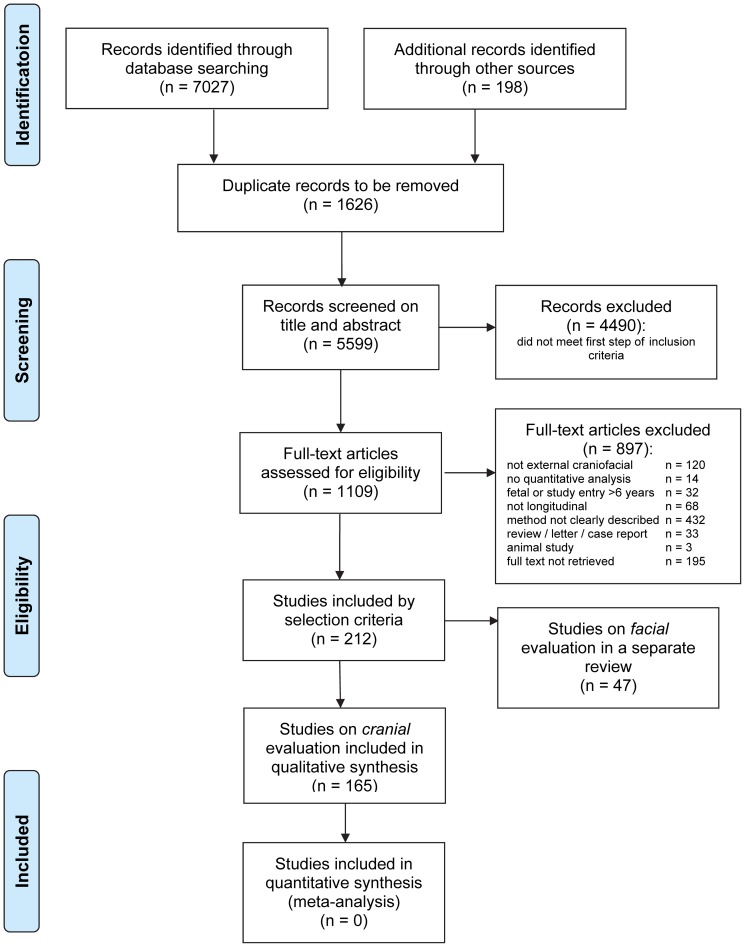
Study selection: Primary publications on facial growth and treatment outcomes in children younger than age 6 years were included.
Data extraction: Independent data extraction was performed by two observers. A quality assessment instrument was used to determine methodological quality. Methods used in studies with good methodological quality were assessed for reliability expressed as the magnitude of the measurement error and the correlation coefficient between repeated measurements.
Results: In total, 165 studies were included, forming three groups of methods: head circumference anthropometry, direct anthropometry, and 2D photography and 3D imaging techniques (surface laser scanning and stereophotogrammetry). In general, the measurement error was below 2 mm, and correlation coefficients were very good.
Conclusion: Various methods for measuring cranial dimensions have shown to be reliable. Stereophotogrammetry is the most versatile method for quantitative longitudinal assessment of cranial dimensions and shapes in children. However, direct anthropometry continues to be the best method for routine clinical assessments of linear cranial dimensions in growing children until age 6 years.
Conflict of interest statement
Figures
References
-
- McGarry A, Dixon MT, Greig RJ, Hamilton DR, Sexton S, et al. (2008) Head shape measurement standards and cranial orthoses in the treatment of infants with deformational plagiocephaly. Dev Med Child Neurol 50: 568–576. - PubMed
-
- Chan FC, Kawamoto HK, Federico C, Bradley JP (2013) Soft-tissue volumetric changes following monobloc distraction procedure: analysis using digital three-dimensional photogrammetry system (3dMD). J Craniofac Surg 24: 416–420. - PubMed
-
- Morhardt DR, Barrow W, Jaworski M, Accardo PJ (2013) Head Circumference in Young Children With Autism: The Impact of Different Head Circumference Charts. J Child Neurol. - PubMed
-
- Boros CA, Spence D, Blaser S, Silverman ED (2007) Hydrocephalus and macrocephaly: new manifestations of neonatal lupus erythematosus. Arthritis Rheum 57: 261–266. - PubMed
-
- Farkas LG (1994) Anthropometry of the head and face; kj, editor. New York: Raven Press. 405 p.
Publication types
MeSH terms
LinkOut - more resources
Full Text Sources
Other Literature Sources
