. 2014 Feb 21;9(2):e89630.
doi: 10.1371/journal.pone.0089630. eCollection 2014.

Aeromonas hydrophila flagella glycosylation: involvement of a lipid carrier

Affiliations

Aeromonas hydrophila flagella glycosylation: involvement of a lipid carrier

Susana Merino et al. PLoS One. .

Retraction in

Abstract

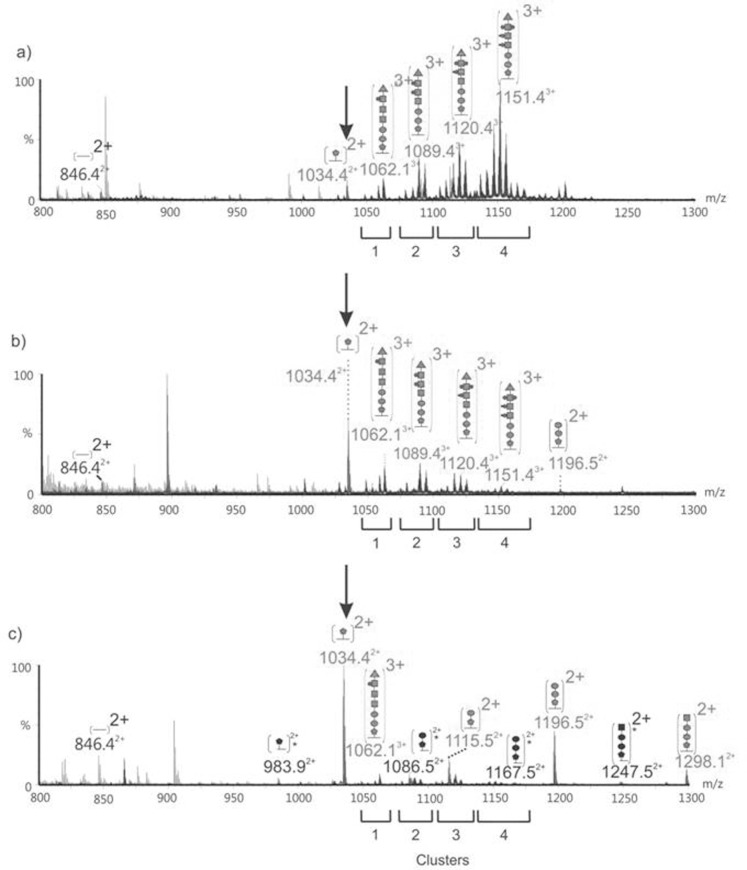
Polar flagellin proteins from Aeromonas hydrophila strain AH-3 (serotype O34) were found to be O-glycosylated with a heterogeneous glycan. Mutants unable to produce WecP or Gne enzymes showed altered motility, and the study of their polar flagellin glycosylation showed that the patterns of glycosylation differed from that observed with wild type polar flagellin. This suggested the involvement of a lipid carrier in glycosylation. A gene coding for an enzyme linking sugar to a lipid carrier was identified in strain AH-3 (WecX) and subsequent mutation abolished completely motility, flagella production by EM, and flagellin glycosylation. This is the first report of a lipid carrier involved in flagella O-glycosylation. A molecular model has been proposed. The results obtained suggested that the N-acetylhexosamines are N-acetylgalactosamines and that the heptasaccharide is completely independent of the O34-antigen lipopolysaccharide. Furthermore, by comparing the mutants with differing degrees of polar flagellin glycosylation, we established their importance in A. hydrophila flagella formation and motility.


Conflict of interest statement

Competing Interests: The authors have declared that no competing interests exist.

Figures

Figure 1
Figure 1. Motility phkenotypes exhibited in swim (0.25%) agar by A. hydrophila AH-3 (A), AH-2767 gne mutant (B), AH-3ΔWecP mutant (C), AH-2767+ pACYC-Gne (D), and AH-3ΔWecP pBAD-WecP (E).
Figure 2
Figure 2. Purified polar flagellins from several A. hydrophila strains obtained according to Materials and Methods.
A/SDS-PAGE and B/Western blot using specific antiserum against purified polar flagellins. St, size standard (14, 20, 30, 45, 60, and 94 kDa). 1, A. hydrophila AH-3 (wild type); 2, A. hydrophila AH-3ΔWecP mutant and 3, A. hydrophila AH-3ΔWecP complemented with plasmid pBAD33-WecPAh (18). Figure 2A lane 2 shows a clear reduction in the AH-3ΔWecP mutant polar flagellin MW. In Figure 2B the AH-3ΔWecP mutant polar flagellin shows several minor bands with reduced MW (lane 2) not observed neither in lane 1 (wild type) or lane 3 (complemented mutant).
Figure 3
Figure 3. nLC-MS alignment of the AH-3 polar flagellin FlaA/FlaB glycopeptide.
The 1691 Da FlaA/FlaB peptide TLAQQSANGSNNTDDR (eluting between 13.1 and 13.8 minutes) was modified by a heptasaccharide glycan with HexNAc residues additionally variably modified by 0–2 phosphate and 0–2 methyl groups . These additional variable modifications resulted in four major glycopeptide ion cluster regions, as indicated below each spectrum. The cluster number corresponds to the number of additional phosphate groups modifying the heptasaccharide, and peaks within that cluster possess a variable number of additional methyl groups. Only the most prominent peak within each cluster is annotated. a) Wild type polar flagellin. Glycoforms of the peptide modified with the heptasaccharide were more abundant, compared with the peptide modified with only a single 376 Da (this form of the glycopeptide is indicated in the Figure with an arrow at m/z 1034.4). The glycopeptide peak intensity increased with the addition of each phosphate group. b) AH-3ΔWecP mutant polar flagellin. In contrast to wild type, the predominant modification in AH-3ΔwecP was a single 376 Da sugar. The glycopeptide clusters resulting from the heptasaccharide glycan and additional variable modification were observed, but with decreased intensity as compared to the monosaccharide modification. c) AH-2767 (gne mutant) polar flagellin. A decrease in the complexity of glycan modification was observed. The most abundant modification observed was the single 376 Da glycan. Glycopetpide clusters were not observed in the same manner when compared to wild type and the longer chain modifying glycans were not observed. In addition, the unmodified peptide was also observed at m/z 846.42+. This was not detectable in wild type, Figure legend: solid pentagon = 376 Da sugar; • = hexose; ▪ = HexNAc; ▴ = 102 Da unknown moiety; • = phosphorylation; ▪ = methylation; * = truncated peptide.
Figure 4
Figure 4. A/Electron microscopy of whole cells from A. hydrophila strains stained according to Experimental Procedures.
Bar represents 1μ. B/SDS-PAGE of purified polar flagellins. C/Western blot using specific antiserum against purified polar flagellins. St, size standard. 1, A. hydrophila AH-3 (wild type); 2, A. hydrophila AH-2767 gne mutant; and 3, A. hydrophila AH-2767 mutant complemented with plasmid pACYC-GNE (19). D/Western blot using specific antiserum against GlcNAc. Lanes: 1, A. hydrophila AH-2767 gne mutant; 2, A. hydrophila AH-3 (wild type); and 3, AH-3ΔWecP mutant. The arrows in 2A pointed out the more short and fragile flagellum of the mutant versus the wild type flagellum.
Figure 5
Figure 5. nLC-MS/MS AH-2767 gne mutant polar flagellin glycopeptide spectrum.
nLC-MS/MS spectrum of the doubly charged ion at m/z 1034.4 eluting between 13.1 and 13.8 minutes. The peptide sequence was identified by y and b ion series as 94TLAQQSANGSNNTDDR109 which corresponds to both the FlaA and FlaB sequences. The unmodified singly charged peptide ion can be seen at m/z 1691.7, giving a mass excess of 376 Da (pseudaminic acid derivative). The prominent peaks at m/z 377 and 359 correspond to the glycan oxonium ion and its dehydrated form, respectively.
Figure 6
Figure 6. nLC-MS alignment of AH-3 polar flagellin FlaB glycopeptide.
The 1501 Da FlaB peptide MTSAFTISGIASSTK (eluting between 17.9 and 18.9 minutes) was modified by the same heptasaccharide glycan with HexNAc residues additionally variably modified by phosphate and methyl groups . These additional variable modifications resulted in four major glycopeptide ion cluster regions, as indicated below each spectrum. The cluster number corresponds to the number of additional phosphate groups modifying the heptasaccharide, and peaks within that cluster possess a variable number of additional methyl groups. Only the most prominent peak within each cluster is annotated. a) Wild type polar flagellin. The heptasaccharide modification was the most abundant form of modification, with only minor amounts of peptide harbouring the 376 Da sugar were observed. The glycopeptide peak intensity increases with the addition of each phosphate group to the glycans. b) AH-2767 gne mutant polar flagellin. In this mutant, the FlaB peptide showed only minor modification with intermediate glycan chains. The unmodified peptide and peptide modified only with the 376 Da sugar were also not observed at high levels. c) AH-3ΔWecP mutant polar flagellin. In contrast to wild type, the predominant modification in AH-3ΔWecP was a single 376 Da sugar. Clustering resulting from the heptasaccharide and additional variable modification similar to that observed in wild type polar flagellin was observed, but with decreased intensity as compared to the monosaccharide modification. Figure legend: solid pentagon = 376 Da sugar; • = hexose; ▪ = HexNAc; ▴ = 102 Da unknown moiety; • = phosphorylation; ▪ = methylation; * = truncated peptide.
Figure 7
Figure 7. A/Motility phenotypes exhibited in swim (0.25%) agar.
B/Electron microscopy of whole cells from A. hydrophila strains stained according to Experimental Procedures. Bar represents 1μ. 1, A. hydrophila AH-3 (wild type); 2, A. hydrophila O mutant AH-3ΔManC; 3, A. hydrophila O mutant AH-3ΔWaaL , . Panel 3 of Figure 7A is excluded from this article's CC BY license. See the accompanying retraction notice for more information.
Figure 8
Figure 8. Purified polar flagellins from several A. hydrophila strains obtained according to Materials and Methods.
A, LPS profiles of 1, A. hydrophila AH-3 (wild type); 2, A. hydrophila O mutant AH-3ΔManC; 3, A. hydrophila O mutant AH-3ΔWaaL , ; 4, AH-3ΔManC+pBAD-ManC and 5, AH-3ΔWaaL+pBAD-WaaL. B/SDS-PAGE and C/Western blot using specific antiserum against purified polar flagellins. St, size standard (14, 20, 30, 45, 60, and 94 kDa). 1, A. hydrophila AH-3 (wild type); 2, A. hydrophila O mutant AH-3ΔManC; and 3, A. hydrophila O mutant AH-3ΔWaaL , .
Figure 9
Figure 9. A/Motility phenotypes exhibited in swim (0.25%) agar by A. hydrophila AH-3 (1), AH-3:WecX mutant (2) and the mutant complemented with pBAD-WecX (3).
B/LPS profiles of 1, A. hydrophila AH-3 (wild type); 2, AH-3:WecX mutant; 3, AH-3ΔWecP:WecX double mutant; and 4, AH-3ΔWecP:WecX with pBAD-WecX.
Figure 10
Figure 10. A/Electron microscopy of whole cells from A. hydrophila AH-3:WecX mutant (1) and the mutant complemented with pBAD-WecX (2) stained according to Experimental Procedures.
Bar represents 1μ. B/Western blots using specific antiserum against purified polar flagellins. Molecular weights are indicated. Cytoplasmic fractions (C), Whole membrane fractions (M), and partially purified polar flagellins (F), the fractions were separated as indicated in Materials and Methods section. Strains: 1, A. hydrophila AH-3 (wild type); 2, AH-3:WecX mutant, and 3 the mutant complemented with pBAD33-WecX.
Figure 11
Figure 11. Molecular model proposed for the polar flagellum glycosylation.
We suggest that WecX is able to link CMP-Pselike to Und-P, and several different glycosyltransferases are able to form sequentially the heptasaccharide linked to Und-P, being first the glycosyltransferases that add Hex (two of them), then the glycosyltransferases that add GalNAc or a derivate of it (three of them), and finally a putative transferase for the unknown glycan of 102 Da. Then an enzyme, that we named OTase like, transfers in O-glycosylation basis these heptasaccharide to the threonine/serine amino acids of the polar flagellin. Once the flagellins molecules are glycosylated could be transported to produce the polar flagellum. Gne is the enzyme that converts UDP-GlcNAc in UDP-GalNAc, and their lack jeopardizes the UDP-GalNAc formation.

Similar articles

Cited by

References

    1. Janda JM, Abbott SL (2010) The genus Aeromonas: taxonomy, pathogenicity, and infection. Clinical Microbiol. Rev. 23: 35–73. - PMC - PubMed
    1. Kirov SM, Tassell BC, Semmler AB, O’Donovan LA, Rabaaan AA, et al.. (2002) Lateral flagella and swarming motility in Aeromonas species. J. Bacteriol. 184: 547–555. - PMC - PubMed
    1. Shimada T, Sakazaki R, Suzuki K (1985) Peritrichous flagella in mesophilic strains of Aeromonas. Jpn. J. Med. Sci. Biol. 38: 141–145. - PubMed
    1. Kirov SM, Castrisios M, Shaw JG (2004) Aeromonas flagella (polar and lateral) are enterocyte adhesins that contribute to biofilm formation on surfaces. Infect. Immun. 72: 1939–1945. - PMC - PubMed
    1. Frenchel T (2002) Microbial behavior in a heterogeneous world. Science 296, 1068–1071. - PubMed

Publication types