. 2014 Feb 24;9(2):e89652.
doi: 10.1371/journal.pone.0089652. eCollection 2014.

Multiple renal cyst development but not situs abnormalities in transgenic RNAi mice against Inv::GFP rescue gene

Affiliations

Multiple renal cyst development but not situs abnormalities in transgenic RNAi mice against Inv::GFP rescue gene

Yuki Kamijho et al. PLoS One. .

Abstract

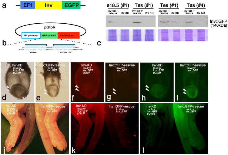
In this study we generated RNA interference (RNAi)-mediated gene knockdown transgenic mice (transgenic RNAi mice) against the functional Inv gene. Inv mutant mice show consistently reversed internal organs (situs inversus), multiple renal cysts and neonatal lethality. The Inv::GFP-rescue mice, which introduced the Inv::GFP fusion gene, can rescue inv mutant mice phenotypes. This indicates that the Inv::GFP gene is functional in vivo. To analyze the physiological functions of the Inv gene, and to demonstrate the availability of transgenic RNAi mice, we introduced a short hairpin RNA expression vector against GFP mRNA into Inv::GFP-rescue mice and analyzed the gene silencing effects and Inv functions by examining phenotypes. Transgenic RNAi mice with the Inv::GFP-rescue gene (Inv-KD mice) down-regulated Inv::GFP fusion protein and showed hypomorphic phenotypes of inv mutant mice, such as renal cyst development, but not situs abnormalities or postnatal lethality. This indicates that shRNAi-mediated gene silencing systems that target the tag sequence of the fusion gene work properly in vivo, and suggests that a relatively high level of Inv protein is required for kidney development in contrast to left/right axis determination. Inv::GFP protein was significantly down-regulated in the germ cells of Inv-KD mice testis compared with somatic cells, suggesting the existence of a testicular germ cell-specific enhanced RNAi system that regulates germ cell development. The Inv-KD mouse is useful for studying Inv gene functions in adult tissue that are unable to be analyzed in inv mutant mice showing postnatal lethality. In addition, the shRNA-based gene silencing system against the tag sequence of the fusion gene can be utilized as a new technique to regulate gene expression in either in vitro or in vivo experiments.


Conflict of interest statement

Competing Interests: The authors have declared that no competing interests exist.

Figures

Figure 1
Figure 1. shRNA-based gene silencing of the Inv::GFP fusion gene.
a: Construct of the Inv::GFP fusion gene introduced into Inv::GFP-rescue mice. EGFP coding cDNA was fused to the C terminus of Inv coding cDNA and inserted downstream of the EF1αpromoter . b: Construct of the pH1/siRNAEGFP-CAG/HcRed1 (pGtoR) transgene. Two oligonucleotides containing sense and antisense 21 nt sequences from the EGFP coding region and a spacer sequence that provided a loop structure were inserted downstream of the H1 promoter . HcRed1 coding cDNA was inserted into the vector downstream of the CAG promoter. c: Western blot analysis of Inv::GFP protein of embryonic day 18.5 (e18.5) embryos and adult testis. The lysates of e18.5 embryos (#1 line) and adult testis (#1 and #4 line) of pGtoR-integrated Inv-KD mice (inv/inv, Inv::GFP, pGtoR) and control litter mate Inv::GFP-rescue mice (inv/inv, inv::GFP) were analyzed by western blotting with anti-GFP antibodies. A band of approximately 140 kDa of Inv::GFP fusion protein was detected in Inv::GFP-rescue mice, but was specifically down-regulated in the pGtoR integrated Inv-KD mice embryos and testis. Accuracy of sample loading is indicated by the Coomassie-stained gel shown below. Bright field (d, e and j) and Hc-Red (f, g and k) or GFP (h, i and l) fluorescent observation of e8.0 embryos (d–i, arrowheads) and new bone (j–l) of Inv-KD mice (d, f, h and j–l, left) and control Inv::GFP-rescue mice (e, g, i and j–l, right) are shown. Hc-Red fluorescent was specifically expressed in Inv-KD mice but not control Inv::GFP-rescue mice (f, g and k). Inv::GFP fluorescent is specifically down-regulated in Inv-KD mice compared with Inv::GFP-rescue mice (h, i and l).
Figure 2
Figure 2. Anatomical analysis of adult Inv-KD mice.
Anatomical analysis of pGtoR integrated adult (18 months old) Inv-KD mice (b and c) and control litter mate Inv::GFP-rescue mice (a). High magnification images of the kidney of (a–c) are shown in panels (d–f) and (g–i). Inv-KD mice developed fibrotic kidney with multiple cysts, but this was not observed in Inv::GFP-rescue mice. Highly developed multiple cysts were observed on the surface of Inv-KD mouse kidneys (h and i, arrowheads). Enlarged spleens were observed in adult Inv-KD mice (b and c, arrows).
Figure 3
Figure 3. Multiple renal cyst development in Inv-KD mice.
Cross sections prepared from adult (12 months old) Inv-KD mice (b) and control litter mate Inv::GFP-rescue mouse kidneys (a). High magnification image of the boxed region of (b) is shown in panel (c). Multiple renal cysts were observed in Inv-KD mice. The average number of cysts per single kidney cross section at 1–1.5 months old (lanes 1–4) or 12 months old (lanes 5–8) of kidneys in Inv-KD mice (lanes 2, 4, 6 and 8) and control litter mate Inv::GFP-rescue mice (lanes 1, 3, 5 and 7), diameter 400–800 µm (lanes 1, 2, 5 and 6) and over 800 µm (lanes 3, 4, 7 and 8) are shown (d). Average percentage of cyst area per single kidney cross section of adult (12 months old) Inv::GFP-rescue and Inv-KD mice are shown (e). Error bars represent S.E.M. (*) and (**) indicate Student’s t-test values <0.05 and <0.001, respectively. Scale bars: 1 mm.
Figure 4
Figure 4. Down-regulation of Inv::GFP protein in Inv-KD mouse kidneys.
Cross sections prepared from adult (12 month-old) kidneys of Inv-KD mice (g–l) and control Inv::GFP-rescue mice (a–f) were double-stained with anti-GFP antibodies (a–c, red) and PNA-FITC (b, green) or anti-GFP (d, g and j, red) and anti-acetylated tubulin antibodies (e, h and k, green). Nuclei were counterstained with Hoechst dye (a and c). High magnification image of the boxed region (a and b) are shown in panel (c). Merged and high magnification image of (de, gh and jk) are shown in panel (f, i and l) respectively. Inv::GFP protein was detected in proximal (PNA-negative) and distal (PNA-positive) tubules of Inv::GFP-rescue mice (a and b). Localization of Inv::GFP protein was detected in the cilia of both proximal and distal tubules (c, arrowhead).Inv::GFP protein was specifically down-regulated in the renal tubules of Inv-KD mice (g and j) compared with Inv::GFP-rescue mice (a and d). Inv::GFP fusion proteins were localized in the proximal region of anti-acetylated tubulin positive cilia of the renal tubules of Inv::GFP-rescue mice (d–f, arrowheads), but were down-regulated in Inv-KD mice (g–i, arrowheads). Inv::GFP protein was significantly down-regulated in the cystic tubules (j–l) compared with the non-cystic tubules (g–i). Images were taken at 10×40 (a and b) and 10×60 (d, e, g, h, j and k) magnification. Scale bars: 50 µm (a, d, g and j); 5 µm (c, f, i and l).
Figure 5
Figure 5. Testicular germ cell-specific elimination of Inv::GFP protein in Inv-KD mice.
Testicular sections prepared from adult (a–f and i–p) or postnatal (3 day) (g and h) Inv-KD mice (b, d, f, h, j, l, n and p) or control Inv::GFP-rescue mice (a, c, e, g, i, k, m and o) were stained with anti-GFP antibodies (g, h, m and n, red) or double-stained with anti-GFP antibodies (c, d, i–l, o and p, red) and testicular germ cell-specific anti-calmegin antibodies (e, f, k and l, green) or vascular endothelial cell-specific PECAM-1 antibodies (o and p, green). Hoechst-stained images are shown to identify the cell types of the testis (a, b, g–i and m–p, blue). Inv::GFP protein was mainly observed in the spermatid cells of testicular germ cells (a, c, e, i and k, arrowhead) and somatic cells, such as connective tissue cells and Leydig cells (m, double arrows and arrow) and vascular endothelial cells (o, arrow) in Inv::GFP-rescue mice. Inv::GFP protein was specifically eliminated in the germ cells of Inv-KD mice (b, d, f, j, and l), and was down-regulated in connective tissue and Leydig cells (n, double arrow and arrow) and vascular endothelial cells (p, arrow). In Inv-KD mice, Inv::GFP protein was eliminated in anti-calmegin antibody-positive germ cells in adult testis, and in spermatogonia cells in postnatal mice (day 3) testis (h, arrowhead) but not in Inv::GFP-rescue mice (g, arrowhead). Scale bars: 200 µm (a and b); 50 µm (g−j and m–p).

Similar articles

References

    1. Brummelkamp TR, Bernards R, Agami R (2002) A system for stable expression of short interfering RNAs in mammalian cells. Science 296: 550–553. - PubMed
    1. McManus MT, Petersen CP, Haines BB, Chen J, Sharp PA (2002) Gene silencing using micro-RNA designed hairpins. RNA 8: 842–850. - PMC - PubMed
    1. Miyagishi M, Taira K (2002) U6 promoter-driven siRNAs with four uridine 3' overhangs efficiently suppress targeted gene expression in mammalian cells. Nat Biotechnol 20: 497–500. - PubMed
    1. Sui G, Soohoo C, Affar el B, Gay F, Shi Y, et al. (2002) A DNA vector-based RNAi technology to suppress gene expression in mammalian cells. Proc Natl Acad Sci U S A 99: 5515–5520. - PMC - PubMed
    1. Paul CP (2005) Subcellular distribution of small interfering RNA: Directed delivery through RNA polymerase III expression cassettes and localization by in situ hybridization. Methods Enzymol 392: 125–145. - PubMed

Publication types