. 2014 Feb 20;9(2):e89670.
doi: 10.1371/journal.pone.0089670. eCollection 2014.

Remote control of respiratory neural network by spinal locomotor generators

Affiliations

Remote control of respiratory neural network by spinal locomotor generators

Jean-Patrick Le Gal et al. PLoS One. .

Abstract

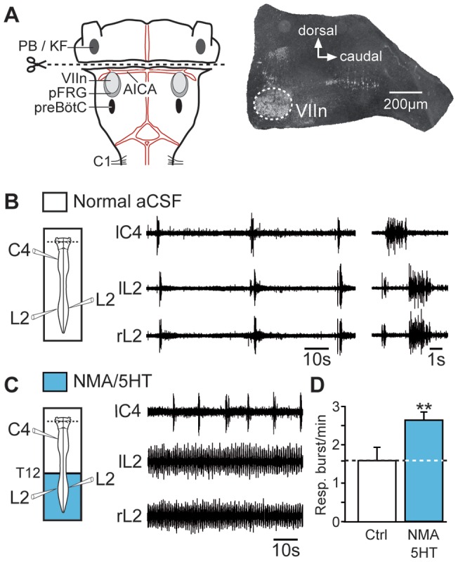
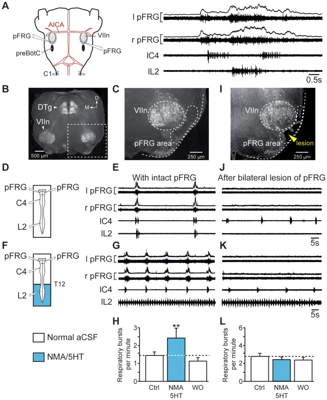
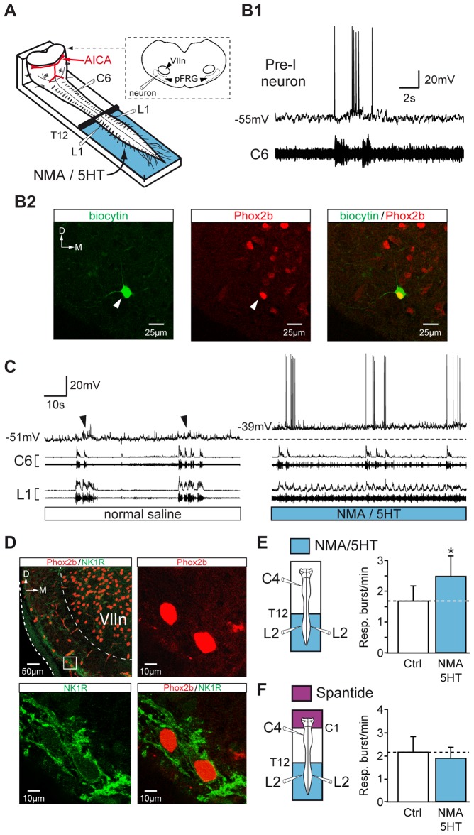
During exercise and locomotion, breathing rate rapidly increases to meet the suddenly enhanced oxygen demand. The extent to which direct central interactions between the spinal networks controlling locomotion and the brainstem networks controlling breathing are involved in this rhythm modulation remains unknown. Here, we show that in isolated neonatal rat brainstem-spinal cord preparations, the increase in respiratory rate observed during fictive locomotion is associated with an increase in the excitability of pre-inspiratory neurons of the parafacial respiratory group (pFRG/Pre-I). In addition, this locomotion-induced respiratory rhythm modulation is prevented both by bilateral lesion of the pFRG region and by blockade of neurokinin 1 receptors in the brainstem. Thus, our results assign pFRG/Pre-I neurons a new role as elements of a previously undescribed pathway involved in the functional interaction between respiratory and locomotor networks, an interaction that also involves a substance P-dependent modulating mechanism requiring the activation of neurokinin 1 receptors. This neurogenic mechanism may take an active part in the increased respiratory rhythmicity produced at the onset and during episodes of locomotion in mammals.


Conflict of interest statement

Competing Interests: The authors have declared that no competing interests exist.

Figures

Figure 1
Figure 1. Activation of the hindlimb locomotor CPG increases the respiratory burst frequency.
A, left, Schematic of the experimental procedure. middle, Raw spontaneous respiratory activity recorded from cervical (C4, inspiratory-like) and left (l) and right (r) lumbar (L2, expiratory-like) ventral roots. Note that under the control conditions, L5 motor output remained silent. right, Expanded traces of averaged integrated (9 sweeps) and raw activity recorded from the same ventral roots. B, Increase of the respiratory burst frequency during NMA/5HT-induced fictive locomotion (schematic at left). The layout is the same as that in (A). Expanded traces of the respiratory and locomotor bursts recorded from cervical (C4) and lumbar (L2 and L5) ventral roots, respectively, are shown at right. C, Recordings of respiratory activity during wash-out of NMA/5HT (schematic at left). D, Time course of the left–right (right L2 versus left L2, top) and flexor–extensor (left L2 versus left L5, bottom) phase coordination during fictive locomotion. Recordings and analysis shown in A to D are from the same preparation. E, F, left, Schematics of experimental setup. right, Bar charts showing the spontaneous respiratory burst frequency (mean ± SEM) under control (Ctrl) saline conditions (left, open bar), during NMA/5HT application to the lumbar region (blue bar), and during NMA/5HT wash-out (WO; right, open bar) when the lumbar segments of the cord were connected to the upper spinal and brainstem regions (E, n = 14 preparations) or disconnected by sucrose blockade of the thoracic (T2–T12) segments (F, n = 7 preparations). ***P<0.001 (repeated measures ANOVA, Tukey's post hoc tests).
Figure 2
Figure 2. Phase relationship between respiratory and locomotor bursts.
A, C, Schematics of experimental setup. B, D, upper and middle traces, Raw respiratory and locomotor activity recorded from right cervical (rC4) and left-right lumbar (rL2 and lL2) ventral roots, respectively, when lumbar spinal region was connected to the upper spinal and brainstem structures (B) or disconnected by a sucrose blockade of thoracic segments (D). Yellow lines superimposed on rL2 and lL2 discharges are an averaging (30 successive locomotor cycles) of integrated locomotor ventral root activity. lower diagrams, Phase diagrams illustrating normalized locomotor step cycles during pharmacologically evoked fictive locomotion. Note that cervical respiratory (resp.) bursts (thin horizontal lines) occur preferentially in-phase with lumbar locomotor (loco.) discharges (blue horizontal bars) when hindlimb locomotor CPG are connected to the medullary respiratory generators (B). Recordings and analysis shown in B and D are from the same preparation.
Figure 3
Figure 3. Electrical stimulation of SCA activates the lumbar locomotor CPG and increases the respiratory rate.
A, left, Schematic of experimental procedure. right, Spontaneous respiratory activity from cervical (C4) and right (r) and left (l) lumbar (L2) ventral roots. White arrowheads denote detected respiratory bursts. B, Fictive locomotion and increase in respiratory burst frequency induced during the tonic stimulation of SCA. The layout is the same as that in (A). Expanded traces of fictive locomotor burst activity recorded from bilateral L2 ventral roots in (B). D, Plot showing left-right locomotor phase coordination occurring between bilateral flexor (L2) ventral roots during 3 successive episodes of SCA stimulation–induced locomotion. E, Bar charts illustrating changes in respiratory burst frequency (Resp.; mean ± SEM; n = 7 preparations) following SCA stimulation. Recordings and analysis shown in A to D are from the same preparation. Ctrl, control. *** P<0.001 (Student's paired t test).
Figure 4
Figure 4. Excitatory influence of lumbar locomotor networks on respiratory centers does not require supralumbar spinal synaptic relays.
A, B, C, left, Schematics of experimental setup. middle, Raw spontaneous respiratory activity recorded from cervical (C1, inspiratory-like) and lumbar (L2, expiratory-like) ventral roots under control conditions (A), during the application of a low-Ca2+/high-Mg2+ saline solution (Low Ca2+) to the cervicothoracic (C3–T12) segments (B), and during subsequent activation of the lumbar locomotor CPG with a mixture of NMA and 5HT (C). Note that the respiratory pattern remained unchanged after the pharmacological blockade of cervicothoracic synaptic transmission (expanded traces at right in A and B) with the low-Ca2+/high-Mg2+ saline solution. D, Bar charts illustrating changes in respiratory burst frequency (Resp.) under these 3 experimental conditions (mean ± SEM; n = 8 preparations). Recordings shown in A to C are from the same preparation. Ctrl, control conditions. ***P<0.001 (repeated measures ANOVA, Tukey's post hoc tests).
Figure 5
Figure 5. Pontine structures are not engaged in the accelerated respiration induced by activation of the lumbar locomotor CPG.
A, left, Schematic drawing showing the level of transection (dotted line) performed rostral to the AICA. Note that this transection allowed removal of pontine structures of the brainstem while preserving medullary nuclei. right, Histological control of the brainstem transection. The parasagittal section, which was immunostained for acetylcholinesterase, clearly shows the transection rostral to the facial nucleus (VIIn). B, C, left, Schematic of experimental procedures. middle, Raw spontaneous respiratory activity recorded from cervical (C4, inspiratory-like) and left (l) and right (r) lumbar (L2, expiratory-like) ventral roots under control conditions (B, with expanded traces shown at right) and during the pharmacological activation of the lumbar locomotor generators with NMA/5HT (C). D, Bar charts showing changes in respiratory burst frequency (Resp.; mean ± SEM; n = 7 preparations) under control saline conditions (open bar) and during NMA/5HT application to the lumbar spinal region (blue bar). Ctrl, control conditions; PB/KF, parabrachial/Kölliker–Fuse complex. **P<0.01 (Student's paired t test).
Figure 6
Figure 6. Bilateral lesion of the pFRG suppresses the locomotion-induced accelerated respiration.
A, left, Representation of the brainstem–spinal cord preparation with medullary recording sites. right, Averaged integrated (6 sweeps) and raw respiratory burst activity recorded from the left (l) and right (r) pFRG and from spinal (cervical C4 and lumbar L2) ventral roots. B, C, I, Transverse acetylcholinesterase-stained sections as histological control in intact (B, C) and lesioned (I) brainstem preparations. The position of the lesion ventral to the facial motor nucleus corresponds to the location of the pFRG (I). D, F, Schematics of experimental setup. E, J, Spontaneous respiratory activity recorded bilaterally from the pFRG and homolaterally from cervical (C4) and lumbar (L2) ventral roots under control conditions in intact preparation (E) and after the bilateral lesion of the pFRG (J). G, K, Respiratory effects of lumbar cord activation with NMA/5HT in intact (G) and pFRG-lesioned (K) brainstem-spinal cord preparations. H, L, Bar charts showing variation in the respiratory burst frequency (Resp.; mean ± SEM) under control conditions (open bar, left), during pharmacological activation of lumbar locomotor CPG (blue bar), and after wash-out (open bar, right) in intact (H, n = 7 preparations) and pFRG-lesioned (L, n = 7 preparations) preparations. Recordings shown in E, G, J and K are from the same preparation. VIIn, facial motor nucleus; Ctrl, control conditions; DTg, dorsal tegmental nucleus; D, dorsal; M, medial; WO, wash-out. **P<0.01 (repeated measures ANOVA, Tukey's post hoc tests).
Figure 7
Figure 7. Depolarization of pFRG neurons during pharmacologically induced locomotion in the lumbar spinal cord: involvement of an SP pathway.
A, Schematic representation of the preparation. The brainstem was transected rostrally to the AICA to allow direct access to pFRG neurons for patch-clamp recording. B1, Simultaneous whole-cell patch-clamp recording of a pFRG/Pre-I neuron and raw activity of the cervical (C6, inspiratory-like) ventral root under control conditions. B2, Photomicrographs (z-stack of 3 images, 1.8 µm in total thickness) of immunolabeling for Phox2B (middle, red) in a biocytin-filled neuron (left, green). The white arrowhead indicates the neuron recorded in B1, and the merged image (right) confirms that the double-labeled (yellow) cell was a functionally identified Pre-I neuron. The dashed line shows the ventrolateral edge of the medulla. C, Simultaneous whole-cell patch-clamp recording of a pFRG/Pre-I neuron (black arrowhead indicating spontaneous rhythmic respiratory depolarization) and integrated and raw activity of cervical (C6) and lumbar (L1) ventral roots under control saline conditions (left) and during NMA/5HT application to lumbar region (right). The dashed line indicates the resting membrane potential level under control conditions. D, Left, top, Distribution of NK1R (green) immunoreactivity and Phox2B-positive cells (red) in the ventrolateral part of the medulla (this photomicrograph corresponds to a z-stack of 18 images, 18 µm in total thickness). The inset (white rectangle) shows the ventrolateral aspect of the VIIn at a higher magnification. Note that cells located in the area corresponding to the pFRG networks are immunopositive for Phox2b (right, top, red; z-stack of 3 images, 2.4 µm in total thickness) and for NK1R (left, bottom, green), as confirmed by the merged image (right, bottom). E, F, left, Schematics of experimental procedures. right, Bar charts showing variation in the respiratory burst frequency (Resp.; mean ± SEM) under control saline conditions (open bar) and during NMA/5HT application to the lumbar spinal cord (blue bar) during perfusion of the brainstem with normal saline (E, n = 7 preparations) or with a medium containing the SP antagonist spantide (F, n = 7 preparations). Ctrl, control conditions; D, dorsal; M, medial. *P<0.05 (Student's paired t test).

Similar articles

Cited by

References

    1. Mateika JH, Duffin J (1995) A review of the control of breathing during exercise. Eur J Appl Physiol Occup Physiol 71: 1–27. - PubMed
    1. Haouzi P, Hirsch JJ, Marchal F, Huszczuk A (1997) Ventilatory and gas exchange response during walking in severe peripheral vascular disease. Respir Physiol 107: 181–190. - PubMed
    1. Tobin MJ, Perez W, Guenther SM, D'Alonzo G, Dantzker DR (1986) Breathing pattern and metabolic behavior during anticipation of exercise. J Appl Physiol 60: 1306–1312. - PubMed
    1. Gravel J, Brocard F, Gariepy JF, Lund JP, Dubuc R (2007) Modulation of respiratory activity by locomotion in lampreys. Neuroscience 144: 1120–1132. - PubMed
    1. Bell HJ (2006) Respiratory control at exercise onset: an integrated systems perspective. Respir Physiol Neurobiol 152: 1–15. - PubMed

Publication types

LinkOut - more resources