. 2014 Feb 28;9(2):e89690.
doi: 10.1371/journal.pone.0089690. eCollection 2014.

Molecular cloning of a novel glucuronokinase/putative pyrophosphorylase from zebrafish acting in an UDP-glucuronic acid salvage pathway

Affiliations

Molecular cloning of a novel glucuronokinase/putative pyrophosphorylase from zebrafish acting in an UDP-glucuronic acid salvage pathway

Roman Gangl et al. PLoS One. .

Abstract

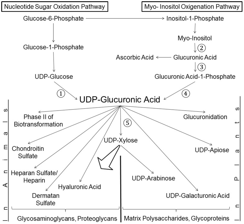
In animals, the main precursor for glycosaminoglycan and furthermore proteoglycan biosynthesis, like hyaluronic acid, is UDP-glucuronic acid, which is synthesized via the nucleotide sugar oxidation pathway. Mutations in this pathway cause severe developmental defects (deficiency in the initiation of heart valve formation). In plants, UDP-glucuronic acid is synthesized via two independent pathways. Beside the nucleotide sugar oxidation pathway, a second minor route to UDP-glucuronic acid exist termed the myo-inositol oxygenation pathway. Within this myo-inositol is ring cleaved into glucuronic acid, which is subsequently converted to UDP-glucuronic acid by glucuronokinase and UDP-sugar pyrophosphorylase. Here we report on a similar, but bifunctional enzyme from zebrafish (Danio rerio) which has glucuronokinase/putative pyrophosphorylase activity. The enzyme can convert glucuronic acid into UDP-glucuronic acid, required for completion of the alternative pathway to UDP-glucuronic acid via myo-inositol and thus establishes a so far unknown second route to UDP-glucuronic acid in animals. Glucuronokinase from zebrafish is a member of the GHMP-kinase superfamily having unique substrate specificity for glucuronic acid with a Km of 31 ± 8 µM and accepting ATP as the only phosphate donor (Km: 59 ± 9 µM). UDP-glucuronic acid pyrophosphorylase from zebrafish has homology to bacterial nucleotidyltransferases and requires UTP as nucleosid diphosphate donor. Genes for bifunctional glucuronokinase and putative UDP-glucuronic acid pyrophosphorylase are conserved among some groups of lower animals, including fishes, frogs, tunicates, and polychaeta, but are absent from mammals. The existence of a second pathway for UDP-glucuronic acid biosynthesis in zebrafish likely explains some previous contradictory finding in jekyll/ugdh zebrafish developmental mutants, which showed residual glycosaminoglycans and proteoglycans in knockout mutants of UDP-glucose dehydrogenase.


Conflict of interest statement

Competing Interests: The authors have declared that no competing interests exist.

Figures

Figure 1
Figure 1. Biosynthesis of UDP-GlcA in Lower Animals.
Two pathways for the biosynthesis of UDP-GlcA are present in plants and lower animals. The nucleotide sugar oxidation pathway with the enzyme UDP-glucose dehydrogenase (1) is predominant in both plant and animal kingdoms. Glucuronokinase (3) and UDP-glucuronic acid pyrophosphorylase (4) are part of the myo-Inositol oxygenation pathway starting with myo-inositol oxygenase (2). UDP-xylose synthase (5) converts UDP-GlcA into UDP-xylose for GAG and furthermore PG biosynthesis. Note that GlcA is also the precursor for ascorbic acid in animals. Thus the salvage pathway to UDP-GlcA and ascorbic acid biosynthesis compete for the same substrate GlcA.
Figure 2
Figure 2. Sequence and Phylogenetic Analysis of DrGKUP.
(A) shows a bifunctional enzyme comprised of a putative kinase domain (DrGK; 357 amino acids; 40 kDa) at the C-terminal end and a putative pyrophosphorylase domain (DrUP; 260 amino acids; 30 kDa) at the N-terminal end. For the pyrophosphorylase domain analysis Danio rerio (XP_005157584.1), Xenopus tropicalis (XP_002940431.1), Ruminococcus_sp. (WP_021925996.1) and Firmicutes_bacterium (WP_022231022.1) were aligned and for glucuronokinase domain analysis Danio rerio (NP_001107088.1), Xenopus tropicalis (AAI55522.1), Arabidopsis thaliana (AAV74231.1) and Glycine max (NP_001242154.1) were aligned and with the pairwise method using Clustal Omega. (C) Identity and Similarity were calculated with GENEDOC and (B) phylogenetic distance trees were calculated with JalView. Neither a homologous glucuronokinase nor a pyrophosphorylase sequence could be found in higher animals or humans.
Figure 3
Figure 3. Distance tree of one group of pyrophosphorylases from pro- and eukaryotes calculated from an alignment with CLUSTAL.
In bacteria and archaea the proteins only encode a pyrophosphorylase whereas in eukaryotes a fusion protein with glucuronokinase in typically found. For the latter group only the pyrophosphorylase domain was used in the alignment. Each species name has a prefix (E: eukaryote; P: prokaryote) followed by the type of protein (PP: only pyrophosphorylase; K_PP: glucuronokinase and pyrophosphorylase). The fusion proteins are found e.g. in fungi, tunicates, polychataea, amphibian and fish. Danio rerio (gi|528501576), Oreochromis niloticus (gi|542261080), Maylandia zebra (gi|498923801), Haplochromis burtoni (gi|554854526), Oryzias latipes (gi|432895574), Takifugu rubripes (gi|410910724), Xenopus tropicalis (gi|301622206), Strongylocentrotus purpuratus (gi|390355576), Branchiostoma floridae (gi|260786665), Nematostella vectensis (gi|156391024), Capitella teleta (gi|443696256), Salpingoeca rosetta (gi|514691982), Tetraodon nigroviridis (gi|47224901), Monosiga brevicollis MX1 (gi|167517681), Aureococcus anophagefferens (gi|323454734), Oikopleura dioica (gi|313221557), Acanthamoeba castellanii (gi|470509640), Mucor circinelloides (gi|510999707), Xenopus laevis (gi|163914505), Amphimedon queenslandica (gi|340375174), Ruminococcus sp. (gi|547183951), Latimeria chalumnae (gi|556979378), Firmicutes bacterium (gi|547822613), Pundamilia nyererei (gi|548393190), Clostridium sp. (gi|547295929), Gracilibacillus lacisalsi (gi|517760452), Eubacterium sp. CAG:274 (gi|547841474), Methylacidiphilum infernorum V4 (gi|189220259), Methylacidiphilum fumariolicum (gi|496352483), Rhizophagus irregularis (gi|552908620), Natrialba asiatica (gi|493050309), Natronorubrum bangense (gi|492959802), Paenibacillus sp. (gi|518757082), OP3 bacterium SCGC (gi|551125677), Halalkalicoccus jeotgali (gi|300711896), Natrinema pellirubrum (gi|433593563), Natrialba chahannaoensis (gi|493163024), Aerococcus viridans (gi|515407717), Alistipes sp. (gi|546342953), Halogeometricum borinquense (gi|313127001), Haloferax mediterranei (gi|389846545), Sphaerochaeta globosa (gi|325970222), Halopiger xanaduensis (gi|336255356), Prevotella sp. (gi|547906812), Natronomonas pharaonis (gi|76800846), Haloferax mucosum (gi|495596680), Haloterrigena thermotolerans (gi|493700315), Haloterrigena salina (gi|496171985), Halosarcina pallida (gi|495664926), Butyrivibrio proteoclasticus (gi|302671768), Haplochromis burtoni (gi|554859531), Natrinema altunense (gi|494168028), Thermococcus litoralis (gi|530548455), Halophilic archaeon DL31 (gi|345004592), Dorea formicigenerans (gi|547873312), Halobiforma nitratireducens (gi|493722014), Chondrus crispus (gi|546322381), Haloquadratum walsbyi (gi|544617810), Verrucomicrobium sp. (gi|521982779) and Haladaptatus paucihalophilus (gi|495256835).
Figure 4
Figure 4. SDS-PAGE of DrGKUP Purification Expressed in N. benthamiana.
Tobacco leaf crude extract (A, 1) and purified recombinant DrGKUP (A, 2). Crude extract (B, 1) and purified recombinant DrGK (B, 3) and crude extract (C, 1) and purified recombinant DrUP (C, 4).
Figure 5
Figure 5. Coupled HPLC Enzyme Assay of DrGK.
(A) Enzyme activity of DrGK was tested. Peak (3) indicates produced UDP-GlcA during coupled enzyme reaction. Peak (1) UMP, (2) AMP, (4) UDP, (5) ADP, (6) UTP and (7) ATP. (B) Control without DrGKUP. (C) 50 µM UDP-GlcA as reference compound.
Figure 6
Figure 6. pH-Optimum and Temperature-Optimum of DrGK.
(A) The effect of different pH values on DrGK activity was measured with coupled HPLC enzyme assay at different pH values dependent on the used buffers (50 mM). ▪potassium-Acetat: pH 4–5; ♦MES-KOH: pH 5.5–6.5; ▴MOPS-KOH: pH 7–7.5; •Tris-HCl: pH 8–9.5. UDP-GlcA level was determined using coupled HPLC enzyme assay. Values are averages of three independently performed assays (+/− SD). (B) The dependence of DrGK activity on reaction temperature was measured with coupled HPLC enzyme assay at different temperatures within a range from 10°C to 60°C. UDP-GlcA level was determined using coupled HPLC enzyme assay. Values are averages of three independently performed assays (+/− SD).
Figure 7
Figure 7. Kinetics of DrGK for ATP and GlcA.
(A) Substrate saturation by Michaelis-Menten curve for ATP is shown. Activity of DrGK was measured with varying concentrations of ATP (0.01–1.4 mM) under standard conditions for 20 min with coupled HPLC enzyme assay. Km with 59.3 µM and Vmax 132.1 pkat/µg protein was calculated. Values are averages of three independently performed assays (+/− SD). (B) Substrate saturation by Michaelis-Menten curve for GlcA is shown. Activity of DrGK was measured with varying concentrations of GlcA (0.01–1.4 mM) under standard conditions for 20 min with coupled HPLC enzyme assay. Km with 31.3 µM and Vmax 137.6 pkat/µg protein was calculated. Values are averages of three independently performed assays (+/− SD).
Figure 8
Figure 8. Metabolic pathway design experiments in S. cerevisiae cells.
Metabolite profiles from yeast expressing recombinant proteins. (A) DrGKUP, (B) empty pToy4 vector control, (C) pToy4 expressing Arabidopsis UDP-glucose dehydrogenase 1 (D) 50 µM UDP-GlcA as reference compound.
Figure 9
Figure 9. Sequence alignment of two bacterial nucleotidyltransferase with known crystal structure (1H5T ; 2PA4 [51]) and the pyrophosphorylase domain from zebrafish DrGKUP.
Note that some of the residues identified in the E. coli enzyme to bind the substrate are conserved in the zebrafish sequence (e.g. Gly11; Asp91; Asp118; Lys163; Arg220; numbers refer to the E. coli sequence 1H5T).
Figure 10
Figure 10. Long time Enzyme Assay.
We performed a long time enzyme assay for 70% Tween20 as enzyme stabilizing factor. Under these conditions, we could detect a small amount of UDP-GlcA signal on HPLC. Two control assays with either DrGK or DrUP alone did not result in any detectable product formation. For comparison and as a positive control, two enzyme assays are shown, in which the recombinant plant pyrophosphorylase (USP) was added.

Similar articles

Cited by

References

    1. Eames BF, Singer A, Smith GA, Wood ZA, Yan YL, et al. (2010) UDP xylose synthase 1 is required for morphogenesis and histogenesis of the craniofacial skeleton. Dev Biol 341: 400–415. - PMC - PubMed
    1. Velleman SG (2000) The role of the extracellular matrix in skeletal development. Poult Sci 79: 985–989. - PubMed
    1. Holt CE, Dickson BJ (2005) Sugar codes for axons? Neuron 46: 169–172. - PMC - PubMed
    1. Kirn-Safran CB, Gomes RR, Brown AJ, Carson DD (2004) Heparan sulfate proteoglycans: coordinators of multiple signaling pathways during chondrogenesis. Birth Defects Res C Embryo Today 72: 69–88. - PubMed
    1. Knudson CB, Knudson W (2001) Cartilage proteoglycans. Semin Cell Dev Biol 12: 69–78. - PubMed

Publication types

MeSH terms

Substances