Regulation of Drosophila eye development by the transcription factor Sine oculis
- PMID: 24586968
- PMCID: PMC3934907
- DOI: 10.1371/journal.pone.0089695
Regulation of Drosophila eye development by the transcription factor Sine oculis
Abstract
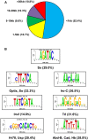
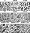
Homeodomain transcription factors of the Sine oculis (SIX) family direct multiple regulatory processes throughout the metazoans. Sine oculis (So) was first characterized in the fruit fly Drosophila melanogaster, where it is both necessary and sufficient for eye development, regulating cell survival, proliferation, and differentiation. Despite its key role in development, only a few direct targets of So have been described previously. In the current study, we aim to expand our knowledge of So-mediated transcriptional regulation in the developing Drosophila eye using ChIP-seq to map So binding regions throughout the genome. We find 7,566 So enriched regions (peaks), estimated to map to 5,952 genes. Using overlap between the So ChIP-seq peak set and genes that are differentially regulated in response to loss or gain of so, we identify putative direct targets of So. We find So binding enrichment in genes not previously known to be regulated by So, including genes that encode cell junction proteins and signaling pathway components. In addition, we analyze a subset of So-bound novel genes in the eye, and find eight genes that have previously uncharacterized eye phenotypes and may be novel direct targets of So. Our study presents a greatly expanded list of candidate So targets and serves as basis for future studies of So-mediated gene regulation in the eye.
Conflict of interest statement
Figures


Similar articles
-
Identification of novel direct targets of Drosophila Sine oculis and Eyes absent by integration of genome-wide data sets.Dev Biol. 2016 Jul 1;415(1):157-167. doi: 10.1016/j.ydbio.2016.05.007. Epub 2016 May 10. Dev Biol. 2016. PMID: 27178668 Free PMC article.
-
Genome-wide DNA binding pattern of the homeodomain transcription factor Sine oculis (So) in the developing eye of Drosophila melanogaster.Genom Data. 2014 Dec 1;2:153-155. doi: 10.1016/j.gdata.2014.06.016. Genom Data. 2014. PMID: 25126519 Free PMC article.
-
Six3, a medaka homologue of the Drosophila homeobox gene sine oculis is expressed in the anterior embryonic shield and the developing eye.Mech Dev. 1998 Jun;74(1-2):159-64. doi: 10.1016/s0925-4773(98)00055-0. Mech Dev. 1998. PMID: 9651515
-
Networks of genes governing the development of optic and otic vesicles: implications for eye and ear development.Invest Ophthalmol Vis Sci. 2015 Feb 5;56(2):892. doi: 10.1167/iovs.15-16420. Invest Ophthalmol Vis Sci. 2015. PMID: 25656090 Review. No abstract available.
-
Vertebrate eye development as modeled in Drosophila.Hum Mol Genet. 2000 Apr 12;9(6):917-25. doi: 10.1093/hmg/9.6.917. Hum Mol Genet. 2000. PMID: 10767315 Review.
Cited by
-
Six1 proteins with human branchio-oto-renal mutations differentially affect cranial gene expression and otic development.Dis Model Mech. 2020 Mar 3;13(3):dmm043489. doi: 10.1242/dmm.043489. Dis Model Mech. 2020. PMID: 31980437 Free PMC article.
-
Multilevel regulation of the glass locus during Drosophila eye development.PLoS Genet. 2019 Jul 12;15(7):e1008269. doi: 10.1371/journal.pgen.1008269. eCollection 2019 Jul. PLoS Genet. 2019. PMID: 31299050 Free PMC article.
-
Conditional knockout of retinal determination genes in differentiating cells in Drosophila.FEBS J. 2016 Aug;283(15):2754-66. doi: 10.1111/febs.13772. Epub 2016 Jun 23. FEBS J. 2016. PMID: 27257739 Free PMC article.
-
New regulators of Drosophila eye development identified from temporal transcriptome changes.Genetics. 2021 Apr 15;217(4):iyab007. doi: 10.1093/genetics/iyab007. Genetics. 2021. PMID: 33681970 Free PMC article.
-
Glass confers rhabdomeric photoreceptor identity in Drosophila, but not across all metazoans.Evodevo. 2019 Mar 5;10:4. doi: 10.1186/s13227-019-0117-6. eCollection 2019. Evodevo. 2019. PMID: 30873275 Free PMC article.
References
-
- Pappu KS, Mardon G (2004) Genetic control of retinal specification and determination in Drosophila. Int J Dev Biol 48: 913–924. - PubMed
-
- Ready DF, Hanson TE, Benzer S (1976) Development of the Drosophila retina, a neurocrystalline lattice. Dev Biol 53: 217–240. - PubMed
-
- Cheyette BN, Green PJ, Martin K, Garren H, Hartenstein V, et al. (1994) The Drosophila sine oculis locus encodes a homeodomain-containing protein required for the development of the entire visual system. Neuron 12: 977–996. - PubMed
-
- Pignoni F, Hu B, Zavitz KH, Xiao J, Garrity PA, et al. (1997) The eye-specification proteins So and Eya form a complex and regulate multiple steps in Drosophila eye development. Cell 91: 881–891. - PubMed
Publication types
MeSH terms
Substances
Grants and funding
LinkOut - more resources
Full Text Sources
Other Literature Sources
Molecular Biology Databases

