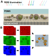
Imaging and identification of waterborne parasites using a chip-scale microscope
- PMID: 24586978
- PMCID: PMC3935895
- DOI: 10.1371/journal.pone.0089712
Imaging and identification of waterborne parasites using a chip-scale microscope
Abstract
We demonstrate a compact portable imaging system for the detection of waterborne parasites in resource-limited settings. The previously demonstrated sub-pixel sweeping microscopy (SPSM) technique is a lens-less imaging scheme that can achieve high-resolution (<1 µm) bright-field imaging over a large field-of-view (5.7 mm×4.3 mm). A chip-scale microscope system, based on the SPSM technique, can be used for automated and high-throughput imaging of protozoan parasite cysts for the effective diagnosis of waterborne enteric parasite infection. We successfully imaged and identified three major types of enteric parasite cysts, Giardia, Cryptosporidium, and Entamoeba, which can be found in fecal samples from infected patients. We believe that this compact imaging system can serve well as a diagnostic device in challenging environments, such as rural settings or emergency outbreaks.
Conflict of interest statement
Figures


Similar articles
-
Evaluation of five commercial methods for the extraction and purification of DNA from human faecal samples for downstream molecular detection of the enteric protozoan parasites Cryptosporidium spp., Giardia duodenalis, and Entamoeba spp.J Microbiol Methods. 2016 Aug;127:68-73. doi: 10.1016/j.mimet.2016.05.020. Epub 2016 May 27. J Microbiol Methods. 2016. PMID: 27241828
-
Performance of microscopy and ELISA for diagnosing Giardia duodenalis infection in different pediatric groups.Parasitol Int. 2016 Dec;65(6 Pt A):635-640. doi: 10.1016/j.parint.2016.08.012. Epub 2016 Aug 30. Parasitol Int. 2016. PMID: 27586394
-
Evaluation of an immunochromatographic dip strip test for simultaneous detection of Cryptosporidium spp, Giardia duodenalis, and Entamoeba histolytica antigens in human faecal samples.Eur J Clin Microbiol Infect Dis. 2012 Aug;31(8):2077-82. doi: 10.1007/s10096-012-1544-7. Epub 2012 Jan 20. Eur J Clin Microbiol Infect Dis. 2012. PMID: 22262367
-
[New methods for the diagnosis of Cryptosporidium and Giardia].Parassitologia. 2004 Jun;46(1-2):151-5. Parassitologia. 2004. PMID: 15305706 Review. Italian.
-
Waterborne protozoan pathogens.Clin Microbiol Rev. 1997 Jan;10(1):67-85. doi: 10.1128/CMR.10.1.67. Clin Microbiol Rev. 1997. PMID: 8993859 Free PMC article. Review.
Cited by
-
Compact and Filter-Free Luminescence Biosensor for Mobile in Vitro Diagnoses.ACS Nano. 2019 Oct 22;13(10):11698-11706. doi: 10.1021/acsnano.9b05634. Epub 2019 Sep 6. ACS Nano. 2019. PMID: 31461265 Free PMC article.
-
Digital diffraction detection of protein markers for avian influenza.Lab Chip. 2016 Apr 21;16(8):1340-5. doi: 10.1039/c5lc01558h. Lab Chip. 2016. PMID: 26980325 Free PMC article.
-
Development of a high-speed imaging system for real time evaluation and monitoring of cardiac engineered tissues.Front Bioeng Biotechnol. 2024 Aug 12;12:1403044. doi: 10.3389/fbioe.2024.1403044. eCollection 2024. Front Bioeng Biotechnol. 2024. PMID: 39188370 Free PMC article.
-
Microfluidic multi-angle laser scattering system for rapid and label-free detection of waterborne parasites.Biomed Opt Express. 2018 Mar 6;9(4):1520-1530. doi: 10.1364/BOE.9.001520. eCollection 2018 Apr 1. Biomed Opt Express. 2018. PMID: 29675299 Free PMC article.
-
Microfluidic-integrated laser-controlled microactuators with on-chip microscopy imaging functionality.Lab Chip. 2014 Oct 7;14(19):3781-9. doi: 10.1039/c4lc00790e. Lab Chip. 2014. PMID: 25099225 Free PMC article.
References
-
- Baldursson S, Karanis P (2011) Waterborne transmission of protozoan parasites: Review of worldwide outbreaks – An update 2004–2010. Water Research 45: 6603–6614. - PubMed
-
- Karanis P, Kourenti C, Smith H (2007) Waterborne transmission of protozoan parasites: a worldwide review of outbreaks and lessons learnt. Journal of Water and Health 5: 1–38. - PubMed
-
- Haque R, Roy S, Siddique A, Mondal U, Rahman SMM, et al. (2007) Multiplex Real-Time Pcr Assay For Detection Of Entamoeba Histolytica, Giardia Intestinalis, And Cryptosporidium Spp. The American Journal of Tropical Medicine and Hygiene 76: 713–717. - PubMed
Publication types
MeSH terms
Grants and funding
LinkOut - more resources
Full Text Sources
Other Literature Sources
Medical

