. 2014 Feb 24;9(2):e89742.
doi: 10.1371/journal.pone.0089742. eCollection 2014.

Lipoxin A₄ prevents the progression of de novo and established endometriosis in a mouse model by attenuating prostaglandin E₂ production and estrogen signaling

Affiliations

Lipoxin A₄ prevents the progression of de novo and established endometriosis in a mouse model by attenuating prostaglandin E₂ production and estrogen signaling

Rajesh Kumar et al. PLoS One. .

Abstract

Endometriosis, a leading cause of pelvic pain and infertility, is characterized by ectopic growth of endometrial-like tissue and affects approximately 176 million women worldwide. The pathophysiology involves inflammatory and angiogenic mediators as well as estrogen-mediated signaling and novel, improved therapeutics targeting these pathways are necessary. The aim of this study was to investigate mechanisms leading to the establishment and progression of endometriosis as well as the effect of local treatment with Lipoxin A4 (LXA₄), an anti-inflammatory and pro-resolving lipid mediator that we have recently characterized as an estrogen receptor agonist. LXA₄ treatment significantly reduced endometriotic lesion size and downregulated the pro-inflammatory cytokines IL-1β and IL-6, as well as the angiogenic factor VEGF. LXA₄ also inhibited COX-2 expression in both endometriotic lesions and peritoneal fluid cells, resulting in attenuated peritoneal fluid Prostaglandin E₂ (PGE₂) levels. Besides its anti-inflammatory effects, LXA₄ differentially regulated the expression and activity of the matrix remodeling enzyme matrix metalloproteinase (MMP)-9 as well as modulating transforming growth factor (TGF)-β isoform expression within endometriotic lesions and in peritoneal fluid cells. We also report for first time that LXA₄ attenuated aromatase expression, estrogen signaling and estrogen-regulated genes implicated in cellular proliferation in a mouse model of disease. These effects were observed both when LXA₄ was administered prior to disease induction and during established disease. Collectively, our findings highlight potential targets for the treatment of endometriosis and suggest a pleotropic effect of LXA₄ on disease progression, by attenuating pro-inflammatory and angiogenic mediators, matrix remodeling enzymes, estrogen metabolism and signaling, as well as downstream proliferative pathways.


Conflict of interest statement

Competing Interests: The authors have declared that no competing interests exist.

Figures

Figure 1
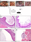
Figure 1. Treatment with LXA4 decreases endometriotic lesion growth.
A. Representative pictures of a mouse with endometriotic lesions treated with vehicle (Veh, left panel) or LXA4 (right panel) for 21 days. Six uterine biopsies were implanted on the peritoneal wall, 3 on each side, in a minimum of 20 mice which were randomly divided into two groups: Veh- and LXA4-treated. Implanted tissues grew into enlarged cyst-like lesions by 21 days after endometriosis induction in the Veh group, while LXA4 treatment significantly reduced lesion size. B. Graphical representation of endometriotic lesions volume, in mm3 ± SEM (**p<0.01). C. Morphology of formalin-fixed, paraffin-embedded lesion sections stained with HE. Upper panels a. and b. Vehicle-treated mouse with endometriosis. Lower panels c. and d. LXA4-treated mouse with endometriosis. Images are representative of ten biological replicates.
Figure 2
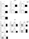
Figure 2. LXA4 significantly attenuates the expression of pro-inflammatory cytokines, angiogenic mediators, COX-2 and the PGE2 transporter MRP4.
A. IL-1β, B. IL-6, C. FPR2, D. HIF-1α, E. COX-2, F. VEGF and G. MRP4 mRNA expression in endometriotic lesions was analyzed by qPCR. H. IL-1β, I. IL-6, J. FPR2, K. HIF-1α, L. COX-2, M. VEGF and N. MRP4 mRNA expression was similarly quantified in PFCs. Ten mice were used per group (Sham, Veh-, LXA4-treatment) and samples analyzed for each individual mouse. Mice were sacrificed 21 days after endometriosis induction and Veh- or LXA4-treatment. Endometriotic lesions and PFCs were collected for RNA isolation and qPCR. Data were normalized to GAPDH and are presented as mean ± SEM (*p<0.05, **p<0.01, ***p<0.001 compared to Veh; ζζζ p<0.001 compared to Sham).
Figure 3
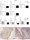
Figure 3. LXA4 reduces peritoneal fluid cytokine and PGE2 levels as well as the expression of COX-2 in endometriotic lesions.
Peritoneal fluid from mice was collected at sacrifice (Sham, Veh-, LXA4; 3–5 mice/group) and ELISAs performed to quantify A. IL-1β, B. VEGF and C. PGE2 levels. Results are expressed as pg mediator/ml for 50 μg of total peritoneal fluid protein and presented as mean ± SEM (*p<0.05, ***p<0.001 compared to Veh; ζ<0.05 compared to Sham). D. Immunohistochemical staining for COX-2 was performed on transverse paraffin-embedded sections of lesions from Veh- and LXA4-treated mice. Representative stainings are shown with the negative control in the inset.
Figure 4
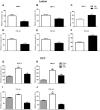
Figure 4. LXA4 significantly inhibits the expression of matrix remodeling genes.
A. MMP-2, B. MMP-3, C. MMP-9, D. TGF-β1, E. TGF-β2, F. TGF-β3, G. MMP-2, H. MMP-9, I. TGF-β1, J. TGF-β2 gene expression in lesions and PFCs. Ten mice were used per group (Sham, Veh-, LXA4-treatment) and sacrificed 21 days after endometriosis induction. Lesions and PFCs were collected for qPCR analyses. Data were normalized to GAPDH and presented as mean ± SEM (***p<0.001 compared to Veh; ζζ p<0.01, ζζζ p<0.001 compared to Sham).
Figure 5
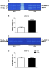
Figure 5. Gelatin zymography analysis of MMP-2 and MMP-9 activity.
A. Representative gel showing gelatinolytic activities of A. MMP-9, and C. MMP-2 in peritoneal fluid of Veh- and LXA4-treated mice (3–4 mice/group). Bar graphs represent the densitometric quantification of B. MMP-9 and D. MMP-2 activity, respectively. Unpaired t-tests were performed for comparison with the corresponding Veh-treatment group (**p<0.01).
Figure 6
Figure 6. Mediators implicated in estrogen production and cellular proliferation in endometriotic lesions and PFCs are inhibited by LXA4 treatment.
mRNA was extracted from endometriotic lesions and PFCs and subjected to qPCR analysis to assess transcript levels of A. CYP19a1, B. ERα, C. GREB1, D. CCND1, E. c-Myc and F. ERα, G. CCND1, H. c-Myc, respectively. Results were normalized to GAPDH. Ten mice were used per group (Sham, Veh-, LXA4-treatment) and sacrificed 21 days after surgical induction of endometriosis. Data are presented as mean ± SEM. (***p<0.001 compared to Veh; ζζζ p<0.001 compared to Sham). I. Immunohistochemical staining for ERα was performed on transverse sections of endometriotic lesions from Veh- (left panel) and LXA4-treated (right panel) mice. The negative control is shown in the inset.
Figure 7
Figure 7. LXA4 reduces the progression of established endometriosis by similar mechanisms.
Peritoneal endometriosis was surgically induced in mice divided into 3 groups (n = 7/group): Veh or LXA4 (5 μg/kg/mouse once daily) was administered from the day prior to surgery D(−1), or started at postoperative day 6 D(+6). Treatment was continued until day 21 after surgery, when the mice were sacrificed. A. Endometriotic lesion volume expressed as mean mm3 ± SEM (**p<0.01). B. a. IL-1β, b. COX-2, c. VEGF, d. MRP4, e. MMP-9, f. TGF-β1, g. TGF-β2, h. TGF-β3, i. CYP19a1, j. ERα, k. GREB1 and l. c-Myc mRNA expression was quantified in endometriotic lesions by qPCR and normalized to GAPDH. C. a. IL-1β and b. VEGF protein concentrations in the peritoneal fluid were measured by ELISA and expressed as pg/ml. All data are presented as mean ± SEM. Unpaired t-tests were performed for comparisons with the corresponding Veh-treated group (*p<0.05 **p<0.01, ***p<0.001, ιp<0.05, ι ιp<0.01, ι ι ιp<0.001).

Similar articles

Cited by

References

    1. Eskenazi B, Warner ML (1997) Epidemiology of endometriosis. Obstet Gynecol Clin North Am 24: 235–258. - PubMed
    1. Bulun SE (2009) Endometriosis. The New England journal of medicine 360: 268–279. - PubMed
    1. Gruppo Italiano per lo Studio dE (2001) Relationship between stage, site and morphological characteristics of pelvic endometriosis and pain. Hum Reprod 16: 2668–2671. - PubMed
    1. Waller KG, Shaw RW (1993) Gonadotropin-releasing hormone analogues for the treatment of endometriosis: long-term follow-up. Fertility and sterility 59: 511–515. - PubMed
    1. Vercellini P, Crosignani P, Somigliana E, Vigano P, Frattaruolo MP, et al. (2011) ‘Waiting for Godot’: a commonsense approach to the medical treatment of endometriosis. Hum Reprod 26: 3–13. - PubMed

Publication types

MeSH terms