. 2014 Feb 24;9(2):e89810.
doi: 10.1371/journal.pone.0089810. eCollection 2014.

CXCR4 inhibition ameliorates severe obliterative pulmonary hypertension and accumulation of C-kit⁺ cells in rats

Affiliations

CXCR4 inhibition ameliorates severe obliterative pulmonary hypertension and accumulation of C-kit⁺ cells in rats

Daniela Farkas et al. PLoS One. .

Abstract

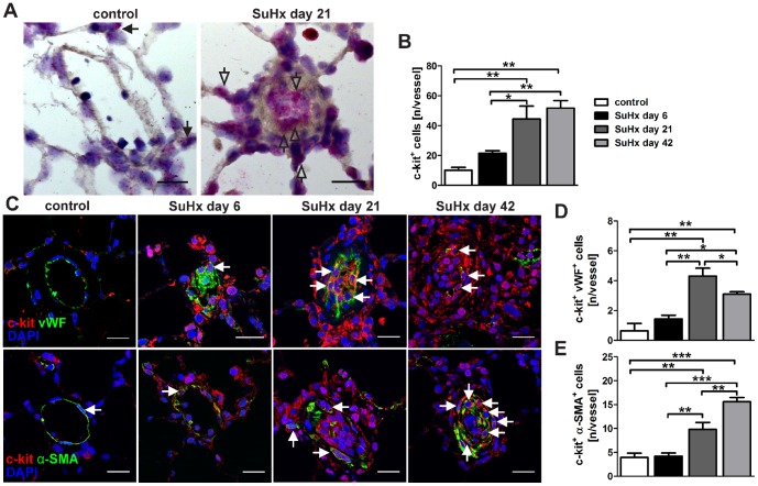
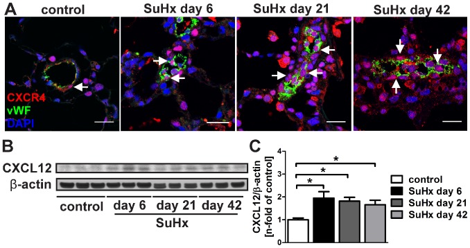
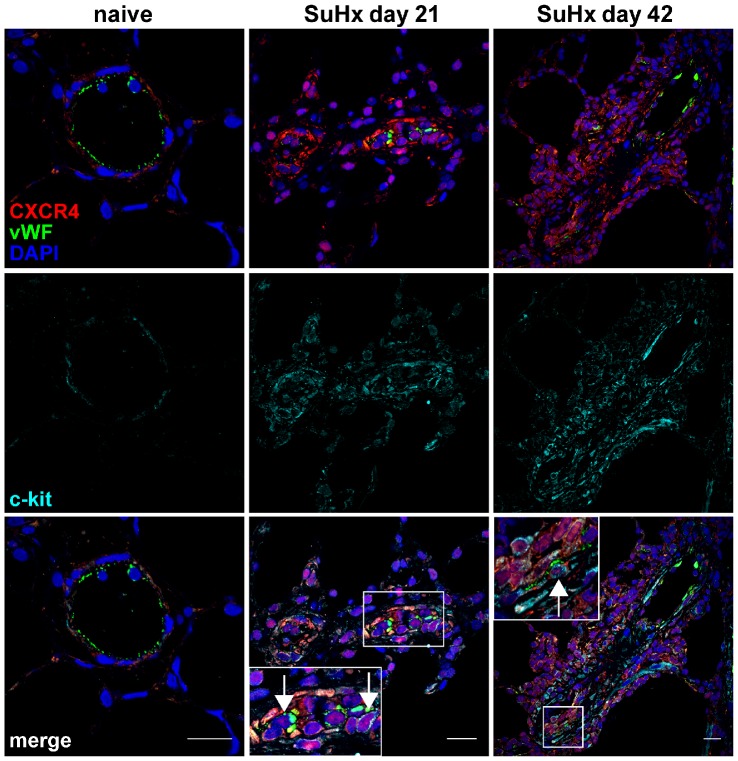
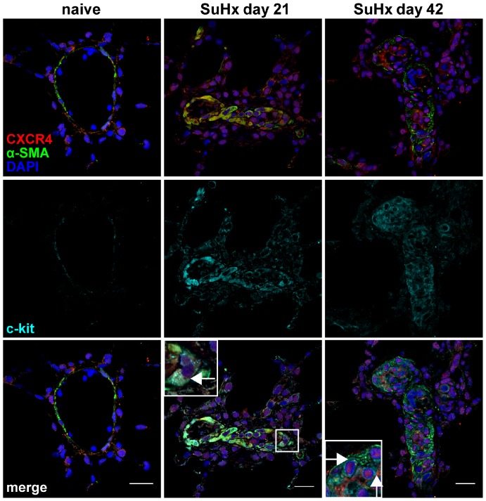
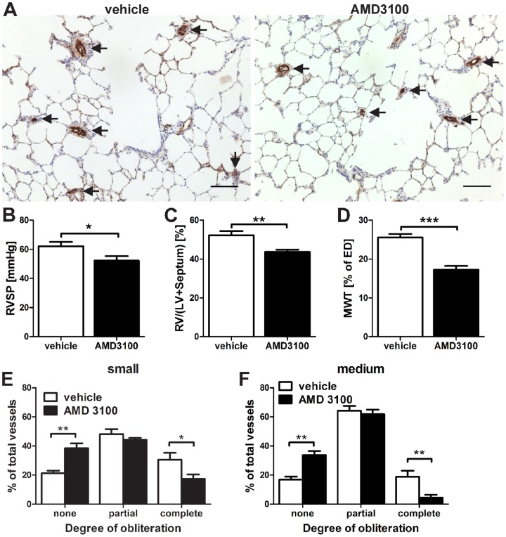
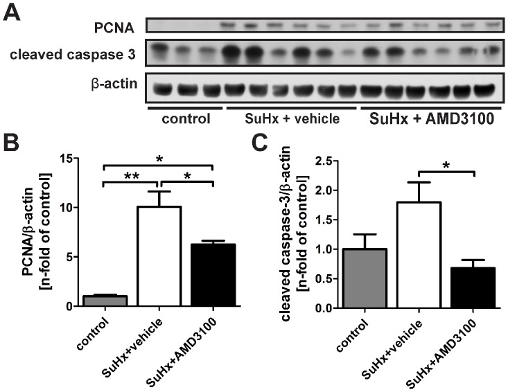
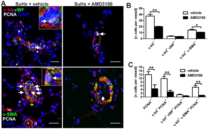
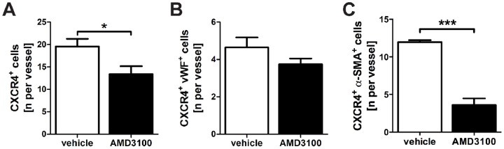
Successful curative treatment of severe pulmonary arterial hypertension with luminal obliteration will require a thorough understanding of the mechanism underlying the development and progression of pulmonary vascular lesions. But the cells that obliterate the pulmonary arterial lumen in severe pulmonary arterial hypertension are incompletely characterized. The goal of our study was to evaluate whether inhibition of CXC chemokine receptor 4 will prevent the accumulation of c-kit⁺ cells and severe pulmonary arterial hypertension. We detected c-kit⁺⁻ cells expressing endothelial (von Willebrand Factor) or smooth muscle cell/myofibroblast (α-smooth muscle actin) markers in pulmonary arterial lesions of SU5416/chronic hypoxia rats. We found increased expression of CXC chemokine ligand 12 in the lung tissue of SU5416/chronic hypoxia rats. In our prevention study, AMD3100, an inhibitor of the CXC chemokine ligand 12 receptor, CXC chemokine receptor 4, only moderately decreased pulmonary arterial obliteration and pulmonary hypertension in SU5416/chronic hypoxia animals. AMD3100 treatment reduced the number of proliferating c-kit⁺ α-smooth muscle actin⁺ cells and pulmonary arterial muscularization and did not affect c-kit⁺ von Willebrand Factor⁺ cell numbers. Both c-kit⁺ cell types expressed CXC chemokine receptor 4. In conclusion, our data demonstrate that in the SU5416/chronic hypoxia model of severe pulmonary hypertension, the CXC chemokine receptor 4-expressing c-kit⁺ α-smooth muscle actin⁺ cells contribute to pulmonary arterial muscularization. In contrast, vascular lumen obliteration by c-kit⁺ von Willebrand Factor⁺ cells is largely independent of CXC chemokine receptor 4.


Conflict of interest statement

Competing Interests: The authors have declared that no competing interests exist.

Figures

Figure 1
Figure 1. Accumulation of c-kit+ cells in the SU5416/chronic hypoxia (SuHx) model.
(A) Representative images of in situ hybridization images demonstrating Kit mRNA expression. In naïve control animals, occasional low Kit expression was found in cells of vessels and alveolar walls, whereas multiple cells in lumen-obliterating regions and vessel wall/perivascular region expressed Kit (granular staining pattern) in the lung of a SuHx animal. The open arrows indicate cells with strong Kit expression. Counterstaining with Gill’s Hematoxylin. Magnification: 400×. Scale bar: 20 µm. (B) Quantification of the number of c-kit+ cells/vessel over time. n = 3 animals per group. * P<0.05, ** P<0.01. (C) Images demonstrate representative optical sections (confocal microscopy). c-kit+, c-kit+ von Willebrand Factor+ (vWF+) and c-kit+ α-smooth muscle actin+ (α-SMA+) cells were occasionally found in alveolar walls and vessel walls of naïve control animals. c-kit+ and c-kit+ α-SMA+ cells accumulated in and around the pulmonary arteries of SuHx animals over time. The number of c-kit+ vWF+ cells also increased until day 21 in and around the pulmonary arteries of SuHx animals and started to decline thereafter. Arrows indicate c-kit+ vWF+ or c-kit+ α-SMA+ cells. Nuclear counterstaining with 4',6-diamidino-2-phenylindole (DAPI). Magnification: 630×. Scale bar: 20 µm. (D-E) Quantification of the number of c-kit+ vWF+ (D) and c-kit+ α-SMA+ (E) cells per vessel. n = 3 animals per group. * P<0.05, ** P<0.01 and *** P<0.0001.
Figure 2
Figure 2. c-kit+ cells in the angioobliterative lesions were not mast cells.
Representative optical section obtained by confocal microscopy demonstrates the presence of mast cells (c-kit+ mast cell tryptase+, arrows) around the airway mucosa of SU5416/chronic hypoxia (SuHx) animal at day 21 (positive control). In a naïve control lung, only one isolated c-kit+ mast cell tryptase cell was present in the pulmonary artery (open arrow). One cell with faint granular green autofluorescence (likely a macrophage) was present in the alveolar space (double-headed arrow). No mast cells were seen in the lumen-obliterating lesions of SuHx animals at day 21, only c-kit+ mast cell tryptase cells (open arrows). A single cell with faint granular green autofluorescence was seen in the alveolar space, likely a macrophage (double-headed arrow). Nuclear counterstaining: 4',6-diamidino-2-phenylindole (DAPI). Magnification: 630×. Scale bar: 20 µm.
Figure 3
Figure 3. CXC chemokine receptor 4 (CXCR4) and CXC chemokine ligand 12 (CXCL12) expression.
(A) Representative immunofluorescence stainings for CXCR4 and von Willebrand Factor (vWF) showing that while CXCR4 was found on vWF+ endothelial cells (arrow), perivascular cells and cells in the alveolar walls in the lungs of control animals, there was an increase in CXCR4 expression in luminal, vessel wall and perivascular cells with multiple CXCR4+ vWF+ cells (arrows) in the lumen of the pulmonary artery in the lungs of SuHx animals. Please note that the vessel shown in the image of SuHx day 6 was sectioned transversally, whereas the vessels in the images of SuHx day 21 and 42 were sectioned in a more longitudinal manner. Nuclear staining with 4',6-diamidino-2-phenylindole (DAPI). Magnification: 630×. Scale bar: 20 µm. (B) Representative Western blot analysis indicates that the protein expression of the CXCR4 ligand CXCL12 was increased in SuHx lung tissue protein lysate as compared to naïve control animals. β-actin was used as loading control. (C) Densitometry of the Western blot in (B). Densitometric values were normalized vs. β-actin and expressed as n-fold of naïve controls. n = 3 animals per group. * P<0.05.
Figure 4
Figure 4. Expression of CXC chemokine receptor 4 (CXCR4) and von Willebrand Factor (vWF) in c-kit+ cells.
Representative optical sections obtained by confocal microscopy demonstrating the co-expression of CXCR4, vWF and c-kit in the pulmonary artery wall of a naïve animal and in pulmonary vascular lesion cells of animals with SU541/chronic hypoxia (SuHx) induced severe PAH. The inserts show the area outlined by a box in more detail. Arrows indicate triple positive cells in the inserts. Nuclear counterstaining: 4’-6-diamidino-2-phenylindole (DAPI). Magnification: 400×. Scale bar: 20 µm.
Figure 5
Figure 5. Expression of CXC chemokine receptor 4 (CXCR4) and α-smooth muscle actin (α-SMA) in c-kit+ cells.
Representative optical sections obtained by confocal microscopy demonstrating the co-expression of CXCR4, α-SMA and c-kit in the pulmonary artery of a naïve animal and in pulmonary vascular lesion cells of animals with SU5416/chronic hypoxia (SuHx) induced severe PAH. The inserts show the area outlined by a box in more detail. Arrows indicate triple positive cells in the inserts. Nuclear counterstaining: 4′-6-diamidino-2-phenylindole (DAPI). Magnification: 400×. Scale bar: 20 µm.
Figure 6
Figure 6. AMD3100 prevented severe pulmonary arterial hypertension (PAH) in the SU5416/chronic hypoxia (SuHx) model.
(A) Representative von Willebrand Factor (vWF) immunohistochemistry indicates the occlusion of pulmonary arteries (arrows). These images demonstrate that treatment with the CXC chemokine receptor 4 inhibitor AMD3100 only partially prevented the obliteration of pulmonary arteries. Counterstaining: Mayer’s Hematoxylin. Magnification: 100×. Scale bar: 100 µm. (B) Reduced right ventricular systolic pressure (RVSP) and (C) decreased right ventricle (RV)/(left ventricle [LV]+Septum) ratio. (D) Reduced pulmonary arterial muscularization (external diameter [ED] <100 µm) was detected after AMD3100 treatment. (E-F) The degree of obliteration of pulmonary arteries in AMD3100-treated SuHx animals was partially reduced for small (E) (25 µm<ED<50 µm) and for medium-sized (F) (50 µm ≤ ED<100 µm) pulmonary arteries. (B-F): n = 6 animals per group. * P<0.05, ** P<0.01, *** P<0.0001.
Figure 7
Figure 7. Proliferation and apoptosis in the lungs of AMD3100-treated SU5416/chronic hypoxia (SuHx) animals.
(A) Representative Western blot for proliferating cell nuclear antigen (PCNA) and cleaved caspase-3 in the lung tissue protein lysate of naïve control animals (n = 3), SuHx + vehicle (n = 6) and SuHx + AMD3100 treated rats (n = 6). β-actin was used as loading control. (B-C) Densitometric analysis indicates increased PCNA (B) and cleaved caspase-3 (C) protein levels in the lungs of SuHx + vehicle animals vs. controls, and that AMD3100 treatment significantly reduced PCNA and cleaved caspase-3 protein levels in the lungs of SuHx animals. Densitometric values were normalized vs. β-actin and expressed as n-fold of naïve controls. n = 3 animals per group for controls and n = 6 animals per group for SuHx + vehicle and SuHx + AMD3100 groups. * P<0.05 and ** P<0.01.
Figure 8
Figure 8. Proliferation of c-kit+ cells in the lungs of AMD3100-treated SU5416/chronic hypoxia (SuHx) animals.
(A) Representative optical sections (confocal microscopy) demonstrate c-kit+ von Willebrand Factor+ (vWF+) proliferating cell nuclear antigen+ (PCNA+) cells (upper row) or c-kit+ α-smooth muscle actin+ (α-SMA+) PCNA+ cells (lower row) in pulmonary arteries of SuHx + vehicle and SuHx + AMD3100 treated animals. The inserts show a triple positive cell (arrow) in more detail. Please note that the bright white stained dots indicate cells with very strong PCNA staining. Nuclear counterstaining: 4',6-diamidino-2-phenylindole (DAPI). Magnification: 630×. Scale bar: 20 µm. (B-C) Quantification of the number of c-kit+, c-kit+ vWF+ and c-kit+ α-SMA+, as well as PCNA+, c-kit+ PCNA+, c-kit+ vWF+ PCNA+ and c-kit+ α-SMA+ PCNA+ cells in and around the pulmonary arteries of SuHx + vehicle and SuHx + AMD3100 animals. n = 3 animals per group. * P<0.05 and ** P<0.01.
Figure 9
Figure 9. Effect of AMD3100 treatment on CXC chemokine receptor 4+ (CXCR4+) cells.
Quantification of the number of total CXCR4+ cells (A), CXCR4+ von Willebrand Factor+ (vWF+) cells (B) and CXCR4+ α-smooth muscle actin+ (α-SMA+) cells (C) per vessel in pulmonary arteries of SuHx animals treated with vehicle or AMD3100. The data indicate that while the number of total CXCR4+ cells and CXCR4+ α-SMA+ cells per vessel was significantly reduced by AMD3100 treatment, there was only a small trend towards decreased number of CXCR4+ vWF+ cells per vessel in AMD3100 treated SuHx animals. n = 4 animals/group. * P<0.05, *** P<0.0001.

Similar articles

Cited by

References

    1. Simonneau G, Robbins IM, Beghetti M, Channick RN, Delcroix M, et al. (2009) Updated Clinical Classification of Pulmonary Hypertension. Journal of the American College of Cardiology 54: S43–S54. - PubMed
    1. Erzurum S, Rounds SI, Stevens T, Aldred M, Aliotta J, et al. (2010) Strategic Plan for Lung Vascular Research: An NHLBI-ORDR Workshop Report. Am J Respir Crit Care Med 182: 1554–1562. - PMC - PubMed
    1. Sakao S, Taraseviciene-Stewart L, Lee JD, Wood K, Cool CD, et al. (2005) Initial apoptosis is followed by increased proliferation of apoptosis-resistant endothelial cells. Faseb J 19: 1178–1180. - PubMed
    1. Taraseviciene-Stewart L, Kasahara Y, Alger L, Hirth P, Mc Mahon G, et al. (2001) Inhibition of the VEGF receptor 2 combined with chronic hypoxia causes cell death-dependent pulmonary endothelial cell proliferation and severe pulmonary hypertension. Faseb J 15: 427–438. - PubMed
    1. Rabinovitch M (2012) Molecular pathogenesis of pulmonary arterial hypertension. The Journal of Clinical Investigation 122: 4306–4313. - PMC - PubMed

Publication types

MeSH terms