

Genistein partly eases aging and estropause-induced primary cortical neuronal changes in rats
- PMID: 24587060
- PMCID: PMC3934964
- DOI: 10.1371/journal.pone.0089819
Genistein partly eases aging and estropause-induced primary cortical neuronal changes in rats
Abstract
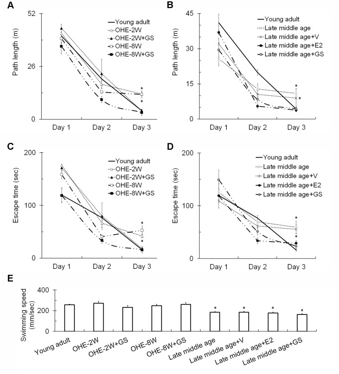
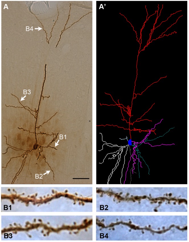
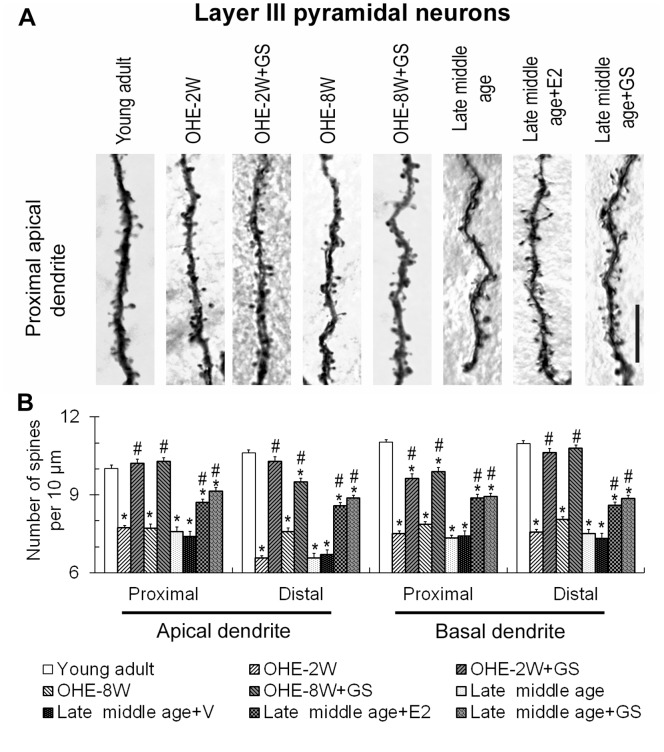
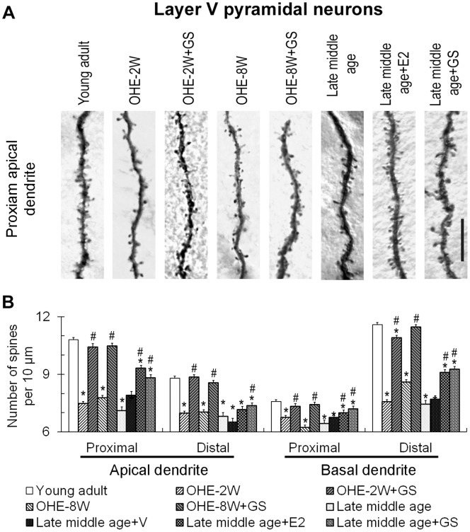
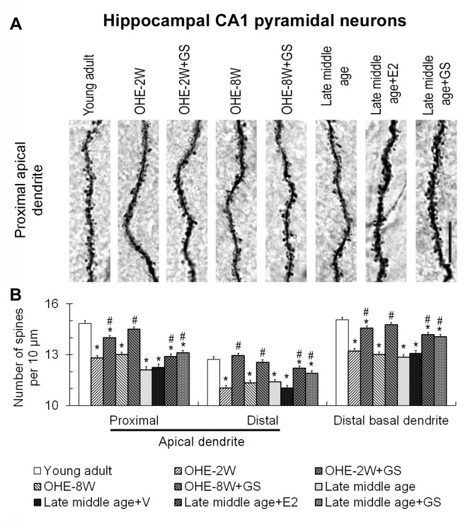
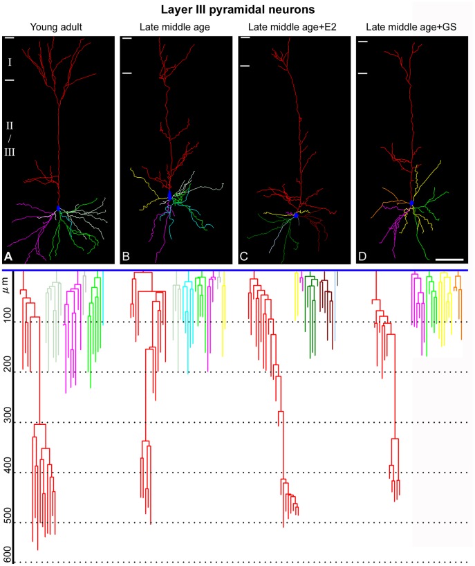
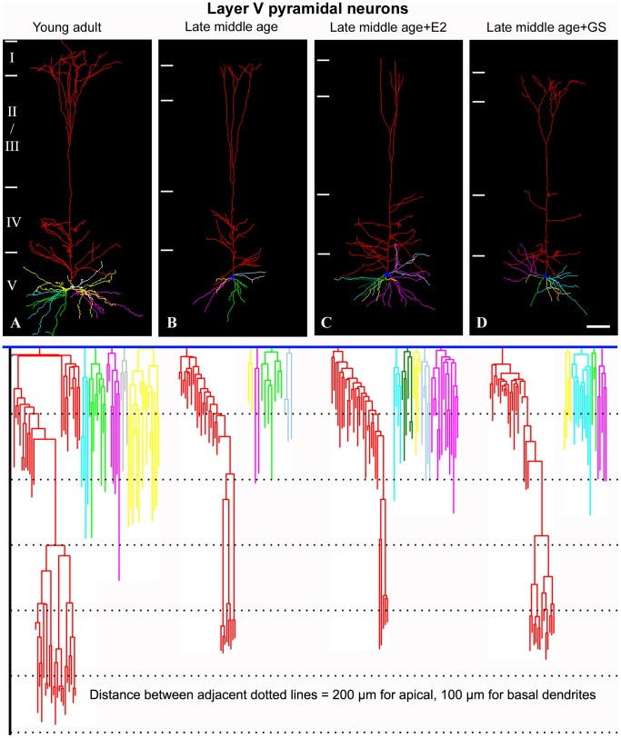
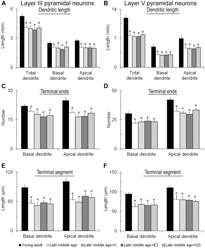
Gonadal hormones can modulate brain morphology and behavior. Recent studies have shown that hypogonadism could result in cortical function deficits. To this end, hormone therapy has been used to ease associated symptoms but the risk may outweigh the benefits. Here we explored whether genistein, a phytoestrogen, is effective in restoring the cognitive and central neuronal changes in late middle age and surgically estropause female rats. Both animal groups showed poorer spatial learning than young adults. The dendritic arbors and spines of the somatosensory cortical and CA1 hippocampal pyramidal neurons were revealed with intracellular dye injection and analyzed. The results showed that dendritic spines on these neurons were significantly decreased. Remarkably, genistein treatment rescued spatial learning deficits and restored the spine density on all neurons in the surgically estropause young females. In late middle age females, genistein was as effective as estradiol in restoring spines; however, the recovery was less thorough than on young OHE rats. Neither genistein nor estradiol rectified the shortened dendritic arbors of the aging cortical pyramidal neurons suggesting that dendritic arbors and spines are differently modulated. Thus, genistein could work at central level to restore excitatory connectivity and appears to be potent alternative to estradiol for easing aging and menopausal syndromes.
Conflict of interest statement
Figures
Similar articles
-
Acute effects of 17 β-estradiol and genistein on insulin sensitivity and spatial memory in aged ovariectomized female rats.Age (Dordr). 2010 Dec;32(4):421-34. doi: 10.1007/s11357-010-9148-6. Epub 2010 May 14. Age (Dordr). 2010. PMID: 20467821 Free PMC article.
-
Reproductive experience modified dendritic spines on cortical pyramidal neurons to enhance sensory perception and spatial learning in rats.Exp Anim. 2017 Jan 27;66(1):61-74. doi: 10.1538/expanim.16-0061. Epub 2016 Oct 25. Exp Anim. 2017. PMID: 27784858 Free PMC article.
-
Exogenous dehydroisoandrosterone sulfate reverses the dendritic changes of the central neurons in aging male rats.Exp Gerontol. 2014 Sep;57:191-202. doi: 10.1016/j.exger.2014.06.010. Epub 2014 Jun 12. Exp Gerontol. 2014. PMID: 24929010
-
Testosterone modulation of dendritic spines of somatosensory cortical pyramidal neurons.Brain Struct Funct. 2013 Nov;218(6):1407-17. doi: 10.1007/s00429-012-0465-7. Epub 2013 Jan 23. Brain Struct Funct. 2013. PMID: 23340667
-
Effects of estrogens and the phytoestrogen genistein on adipogenesis and lipogenesis in males and females.Birth Defects Res A Clin Mol Teratol. 2005 Jul;73(7):472-3. doi: 10.1002/bdra.20142. Birth Defects Res A Clin Mol Teratol. 2005. PMID: 15959885 Review. No abstract available.
Cited by
-
The effects of estrogen depletion in female rats: differential influences on somato-motor and sensory cortices.Biogerontology. 2025 Jan 20;26(1):41. doi: 10.1007/s10522-025-10186-2. Biogerontology. 2025. PMID: 39832048
-
Genistein suppresses the mitochondrial apoptotic pathway in hippocampal neurons in rats with Alzheimer's disease.Neural Regen Res. 2016 Jul;11(7):1153-8. doi: 10.4103/1673-5374.187056. Neural Regen Res. 2016. PMID: 27630702 Free PMC article.
-
Neuromodulating roles of estrogen and phytoestrogens in cognitive therapeutics through epigenetic modifications during aging.Front Aging Neurosci. 2022 Aug 3;14:945076. doi: 10.3389/fnagi.2022.945076. eCollection 2022. Front Aging Neurosci. 2022. PMID: 35992599 Free PMC article. Review.
-
Hyaluronic Acid Conjugated with 17β-Estradiol Effectively Alleviates Estropause-Induced Cognitive Deficits in Rats.Int J Mol Sci. 2023 Oct 25;24(21):15569. doi: 10.3390/ijms242115569. Int J Mol Sci. 2023. PMID: 37958552 Free PMC article.
-
Roles of genistein in learning and memory during aging and neurological disorders.Biogerontology. 2023 Jun;24(3):329-346. doi: 10.1007/s10522-023-10020-7. Epub 2023 Feb 25. Biogerontology. 2023. PMID: 36828983 Review.
References
-
- Horner CH (1993) Plasticity of the dendritic spine. Prog Neurobiol 41: 281–321. - PubMed
-
- Chen JR, Yan YT, Wang TJ, Chen LJ, Wang YJ, et al. (2009) Gonadal hormones modulate the dendritic spine densities of primary cortical pyramidal neurons in adult female rat. Cereb Cortex 19: 2719–2727. - PubMed
-
- Burke SN, Barnes CA (2006) Neural plasticity in the ageing brain. Nat Rev Neurosci 7: 30–40. - PubMed
-
- Wallace M, Frankfurt M, Arellanos A, Inagaki T, Luine V (2007) Impaired recognition memory and decreased prefrontal cortex spine density in aged female rats. Ann N Y Acad Sci 1097: 54–57. - PubMed
Publication types
MeSH terms
Substances
LinkOut - more resources
Full Text Sources
Other Literature Sources
Medical
Miscellaneous
